MAT1A/MAT2A Polyclonal antibody
MAT1A/MAT2A Polyclonal Antibody for IHC, WB, ELISA
Host / Isotype
Rabbit / IgG
Reactivity
human, mouse, rat, arabidopsis
Applications
WB, IHC, ELISA
Conjugate
Unconjugated
Cat no : 12395-1-AP
Synonyms
Validation Data Gallery
 with plant leaf tissue samples.")
 at dilution of 1:1000 incubated at room temperature for 1.5 hours.")
 at dilution of 1:10000 incubated at room temperature for 1.5 hours.")
 at dilution of 1:500 incubated at room temperature for 1.5 hours.")
 at dilution of 1:1000 incubated at room temperature for 1.5 hours.")
 at dilution of 1:500 incubated at room temperature for 1.5 hours.")
 at dilution of 1:200 (under 40x lens). Heat mediated antigen retrieval with Tris-EDTA buffer (pH 9.0).")
Tested Applications
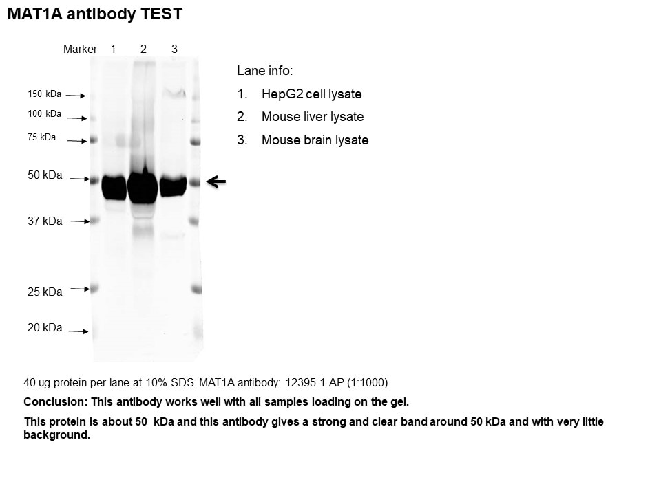
| Positive WB detected in | mouse liver tissue, HepG2 cells, HCT 116 cells, arabidopsis whole plant tissue, rat liver tissue |
| Positive IHC detected in | mouse liver tissue Note: suggested antigen retrieval with TE buffer pH 9.0; (*) Alternatively, antigen retrieval may be performed with citrate buffer pH 6.0 |
Recommended dilution
| Application | Dilution |
|---|---|
| Western Blot (WB) | WB : 1:1000-1:4000 |
| Immunohistochemistry (IHC) | IHC : 1:50-1:500 |
| It is recommended that this reagent should be titrated in each testing system to obtain optimal results. | |
| Sample-dependent, Check data in validation data gallery. | |
Published Applications
| WB | See 5 publications below |
| IHC | See 1 publications below |
Product Information
12395-1-AP targets MAT1A/MAT2A in WB, IHC, ELISA applications and shows reactivity with human, mouse, rat, arabidopsis samples.
| Tested Reactivity | human, mouse, rat, arabidopsis |
| Cited Reactivity | human, mouse, rat |
| Host / Isotype | Rabbit / IgG |
| Class | Polyclonal |
| Type | Antibody |
| Immunogen | MAT1A/MAT2A fusion protein Ag3076 |
| Full Name | methionine adenosyltransferase I, alpha |
| Calculated Molecular Weight | 395 aa, 44 kDa |
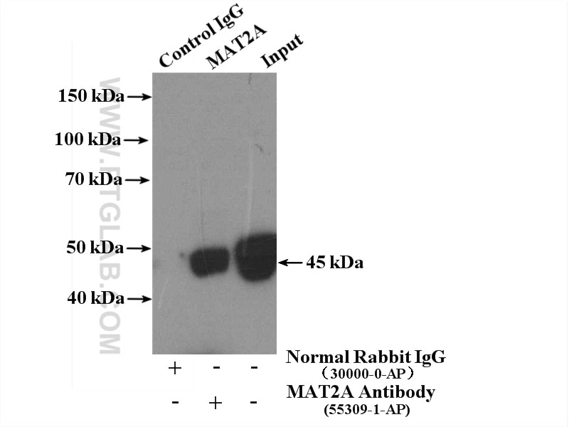
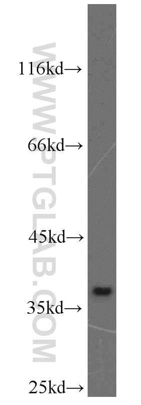
| Observed Molecular Weight | 44-55 kDa |
| GenBank Accession Number | BC018359 |
| Gene Symbol | MAT1A |
| Gene ID (NCBI) | 4143 |
| RRID | AB_2266186 |
| Conjugate | Unconjugated |
| Form | Liquid |
| Purification Method | Antigen affinity purification |
| Storage Buffer | PBS with 0.02% sodium azide and 50% glycerol pH 7.3. |
| Storage Conditions | Store at -20°C. Stable for one year after shipment. Aliquoting is unnecessary for -20oC storage. 20ul sizes contain 0.1% BSA. |
Background Information
Methionine adenosyltransferase (MAT) is the enzyme that catalyzes the synthesis of S-adeno-sylmethionine (AdoMet), the main donor of methyl groups in the cell. In mammals MAT is encoded by two different genes, MAT1A and MAT2A. MAT1A is expressed only in the mature liver whereas fetal hepatocytes, extrahepatic tissues and liver cancer cells express MAT2A. This antibody was raised against the full length of human MAT1A and can recognize both of MAT1A and MAT2A. Western blot analysis using this antibody detected a single band around 44-50 kDa in liver.
Protocols
| Product Specific Protocols | |
|---|---|
| WB protocol for MAT1A/MAT2A antibody 12395-1-AP | Download protocol |
| IHC protocol for MAT1A/MAT2A antibody 12395-1-AP | Download protocol |
| Standard Protocols | |
|---|---|
| Click here to view our Standard Protocols |
Publications
| Species | Application | Title |
|---|---|---|
Am J Pathol Forced expression of methionine adenosyltransferase 1A in human hepatoma cells suppresses in vivo tumorigenicity in mice. | ||
Toxicol Sci Metabolomic Analysis Reveals Metabolic Changes Caused By Bisphenol A in Rats. | ||
Proteomics Early changes in the liver-soluble proteome from mice fed a nonalcoholic steatohepatitis inducing diet. | ||
J Surg Oncol Histological expression of methionine adenosyl transferase (MAT) 2A as a post-surgical prognostic surrogate in patients with hepatocellular carcinoma. |
Reviews
The reviews below have been submitted by verified Proteintech customers who received an incentive forproviding their feedback.
FH Hua (Verified Customer) (03-26-2021) | Great antibody
 |