Kidney Cancer
Investigate Kidney Cancer in depth with our broad range of antibodies and IHC kits
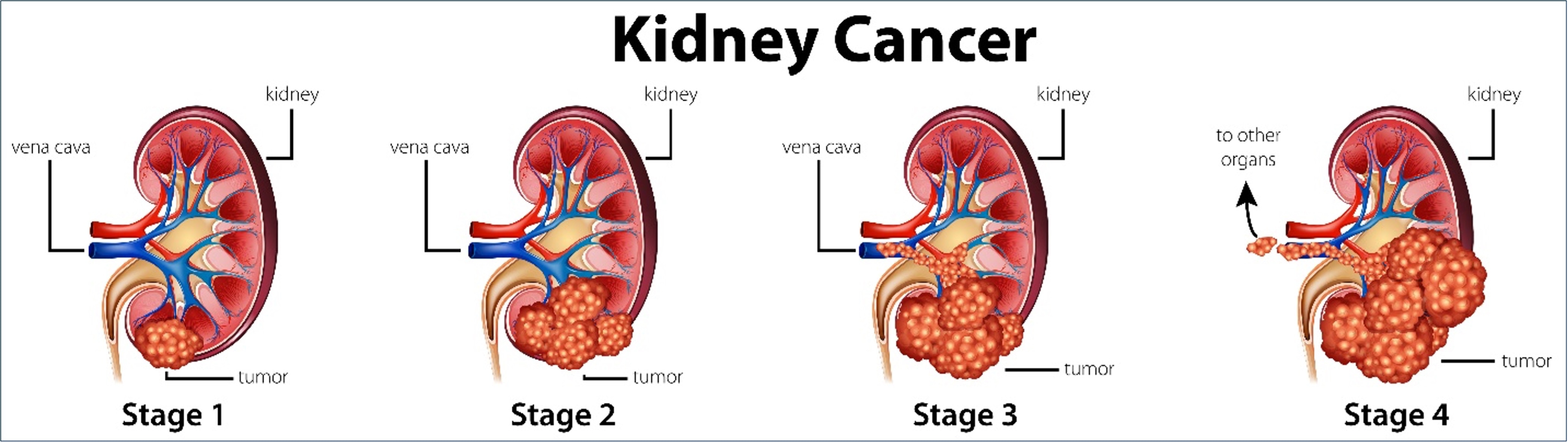
Kidney cancer is the 8th most common form of cancer in the US, with approximately 80,000 new cases diagnosed each year. There are several forms of kidney cancer; renal cell carcinoma (RCC) is the most common. RCC is known for being highly resistant to radiation and chemotherapy. Surgery is usually the best method used to stop tumor progression. To aid researchers studying kidney cancer, Proteintech offers numerous IHC-compatible antibodies and several ready-to-use IHC kits to detect critical biomarkers in kidney tissue samples.
Featured Markers for Kidney Cancer Detection
PAX8
Paired box (PAX) transcription factors typically regulate renal organogenesis and are expressed abundantly in adult kidney tissue. As cancers arising in these areas begin to metastasize, the detection of PAX8 expression in other parts of the body can help to identify these distant tumor growths as renal in origin. Proteintech’s PAX8 antibody has been cited over 450 times, more than any other PAX8 antibody on the market.
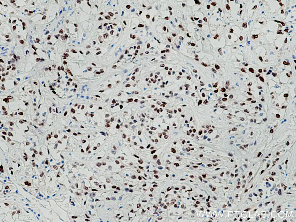
Immunohistochemical analysis of paraffin-embedded human renal cell carcinoma tissue slide using 10336-1-AP (PAX8 antibody) at dilution of 1:5000 (under 10x lens).
CA9
Carbonic anhydrase 9 (CA9) is a transmembrane protein that helps cells to maintain neutral pH levels by catalyzing a reversible reaction which coverts carbon-dioxides and water into bicarbonates and protons. It tends to be overexpressed in renal cell carcinomas as it allows these cells to survive in unfavorable microenvironments. CA9 antibodies can be used to distinguish clear cell renal carcinoma (ccRCC) from other renal carcinoma subtypes because only ccRCC cells will stain positive for this marker.
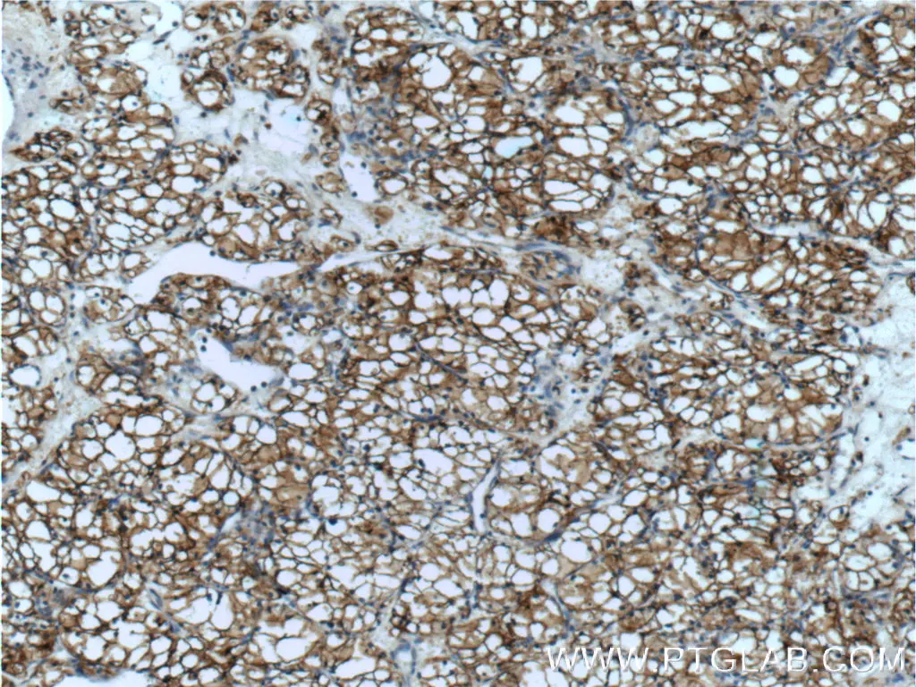
Immunohistochemical analysis of paraffin-embedded human renal cell carcinoma tissue slide using 11071-1-AP (CA9 antibody) at dilution of 1:50 (under 10x lens).
NRF2
Nuclear factor erythroid 2-related factor 2 (NRF2) protects cells from oxidative stress by neutralizing reactive oxygen species (ROS) production and inducing a strong, downstream antioxidant response. It may also be involved in other cellular processes including DNA repair, angiogenesis, and acquired chemoresistance. Many anticancer drugs tend to increase ROS production to kill cancer cells. Hence high levels of NRF2 expression are not only a way to detect renal cell carcinoma, but also a useful indicator of drug resistance. Proteintech’s NRF2 antibody has been cited 1,000 times, far more than any other NRF2 antibody on the market.
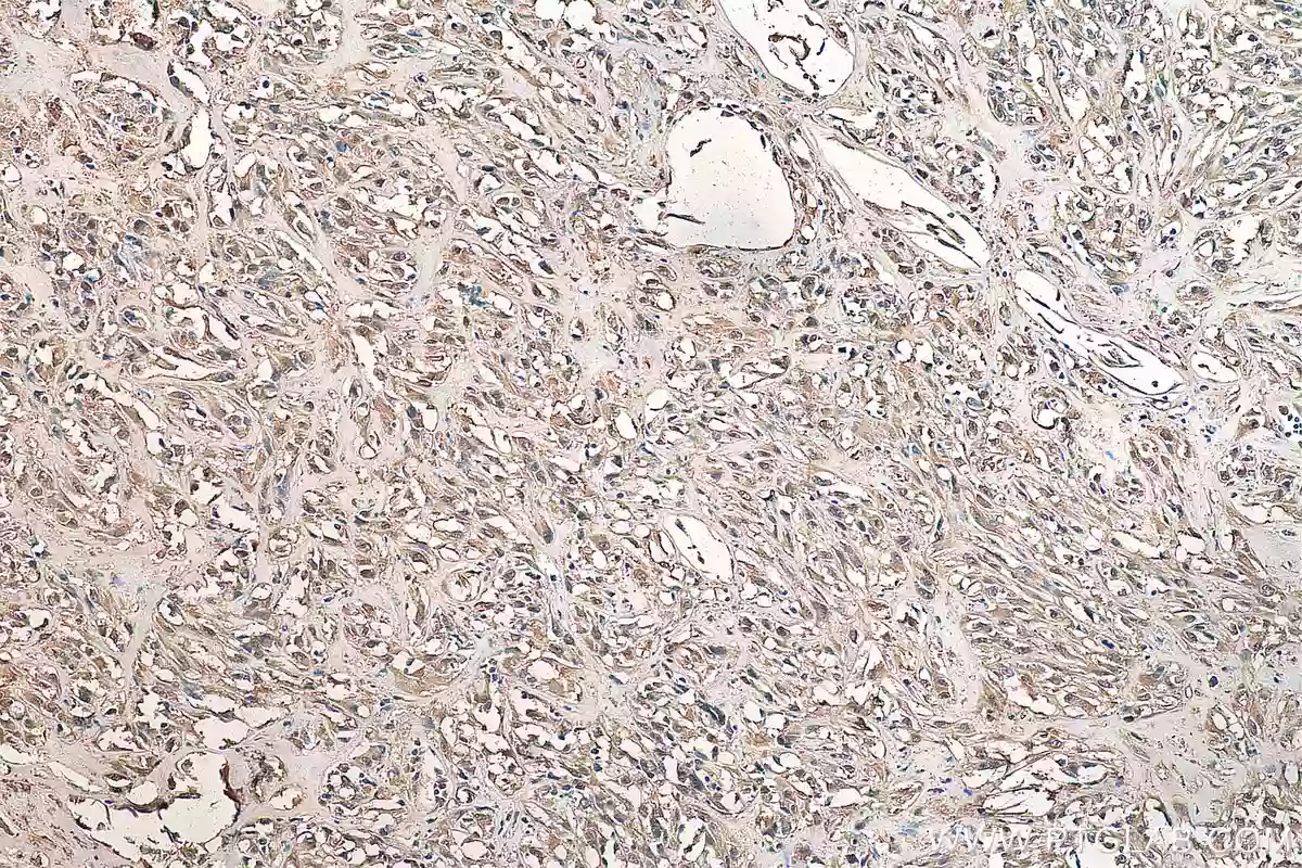
Immunohistochemical analysis of paraffin-embedded human renal cell carcinoma tissue slide using 16396-1-AP (NRF2, NFE2L2 antibody) at dilution of 1:200 (under 10x lens).
Antibodies for Kidney Cancer Research
|
Function |
Marker |
PTG Catalog |
|
Angiogenesis |
19003-1-AP |
|
|
Cell Proliferation |
20960-1-AP |
|
|
18262-1-AP |
||
|
Cell Survival |
11486-2-AP |
|
|
11071-1-AP |
||
|
16225-1-AP |
||
|
16396-1-AP |
||
|
10508-1-AP |
||
|
10037-1-Ig |
||
|
Differentiation |
18008-1-AP |
|
|
18696-1-AP |
||
|
17513-1-AP |
||
|
23614-1-AP |
||
|
26056-1-AP |
||
|
10336-1-AP |
||
|
18150-1-AP |
||
|
10366-1-AP |
IHC Kits for Kidney Cancer Research
|
Function |
Marker |
PTG Catalog |
|
Angiogenesis |
KHC0039 |
|
|
Cell Proliferation |
KHC0190 |
|
|
Cell Survival |
KHC0223 |
|
|
KHC0187 |
||
|
KHC0434 |
||
|
KHC0646 |
||
|
Differentiation |
KHC0386 |
|
|
KHC0204 |
||
|
KHC0544 |
||
|
Metastasis |
KHC0094 |
|
|
KHC0038 |
||
|
KHC0087 |
||
|
KHC0039 |
References
Barr, M. L., Jilaveanu, L. B., Camp, R. L., Adeniran, A. J., Kluger, H. M., & Shuch, B. (2014). PAX-8 expression in renal tumours and distant sites: A useful marker of primary and metastatic renal cell carcinoma? Journal of Clinical Pathology, 68(1), 12–17. https://doi.org/10.1136/jclinpath-2014-202259
Farber, N. J., Kim, C. J., Modi, P. K., Hon, J. D., Sadimin, E. T., & Singer, E. A. (2017). Renal cell carcinoma: the search for a reliable biomarker. Translational Cancer Research, 6(3), 620–632. https://doi.org/10.21037/tcr.2017.05.19
Gudas, L. J., Fu, L., Minton, D. R., Mongan, N. P., & Nanus, D. M. (2014). The role of HIF1α in renal cell carcinoma tumorigenesis. Journal of Molecular Medicine, 92(8), 825–836. https://doi.org/10.1007/s00109-014-1180-z
Kidney Cancer | CDC. (n.d.). https://www.cdc.gov/cancer/kidney/index.htm
Li, F., Aljahdali, I. a. M., Zhang, R., Nastiuk, K. L., Krolewski, J. J., & Ling, X. (2021). Kidney cancer biomarkers and targets for therapeutics: survivin (BIRC5), XIAP, MCL-1, HIF1α, HIF2α, NRF2, MDM2, MDM4, p53, KRAS and AKT in renal cell carcinoma. Journal of Experimental &Amp; Clinical Cancer Research, 40(1). https://doi.org/10.1186/s13046-021-02026-1
Related Content
Cancer stem cells as a key to cure cancer
Molecular markers for liver cancer

Support
Newsletter Signup
Stay up-to-date with our latest news and events. New to Proteintech? Get 10% off your first order when you sign up.