- Featured Product
- KD/KO Validated
COX4I2 Polyclonal antibody
COX4I2 Polyclonal Antibody for IF, IHC, IP, WB, ELISA
Host / Isotype
Rabbit / IgG
Reactivity
human, mouse, rat
Applications
WB, IP, IHC, IF, ELISA
Conjugate
Unconjugated
Cat no : 11463-1-AP
Synonyms
"COX4I2 Antibodies" Comparison
View side-by-side comparison of COX4I2 antibodies from other vendors to find the one that best suits your research needs.
Tested Applications
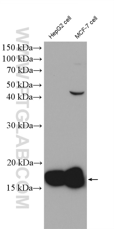
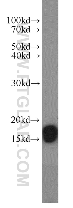
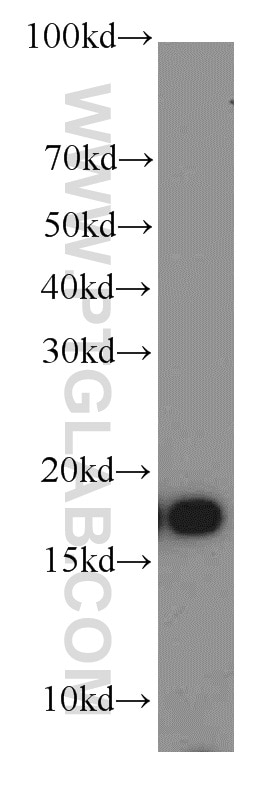
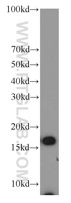
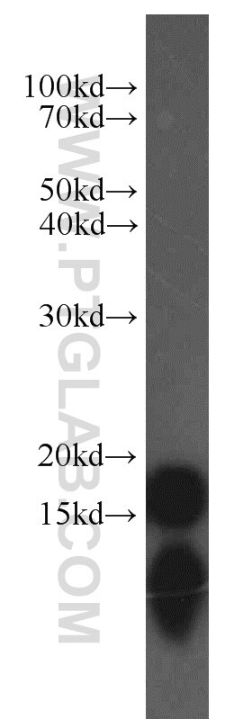
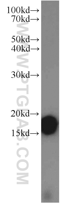
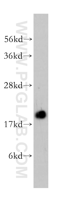
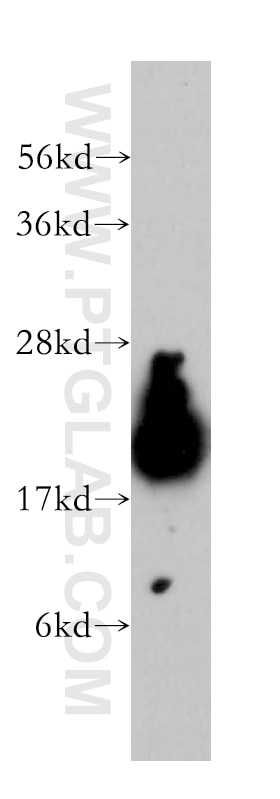
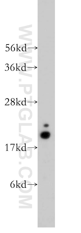
| Positive WB detected in | HepG2 cells, human brain tissue, MCF-7 cells, human heart tissue, human lung tissue, A375 cells |
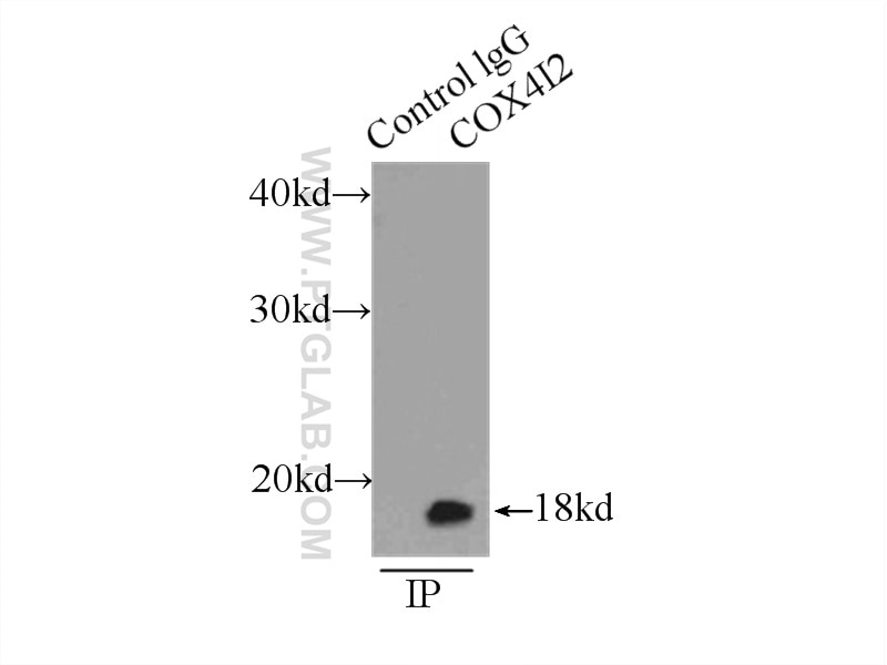
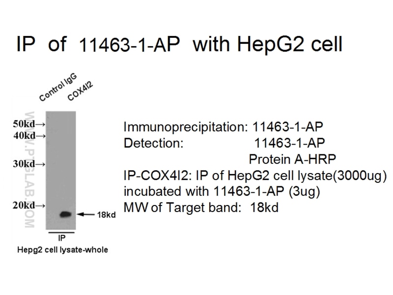
| Positive IP detected in | HepG2 cells |
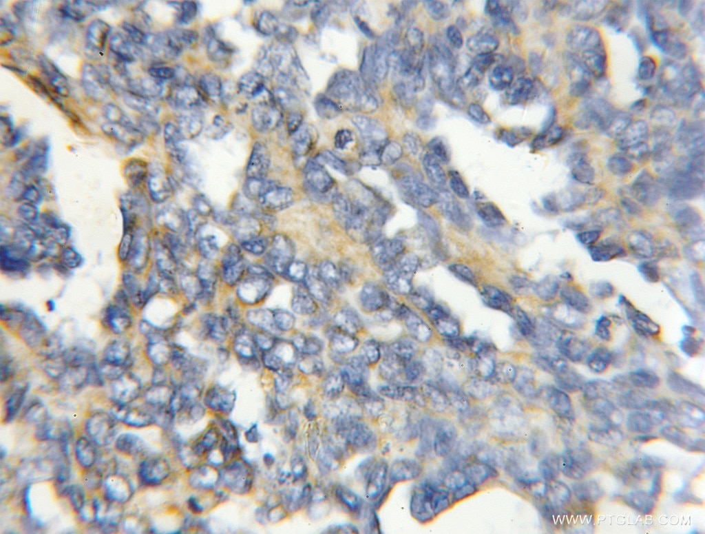
| Positive IHC detected in | human ovary tumor tissue Note: suggested antigen retrieval with TE buffer pH 9.0; (*) Alternatively, antigen retrieval may be performed with citrate buffer pH 6.0 |
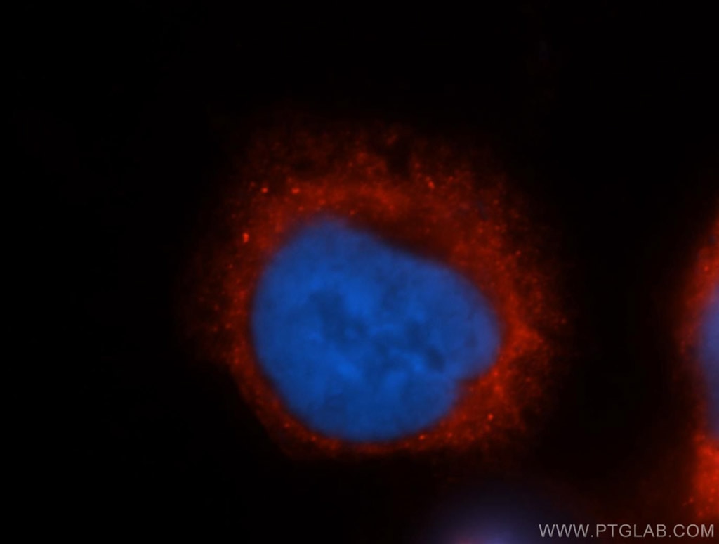
| Positive IF detected in | Hela cells |
Recommended dilution
| Application | Dilution |
|---|---|
| Western Blot (WB) | WB : 1:500-1:3000 |
| Immunoprecipitation (IP) | IP : 0.5-4.0 ug for 1.0-3.0 mg of total protein lysate |
| Immunohistochemistry (IHC) | IHC : 1:20-1:200 |
| Immunofluorescence (IF) | IF : 1:20-1:200 |
| It is recommended that this reagent should be titrated in each testing system to obtain optimal results. | |
| Sample-dependent, Check data in validation data gallery. | |
Published Applications
| KD/KO | See 3 publications below |
| WB | See 11 publications below |
| IHC | See 3 publications below |
| IF | See 4 publications below |
Product Information
11463-1-AP targets COX4I2 in WB, IP, IHC, IF, ELISA applications and shows reactivity with human, mouse, rat samples.
| Tested Reactivity | human, mouse, rat |
| Cited Reactivity | human, mouse, rat |
| Host / Isotype | Rabbit / IgG |
| Class | Polyclonal |
| Type | Antibody |
| Immunogen | COX4I2 fusion protein Ag1987 |
| Full Name | cytochrome c oxidase subunit IV isoform 2 (lung) |
| Calculated Molecular Weight | 171 aa, 20 kDa |
| Observed Molecular Weight | 17-18 kDa |
| GenBank Accession Number | BC057779 |
| Gene Symbol | COX4I2 |
| Gene ID (NCBI) | 84701 |
| RRID | AB_2085287 |
| Conjugate | Unconjugated |
| Form | Liquid |
| Purification Method | Antigen affinity purification |
| Storage Buffer | PBS with 0.02% sodium azide and 50% glycerol pH 7.3. |
| Storage Conditions | Store at -20°C. Stable for one year after shipment. Aliquoting is unnecessary for -20oC storage. 20ul sizes contain 0.1% BSA. |
Background Information
COX4I2, also named as COX4L2 and COXIV-2, belongs to the cytochrome c oxidase IV family. It is one of the nuclear-coded polypeptide chains of cytochrome c oxidase, the terminal oxidase in mitochondrial electron transport. COXIV(cytochrome c oxidase IV) has two isoforms (isoform 1 and 2). Isoform 1(COX4I1) is ubiquitously expressed and isoform 2 is highly expressed in lung tissues. This antibody was generated against full length COX4I2 protein.
Protocols
| Product Specific Protocols | |
|---|---|
| WB protocol for COX4I2 antibody 11463-1-AP | Download protocol |
| IHC protocol for COX4I2 antibody 11463-1-AP | Download protocol |
| IF protocol for COX4I2 antibody 11463-1-AP | Download protocol |
| IP protocol for COX4I2 antibody 11463-1-AP | Download protocol |
| Standard Protocols | |
|---|---|
| Click here to view our Standard Protocols |
Publications
| Species | Application | Title |
|---|---|---|
Cell Rep Defective Mitochondrial Cardiolipin Remodeling Dampens HIF-1α Expression in Hypoxia. | ||
Oncotarget Nuclear-encoded cytochrome c oxidase subunit 4 regulates BMI1 expression and determines proliferative capacity of high-grade gliomas.
| ||
Sci Signal Acute O2 sensing through HIF2α-dependent expression of atypical cytochrome oxidase subunits in arterial chemoreceptors.
| ||
Free Radic Biol Med Cytochrome c oxidase mediates labile iron level and radioresistance in glioblastoma. | ||
Bioconjug Chem GnRH-Gemcitabine Conjugates for the Treatment of Androgen-Independent Prostate Cancer: Pharmacokinetic Enhancements Combined with Targeted Drug Delivery. | ||
Free Radic Biol Med Differentiation of SH-SY5Y cells to a neuronal phenotype changes cellular bioenergetics and the response to oxidative stress. |
