HPSE Polyclonal antibody
HPSE Polyclonal Antibody for IF, IHC, WB, ELISA
Host / Isotype
Rabbit / IgG
Reactivity
human, mouse and More (1)
Applications
WB, IHC, IF, ELISA
Conjugate
Unconjugated
Cat no : 24529-1-AP
Synonyms
Validation Data Gallery
 at dilution of 1:4000 incubated at room temperature for 1.5 hours.")
 at dilution of 1:50.")
 at dilution of 1:50.")
 at dilution of 1:50.")
 at dilution of 1:50.")
 at dilution of 1:25 and Rhodamine-Goat anti-Rabbit IgG.")
 at dilution of 1:25 and Rhodamine-Goat anti-Rabbit IgG.")
Tested Applications
| Positive WB detected in | HeLa cells, HepG2 cells, Jurkat cells, mouse liver tissue |
| Positive IHC detected in | human liver cancer tissue, human placenta tissue Note: suggested antigen retrieval with TE buffer pH 9.0; (*) Alternatively, antigen retrieval may be performed with citrate buffer pH 6.0 |
| Positive IF detected in | HeLa cells, HepG2 cells |
Recommended dilution
| Application | Dilution |
|---|---|
| Western Blot (WB) | WB : 1:1000-1:8000 |
| Immunohistochemistry (IHC) | IHC : 1:20-1:200 |
| Immunofluorescence (IF) | IF : 1:10-1:100 |
| It is recommended that this reagent should be titrated in each testing system to obtain optimal results. | |
| Sample-dependent, Check data in validation data gallery. | |
Published Applications
| WB | See 4 publications below |
| IHC | See 4 publications below |
Product Information
24529-1-AP targets HPSE in WB, IHC, IF, ELISA applications and shows reactivity with human, mouse samples.
| Tested Reactivity | human, mouse |
| Cited Reactivity | human, mouse, rat |
| Host / Isotype | Rabbit / IgG |
| Class | Polyclonal |
| Type | Antibody |
| Immunogen | HPSE fusion protein Ag21479 |
| Full Name | heparanase |
| Calculated Molecular Weight | 543 aa, 61 kDa |
| Observed Molecular Weight | 60 kDa |
| GenBank Accession Number | BC051321 |
| Gene Symbol | HPSE |
| Gene ID (NCBI) | 10855 |
| RRID | AB_2879591 |
| Conjugate | Unconjugated |
| Form | Liquid |
| Purification Method | Antigen affinity purification |
| Storage Buffer | PBS with 0.02% sodium azide and 50% glycerol pH 7.3. |
| Storage Conditions | Store at -20°C. Stable for one year after shipment. Aliquoting is unnecessary for -20oC storage. 20ul sizes contain 0.1% BSA. |
Background Information
HPSE(Heparanase) is also named as HEP, HPA, HPA1, HPR1, HPSE1, HSE1 and belongs to the glycosyl hydrolase 79 family. It is a endoglycosidase that cleaves heparan sulfate proteoglycans (HSPGs) into heparan sulfate side chains and core proteoglycans. HPSE is essential in the disassembly of the extracellular matrix (ECM) by invading cells. It has 3 isoforms produced by alternative splicing with the molecular weight of 61 kDa, 55 kDa and 53 kDa. The full length protein has six glycosylation sites. The cleavage of the 65 kDa form leads to the generation of a linker peptide, and 8 kDa and 50 kDa products. The active form, the 8/50 kDa heterodimer, is resistant to degradation and glycosylation of the 50 kDa subunit appears to be essential for its solubility.
Protocols
| Product Specific Protocols | |
|---|---|
| WB protocol for HPSE antibody 24529-1-AP | Download protocol |
| IHC protocol for HPSE antibody 24529-1-AP | Download protocol |
| IF protocol for HPSE antibody 24529-1-AP | Download protocol |
| Standard Protocols | |
|---|---|
| Click here to view our Standard Protocols |
Publications
| Species | Application | Title |
|---|---|---|
J Control Release cRGD-targeted heparin nanoparticles for effective dual drug treatment of cisplatin-resistant ovarian cancer | ||
EMBO Rep CircHIPK3 sponges miR-558 to suppress heparanase expression in bladder cancer cells. | ||
Br J Cancer Shed Syndecan-1 is involved in chemotherapy resistance via the EGFR pathway in colorectal cancer. | ||
Neural Regen Res Reducing LncRNA-5657 expression inhibits the brain inflammatory reaction in septic rats. | ||
Diabetes Res Clin Pract Heparanase-driven inflammation from the AGEs-stimulated macrophages changes the functions of glomerular endothelial cells. | ||
Exp Ther Med MicroRNA-219a-2-3p modulates the proliferation of thyroid cancer cells via the HPSE/cyclin D1 pathway. |
Reviews
The reviews below have been submitted by verified Proteintech customers who received an incentive forproviding their feedback.
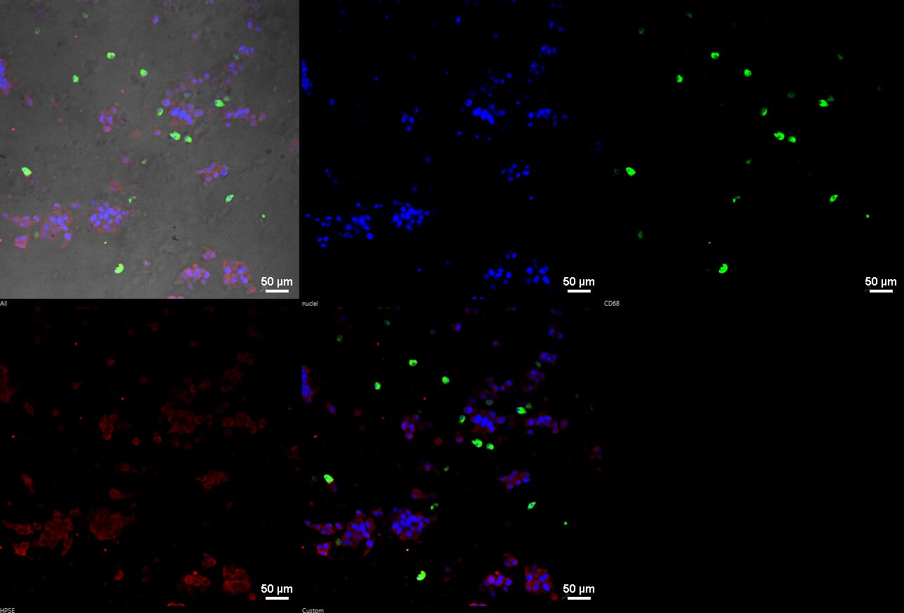
FH Fabio Henrique (Verified Customer) (01-17-2019) | Figure 1. IF of HPSE (red) and CD68 (green) in 3D coculture of C4-2B and primary human macrophages. C4-2B and primary human macrophages were encapsulated in a 3D hydrogel, composed of hyaluronic acid and collagen type 1, and cocultured for 5 days. On day 5, cells were fixed in 4% paraformaldehyde for 10 minutes, washed with PBSTritonX 0.3% for 3x 5min and blocked in PBS 10% Goat serum, 1% BSA, for 1 hr at RT. Anti-HPSE was used at 1:100 in blocking buffer, overnight incubation at 4*C. Secondary antibodies AlexaFluor 488 and 568 were used at 1:500 for 1 hr at RT. Imaged using a Nikon A1 confocal microscope. Figure 2: HPSE (red) and CD68 (green) in Grade 3 Endometrial cancer tissue (Frozen). Staining was performed as described above, except secondary antibodies were used at 1:1000. This antibody did not perform well in WB (data not shown).
 |

