PAX3 Polyclonal antibody
PAX3 Polyclonal Antibody for IHC, WB, ELISA
Host / Isotype
Rabbit / IgG
Reactivity
human, mouse
Applications
WB, IHC, ELISA
Conjugate
Unconjugated
Cat no : 51036-2-AP
Synonyms
Validation Data Gallery
 at dilution of 1:300 incubated at room temperature for 1.5 hours.")
 at dilution of 1:300 incubated at room temperature for 1.5 hours.")
 at dilution of 1:300 incubated at room temperature for 1.5 hours.")
 at dilution of 1:300 incubated at room temperature for 1.5 hours.")
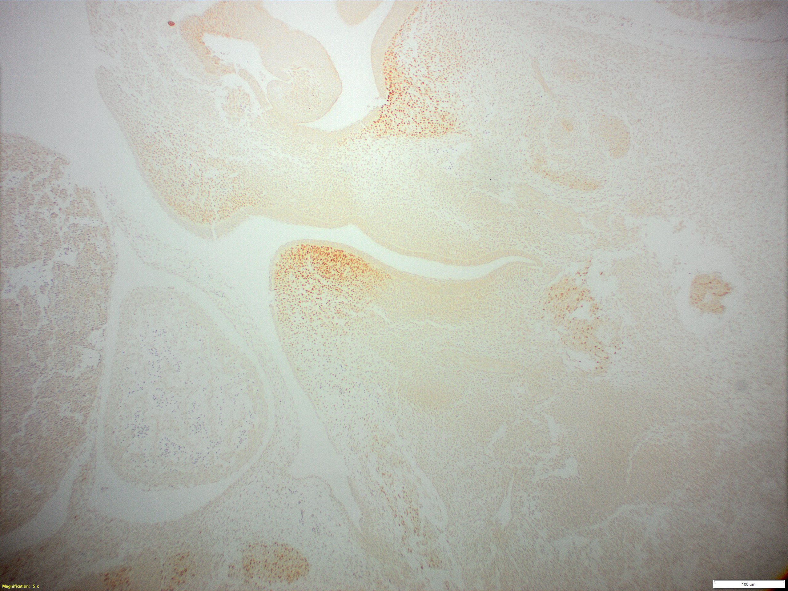
 at dilution of 1:800 (under 10x lens). Heat mediated antigen retrieval with Tris-EDTA buffer (pH 9.0).")
 at dilution of 1:800 (under 40x lens). Heat mediated antigen retrieval with Tris-EDTA buffer (pH 9.0).")
Tested Applications
| Positive WB detected in | mouse skin tissue, A375 cells, HepG2 cells, K-562 cells |
| Positive IHC detected in | rat brain tissue Note: suggested antigen retrieval with TE buffer pH 9.0; (*) Alternatively, antigen retrieval may be performed with citrate buffer pH 6.0 |
Recommended dilution
| Application | Dilution |
|---|---|
| Western Blot (WB) | WB : 1:200-1:1000 |
| Immunohistochemistry (IHC) | IHC : 1:400-1:1600 |
| It is recommended that this reagent should be titrated in each testing system to obtain optimal results. | |
| Sample-dependent, Check data in validation data gallery. | |
Published Applications
| WB | See 2 publications below |
Product Information
51036-2-AP targets PAX3 in WB, IHC, ELISA applications and shows reactivity with human, mouse samples.
| Tested Reactivity | human, mouse |
| Cited Reactivity | mouse |
| Host / Isotype | Rabbit / IgG |
| Class | Polyclonal |
| Type | Antibody |
| Immunogen | PAX3 fusion protein Ag0477 |
| Full Name | paired box 3 |
| Calculated Molecular Weight | 120 kDa |
| Observed Molecular Weight | 56 kDa |
| GenBank Accession Number | BC114363 |
| Gene Symbol | PAX3 |
| Gene ID (NCBI) | 5077 |
| RRID | AB_670479 |
| Conjugate | Unconjugated |
| Form | Liquid |
| Purification Method | Antigen affinity purification |
| Storage Buffer | PBS with 0.02% sodium azide and 50% glycerol pH 7.3. |
| Storage Conditions | Store at -20°C. Stable for one year after shipment. Aliquoting is unnecessary for -20oC storage. 20ul sizes contain 0.1% BSA. |
Background Information
PAX3, a transcription factor and multifunctional regulatory protein, is normally expressed during embryonic development. In the nervous system, PAX3 is involved in neural tube closure, neural crest development, and peripheral neuron differentiation. In the present study, PAX3 was reported as a novel regulator of GFAP transcription, and the overexpression and suppression of PAX3 could inhibit and promote NSC differentiation, respectively. In muscle development, PAX3 ensures the survival of myogenic progenitor cells with Pax3-expressing progenitors giving rise to both skeletal and smooth muscle cells. PAX3 also has a well-established role in the development of melanocytes during embryogenesis, and has recently been characterized as a molecular switch in the mature melanocyte. Mutations in PAX3 can cause Waardenburg syndrome.
Protocols
| Product Specific Protocols | |
|---|---|
| WB protocol for PAX3 antibody 51036-2-AP | Download protocol |
| IHC protocol for PAX3 antibody 51036-2-AP | Download protocol |
| Standard Protocols | |
|---|---|
| Click here to view our Standard Protocols |
Publications
| Species | Application | Title |
|---|---|---|
Food Funct N-3 polyunsaturated fatty acids effectively protect against neural tube defects in diabetic mice induced by streptozotocin. | ||
J Agric Food Chem Synergistic Effects of Folic Acid and n-3 Polyunsaturated Fatty Acid in Preventing Neural Tube Defects in Diabetic Mice |
Reviews
The reviews below have been submitted by verified Proteintech customers who received an incentive forproviding their feedback.
FH Kim (Verified Customer) (12-03-2019) | Tissue fixed in neutral buffered formalin, 24h, 4degC. Paraffin embedded. Citrate pH6.0/0.05% Tween antigen retrieval, 20 min ~90degC. Polymer-HRP secondary. No counterstain. DAB detection.Photos are: snout/tongue and back.
 |

