- Featured Product
- KD/KO Validated
RAB7A Polyclonal antibody
RAB7A Polyclonal Antibody for FC, IHC, IP, WB, ELISA
Host / Isotype
Rabbit / IgG
Reactivity
human, mouse, rat and More (3)
Applications
WB, IP, IHC, FC, ELISA
Conjugate
Unconjugated
Cat no : 55469-1-AP
Synonyms
Validation Data Gallery
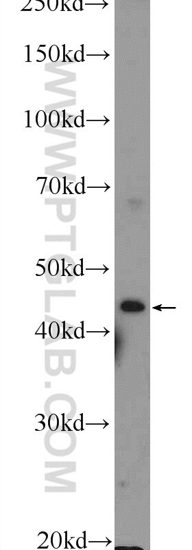
 at dilution of 1:2000 incubated at room temperature for 1.5 hours.")
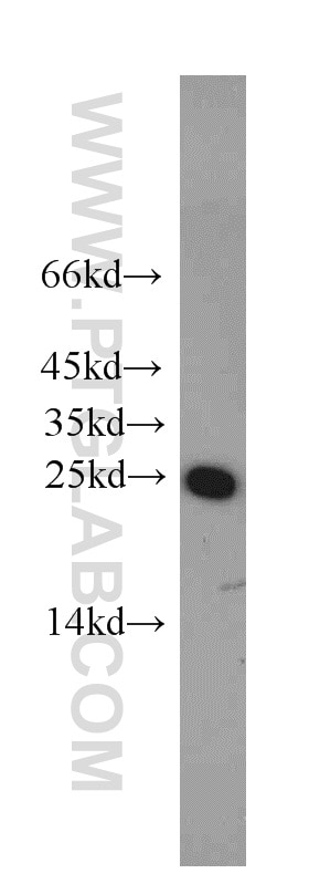
 at dilution of 1:800 incubated at room temperature for 1.5 hours.")
 at dilution of 1:800 incubated at room temperature for 1.5 hours.")
 at dilution of 1:300 incubated at room temperature for 1.5 hours.")
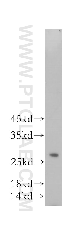
 with SH-SY5Y cells lysate 3360 ug.")
 at dilution of 1:200 (under 10x lens. Heat mediated antigen retrieval with Tris-EDTA buffer (pH 9.0).")
 at dilution of 1:200 (under 40x lens. Heat mediated antigen retrieval with Tris-EDTA buffer (pH 9.0).")
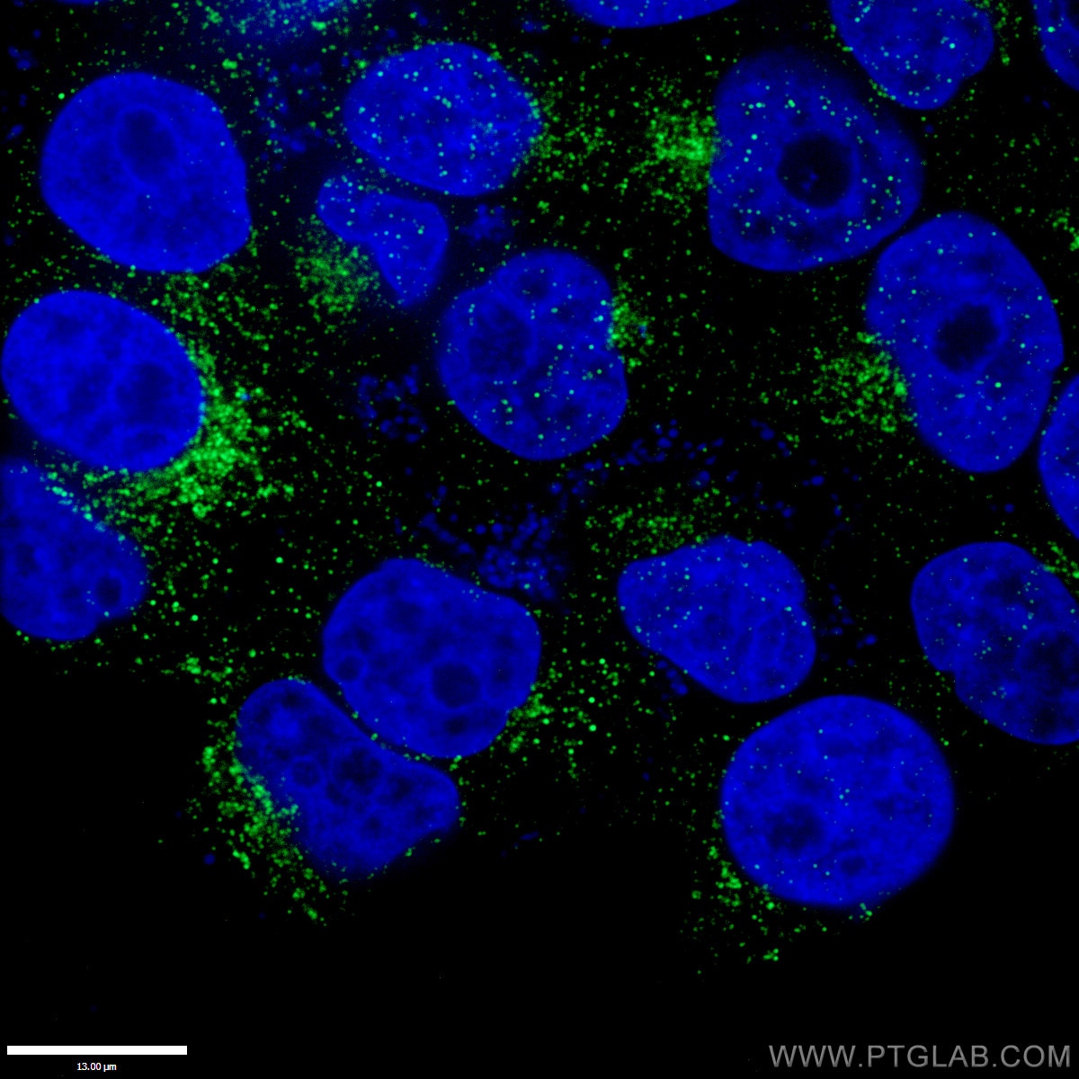
 at dilution of 1:50 (under 10x lens).")
 at dilution of 1:50 (under 40x lens).")
 at dilution of 1:50 (under 10x lens).")
 at dilution of 1:50 (under 40x lens).")
 and CoraLite®488-Conjugated AffiniPure Goat Anti-Rabbit IgG(H+L) at dilution 1:1000 (red), or 0.4 ug Control Antibody. Cells were fixed with 4% PFA and permeabilized with Flow Cytometry Perm Buffer (PF00011-C).")
Tested Applications
| Positive WB detected in | L-929 cells, NIH/3T3 cells, A431 cells, SH-SY5Y cells, RAW 264.7 cells, C6 cells, mouse brain tissue, rat brain tissue |
| Positive IHC detected in | human kidney tissue, human skeletal muscle tissue, human stomach tissue Note: suggested antigen retrieval with TE buffer pH 9.0; (*) Alternatively, antigen retrieval may be performed with citrate buffer pH 6.0 |
| Positive FC detected in | A431 cells |
Recommended dilution
| Application | Dilution |
|---|---|
| Western Blot (WB) | WB : 1:1000-1:4000 |
| Immunohistochemistry (IHC) | IHC : 1:50-1:500 |
| Flow Cytometry (FC) | FC : 0.40 ug per 10^6 cells in a 100 µl suspension |
| It is recommended that this reagent should be titrated in each testing system to obtain optimal results. | |
| Sample-dependent, Check data in validation data gallery. | |
Published Applications
| KD/KO | See 4 publications below |
| WB | See 32 publications below |
| IHC | See 2 publications below |
| IP | See 1 publications below |
Product Information
55469-1-AP targets RAB7A in WB, IP, IHC, FC, ELISA applications and shows reactivity with human, mouse, rat samples.
| Tested Reactivity | human, mouse, rat |
| Cited Reactivity | human, mouse, rat, monkey, Zebrafish, pig |
| Host / Isotype | Rabbit / IgG |
| Class | Polyclonal |
| Type | Antibody |
| Immunogen | Peptide |
| Full Name | RAB7A, member RAS oncogene family |
| Calculated Molecular Weight | 23 kDa |
| Observed Molecular Weight | 23 kDa |
| GenBank Accession Number | NM_004637 |
| Gene Symbol | RAB7A |
| Gene ID (NCBI) | 7879 |
| RRID | AB_11182831 |
| Conjugate | Unconjugated |
| Form | Liquid |
| Purification Method | Antigen affinity purification |
| Storage Buffer | PBS with 0.02% sodium azide and 50% glycerol pH 7.3. |
| Storage Conditions | Store at -20°C. Stable for one year after shipment. Aliquoting is unnecessary for -20oC storage. 20ul sizes contain 0.1% BSA. |
Background Information
RAB7A, also named as RAB7, belongs to the small GTPase superfamily and Rab family. It is involved in late endocytic transport. RAB7A contributes to the maturation of phagosomes (acidification). Defects in RAB7A are the cause of Charcot-Marie-Tooth disease type 2B (CMT2B). This antibody is specific to RAB7A.
Protocols
| Product Specific Protocols | |
|---|---|
| WB protocol for RAB7A antibody 55469-1-AP | Download protocol |
| IHC protocol for RAB7A antibody 55469-1-AP | Download protocol |
| IP protocol for RAB7A antibody 55469-1-AP | Download protocol |
| Standard Protocols | |
|---|---|
| Click here to view our Standard Protocols |
Publications
| Species | Application | Title |
|---|---|---|
Nat Commun C9orf72-catalyzed GTP loading of Rab39A enables HOPS-mediated membrane tethering and fusion in mammalian autophagy | ||
Autophagy SDC1-dependent TGM2 determines radiosensitivity in glioblastoma by coordinating EPG5-mediated fusion of autophagosomes with lysosomes | ||
Nat Commun Dstyk mutation leads to congenital scoliosis-like vertebral malformations in zebrafish via dysregulated mTORC1/TFEB pathway. | ||
Reviews
The reviews below have been submitted by verified Proteintech customers who received an incentive forproviding their feedback.
FH Sophy (Verified Customer) (03-07-2023) | The antibody worked well in western with 1:1000 dilution. The signal was somewhat weak but that could also be due to the cell types that were used.
|
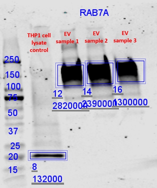
FH Malgorzata (Verified Customer) (08-17-2022) | The antibody worked on my positive control: Lysate of THP1 cell line. Nevertheless, for my samples it gave strong nonspecific signal in high MW
 |
FH Azita (Verified Customer) (06-17-2021) | Immunocytochemistry labelling of (4% PFA) fixed NSC-34 cells by RAB7A Polyclonal antibody at dilution of 1:50 showed strong labelling.
|
FH Laura (Verified Customer) (01-15-2020) | Works well for Western Blot at 1:1000 dilution.
|
FH Aamir (Verified Customer) (01-08-2020) | WB at 1 in 500IF at 1 in 250
|
FH Benjamin (Verified Customer) (12-10-2019) | Has worked well multiple times with dilution of 1:1000 with the RAB7A antibody. I always incubate at 4 degrees overnight in 3% milk TBST solution.
 |