TBP Monoclonal antibody
TBP Monoclonal Antibody for FC, IF, IHC, IP, WB, ELISA
Host / Isotype
Mouse / IgG2a
Reactivity
human, mouse, rat, pig
Applications
WB, IP, IHC, IF, FC, ELISA
Conjugate
Unconjugated
CloneNo.
2H3B2
Cat no : 66166-1-Ig
Synonyms
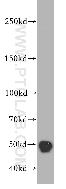
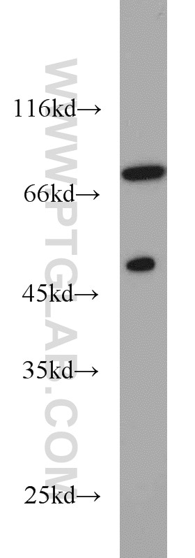
 at dilution of 1:200000 incubated at room temperature for 1.5 hours. The membrane was stripped and reblotted with HRP-conjugated Alpha Tubulin Monoclonal antibody (<a class='green' href='/productredirect?CatalogNo=HRP-66031' target='_blank'>HRP-66031</a>) as loading control.")
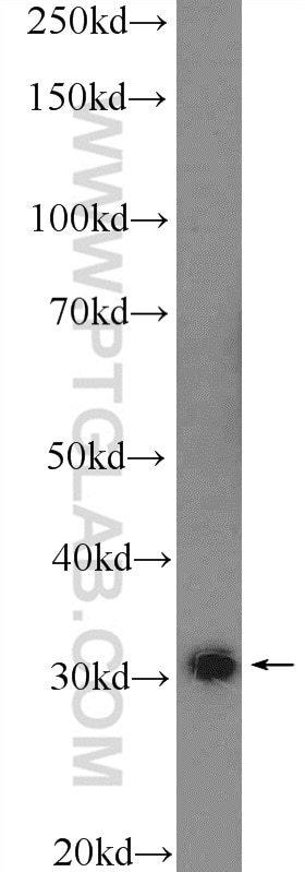
 at dilution of 1:200000 incubated at room temperature for 1.5 hours.")

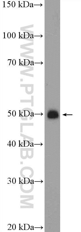
 at dilution of 1:5000 incubated at room temperature for 1.5 hours.")
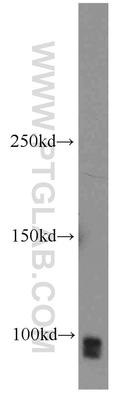
 with <a class='green' href='/productredirect?CatalogNo=HEK-293' target='_blank'>HEK-293</a> cells lysate 1800ug.")
 at dilution of 1:650 (under 40x lens). Heat mediated antigen retrieval with Tris-EDTA buffer (pH 9.0).")
 at dilution of 1:200 (under 10x lens).")
 at dilution of 1:200 (under 40x lens).")
 for 15 mins and 2M HCL treatment for 30mins, Immunohistochemistry of paraffin-embedded human breast cancer tissue slideusing 66166-1-Ig (TBP Antibody) at dilution of 1:400 (under 10x lens).")
 for 15 mins and 2M HCL treatment for 30mins, Immunohistochemistry of paraffin-embedded human breast cancer tissue slideusing 66166-1-Ig (TBP Antibody) at dilution of 1:400 (under 40x lens).")
 at dilution of 1:650 (under 10x lens). Heat mediated antigen retrieval with Tris-EDTA buffer (pH 9.0).")
 fixed A431 cells using TBP antibody (66166-1-Ig, Clone: 2H3B2 ) at dilution of 1:400 and CoraLite®488-Conjugated AffiniPure Goat Anti-Mouse IgG(H+L).")
 fixed HeLa cells using 66166-1-Ig(TBP antibody) at dilution of 1:100 and Alexa Fluor 488-conjugated AffiniPure Goat Anti-Mouse IgG(H+L).")
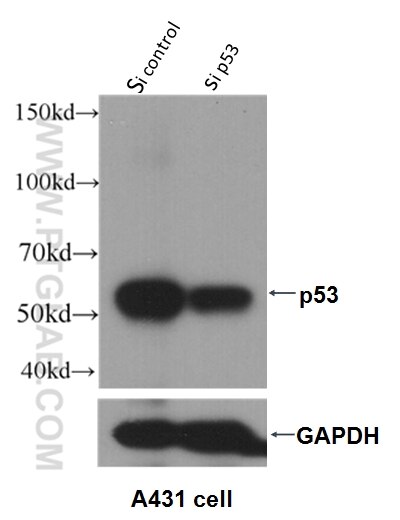
 and control antibody (blue). Fixed with 90% MeOH.")
"TBP Antibodies" Comparison
View side-by-side comparison of TBP antibodies from other vendors to find the one that best suits your research needs.
Tested Applications
| Positive WB detected in | LNCaP cells, HSC-T6 cells, HEK-293 cells, pig liver tissue, HeLa cells, HepG2 cells, Jurkat cells, K-562 cells, NIH/3T3 cells, Neuro-2a cells |
| Positive IP detected in | HEK-293 cells |
| Positive IHC detected in | human liver cancer tissue, human breast cancer tissue Note: suggested antigen retrieval with TE buffer pH 9.0; (*) Alternatively, antigen retrieval may be performed with citrate buffer pH 6.0 |
| Positive IF detected in | A431 cells, HeLa cells |
| Positive FC detected in | HepG2 cells |
Recommended dilution
| Application | Dilution |
|---|---|
| Western Blot (WB) | WB : 1:20000-1:100000 |
| Immunoprecipitation (IP) | IP : 0.5-4.0 ug for 1.0-3.0 mg of total protein lysate |
| Immunohistochemistry (IHC) | IHC : 1:325-1:1300 |
| Immunofluorescence (IF) | IF : 1:200-1:800 |
| Flow Cytometry (FC) | FC : 1:10-1:100 |
| It is recommended that this reagent should be titrated in each testing system to obtain optimal results. | |
| Sample-dependent, Check data in validation data gallery. | |
Published Applications
| WB | See 21 publications below |
| IF | See 1 publications below |
Product Information
66166-1-Ig targets TBP in WB, IP, IHC, IF, FC, ELISA applications and shows reactivity with human, mouse, rat, pig samples.
| Tested Reactivity | human, mouse, rat, pig |
| Cited Reactivity | human, mouse, rat, pig |
| Host / Isotype | Mouse / IgG2a |
| Class | Monoclonal |
| Type | Antibody |
| Immunogen | TBP fusion protein Ag12383 |
| Full Name | TATA box binding protein |
| Calculated Molecular Weight | 338 aa, 38 kDa |
| Observed Molecular Weight | mouse/rat 33-36 kDa and human 37-43kDa |
| GenBank Accession Number | BC110341 |
| Gene Symbol | TBP |
| Gene ID (NCBI) | 6908 |
| RRID | AB_2881562 |
| Conjugate | Unconjugated |
| Form | Liquid |
| Purification Method | Protein A purification |
| Storage Buffer | PBS with 0.02% sodium azide and 50% glycerol pH 7.3. |
| Storage Conditions | Store at -20°C. Stable for one year after shipment. Aliquoting is unnecessary for -20oC storage. 20ul sizes contain 0.1% BSA. |
Background Information
The TATA binding protein (TBP) is a transcription factor that binds specifically to a DNA sequence TATA box. This DNA sequence is found about 25-30 base pairs upstream of the transcription start site in some eukaryotic gene promoters. TBP, along with a variety of TBP-associated factors, make up the TFIID, a general transcription factor that in turn makes up part of the RNA polymerase II preinitiation complex. As one of the few proteins in the preinitation complex that binds DNA in a sequence-specific manner, it helps position RNA polymerase II over the transcription start site of the gene. However, it is estimated that only 10-20% of human promoters have TATA boxes. Therefore, TBP is probably not the only protein involved in positioning RNA polymerase II. This antibody detects human TBP (~40 kDa) and mouse/rat Tbp (~35 kDa).
Protocols
| Product Specific Protocols | |
|---|---|
| WB protocol for TBP antibody 66166-1-Ig | Download protocol |
| IHC protocol for TBP antibody 66166-1-Ig | Download protocol |
| IF protocol for TBP antibody 66166-1-Ig | Download protocol |
| IP protocol for TBP antibody 66166-1-Ig | Download protocol |
| FC protocol for TBP antibody 66166-1-Ig | Download protocol |
| Standard Protocols | |
|---|---|
| Click here to view our Standard Protocols |
Publications
| Species | Application | Title |
|---|---|---|
Genome Res Ligand-induced native G-quadruplex stabilization impairs transcription initiation. | ||
PLoS Biol Loss of adenomatous polyposis coli function renders intestinal epithelial cells resistant to the cytokine IL-22. | ||
Virulence Inhibition of cell proliferation by Tas of foamy viruses through cell cycle arrest or apoptosis underlines the different mechanisms of virus-host interactions. | ||
Biochem Pharmacol PPARα regulates the expression of human arylacetamide deacetylase involved in drug hydrolysis and lipid metabolism. | ||
Front Oncol TFEB Promotes Prostate Cancer Progression via Regulating ABCA2-Dependent Lysosomal Biogenesis. | ||
Mol Cancer Res Identification of Endogenous Adenomatous Polyposis Coli Interaction Partners and β-Catenin-Independent Targets by Proteomics. |