- Featured Product
- KD/KO Validated
TRIB3 Polyclonal antibody
TRIB3 Polyclonal Antibody for WB, ELISA
Host / Isotype
Rabbit / IgG
Reactivity
human and More (1)
Applications
WB,ELISA
Conjugate
Unconjugated
Cat no : 13300-1-AP
Synonyms
Validation Data Gallery
 at dilution of 1:1500 incubated at room temperature for 1.5 hours.")
 fixed HepG2 cells using 13300-1-AP (TRIB3 antibody), at dilution of 1:100 and CoraLite®488-Conjugated AffiniPure Goat Anti-Rabbit IgG(H+L). Phalloidine stains the F-actin.")
Tested Applications
| Positive WB detected in | L02 cells |
Recommended dilution
| Application | Dilution |
|---|---|
| Western Blot (WB) | WB : 1:500-1:3000 |
| It is recommended that this reagent should be titrated in each testing system to obtain optimal results. | |
| Sample-dependent, Check data in validation data gallery. | |
Published Applications
| KD/KO | See 7 publications below |
| WB | See 24 publications below |
| IHC | See 9 publications below |
| IF | See 4 publications below |
| IP | See 1 publications below |
Product Information
13300-1-AP targets TRIB3 in WB,ELISA applications and shows reactivity with human samples.
| Tested Reactivity | human |
| Cited Reactivity | human, monkey |
| Host / Isotype | Rabbit / IgG |
| Class | Polyclonal |
| Type | Antibody |
| Immunogen | TRIB3 fusion protein Ag4085 |
| Full Name | tribbles homolog 3 (Drosophila) |
| Calculated Molecular Weight | 358 aa, 40 kDa |
| Observed Molecular Weight | 40-45 kDa |
| GenBank Accession Number | BC027484 |
| Gene Symbol | TRIB3 |
| Gene ID (NCBI) | 57761 |
| RRID | AB_10641988 |
| Conjugate | Unconjugated |
| Form | Liquid |
| Purification Method | Antigen affinity purification |
| Storage Buffer | PBS with 0.02% sodium azide and 50% glycerol pH 7.3. |
| Storage Conditions | Store at -20°C. Stable for one year after shipment. Aliquoting is unnecessary for -20oC storage. 20ul sizes contain 0.1% BSA. |
Background Information
TRIB3 is a pseudokinase molecule that affects a number of cellular functions. TRIB3 has been reported to be highly activated in the presence of a variety of stressors, including the deprivation of neurotrophic factors, hypoxia, and ER stress.TRIB3 protein is associated with a good prognosis in human breast cancer patients, possibly due to the fact that TRIB3 is involved in hypoxia tolerance.(PMID: 21704407). TRIB3 protein predicted molecular weights is approximately 39~45 kDa.(PMID: 31191318, PMID: 24414038, PMID: 31578236).
Protocols
| Product Specific Protocols | |
|---|---|
| WB protocol for TRIB3 antibody 13300-1-AP | Download protocol |
| IF protocol for TRIB3 antibody 13300-1-AP | Download protocol |
| Standard Protocols | |
|---|---|
| Click here to view our Standard Protocols |
Publications
| Species | Application | Title |
|---|---|---|
J Cachexia Sarcopenia Muscle Sarcopenia is attenuated by TRB3 knockout in aging mice via the alleviation of atrophy and fibrosis of skeletal muscles. | ||
Aging Dis Age-Related Decline in Expression of Molecular Chaperones Induces Endoplasmic Reticulum Stress and Chondrocyte Apoptosis in Articular Cartilage. | ||
Autophagy Cannabidiol inhibits human glioma by induction of lethal mitophagy through activating TRPV4.
| ||
EMBO Rep Wnt regulates amino acid transporter Slc7a5 and so constrains the integrated stress response in mouse embryos. | ||
Oxid Med Cell Longev Hsa_circRNA_0008028 Deficiency Ameliorates High Glucose-Induced Proliferation, Calcification, and Autophagy of Vascular Smooth Muscle Cells via miR-182-5p/TRIB3 Axis
|
Reviews
The reviews below have been submitted by verified Proteintech customers who received an incentive forproviding their feedback.
FH Chun (Verified Customer) (12-05-2019) | a good antibody
|
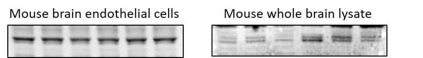
FH KARTHIKEYAN (Verified Customer) (11-20-2019) | I used this antibody in mouse whole brain lysate and in brain endothelial cells, in both the system they work really well. i would definitely recommend this product.
 |
FH Jose (Verified Customer) (11-02-2017) | Bright nuclear staining.
|

