
Tested Applications
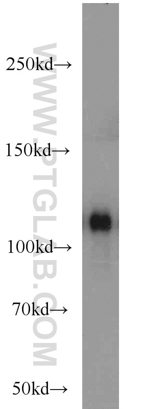
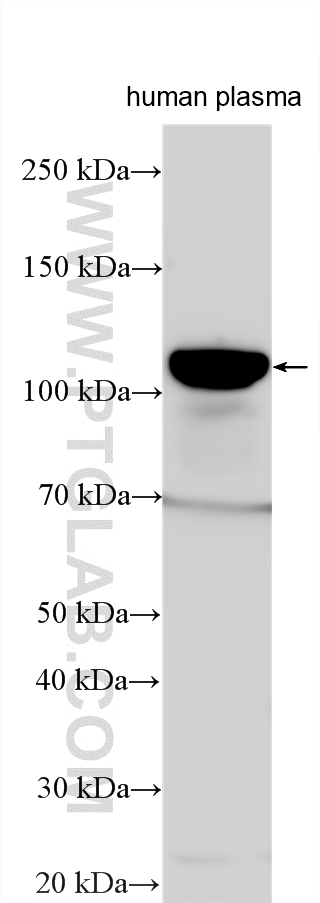
| Positive WB detected in | human plasma |
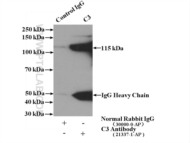
| Positive IP detected in | human plasma tissue |
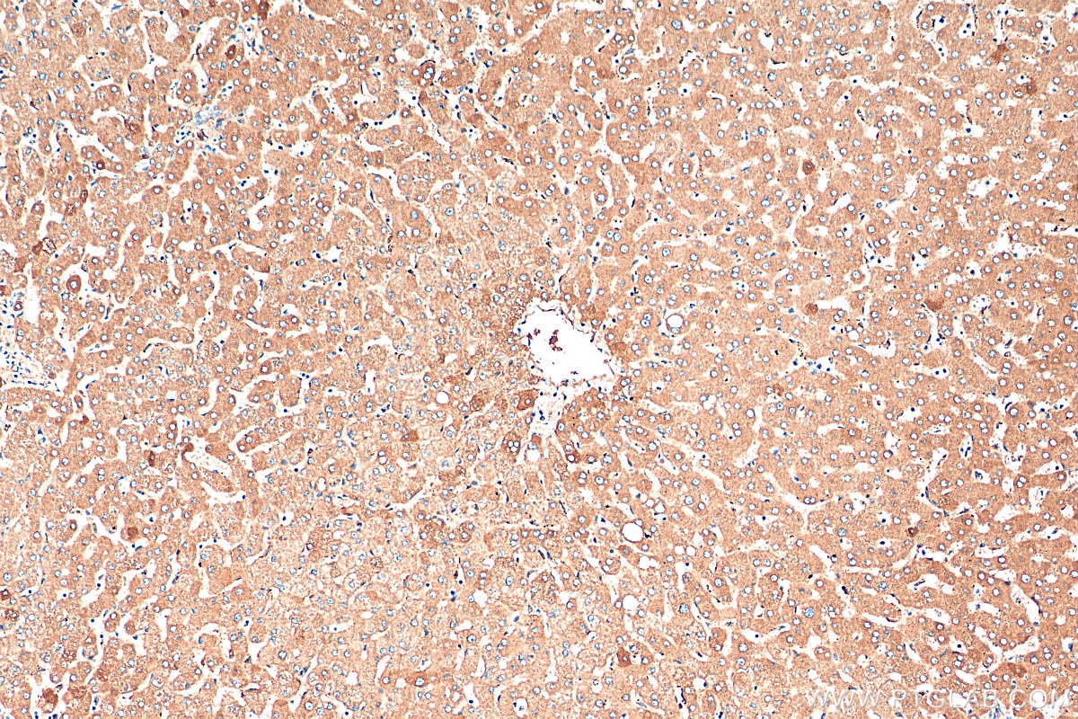
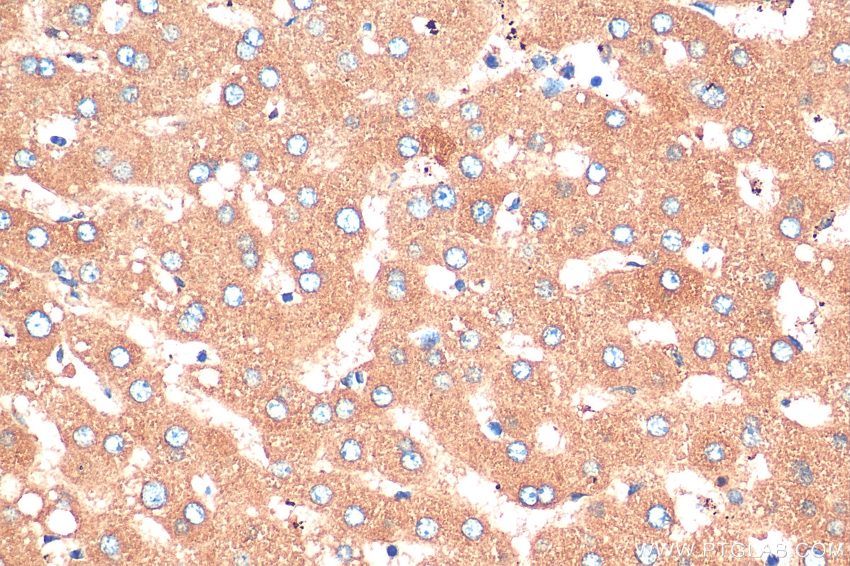
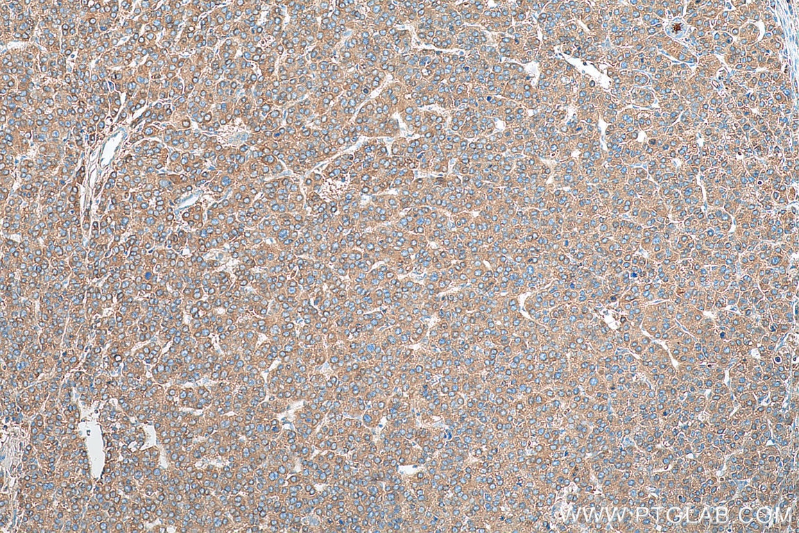
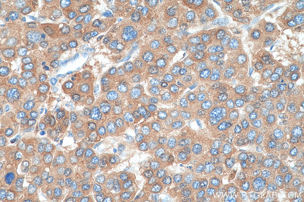
| Positive IHC detected in | human liver tissue, human liver cancer tissue Note: suggested antigen retrieval with TE buffer pH 9.0; (*) Alternatively, antigen retrieval may be performed with citrate buffer pH 6.0 |
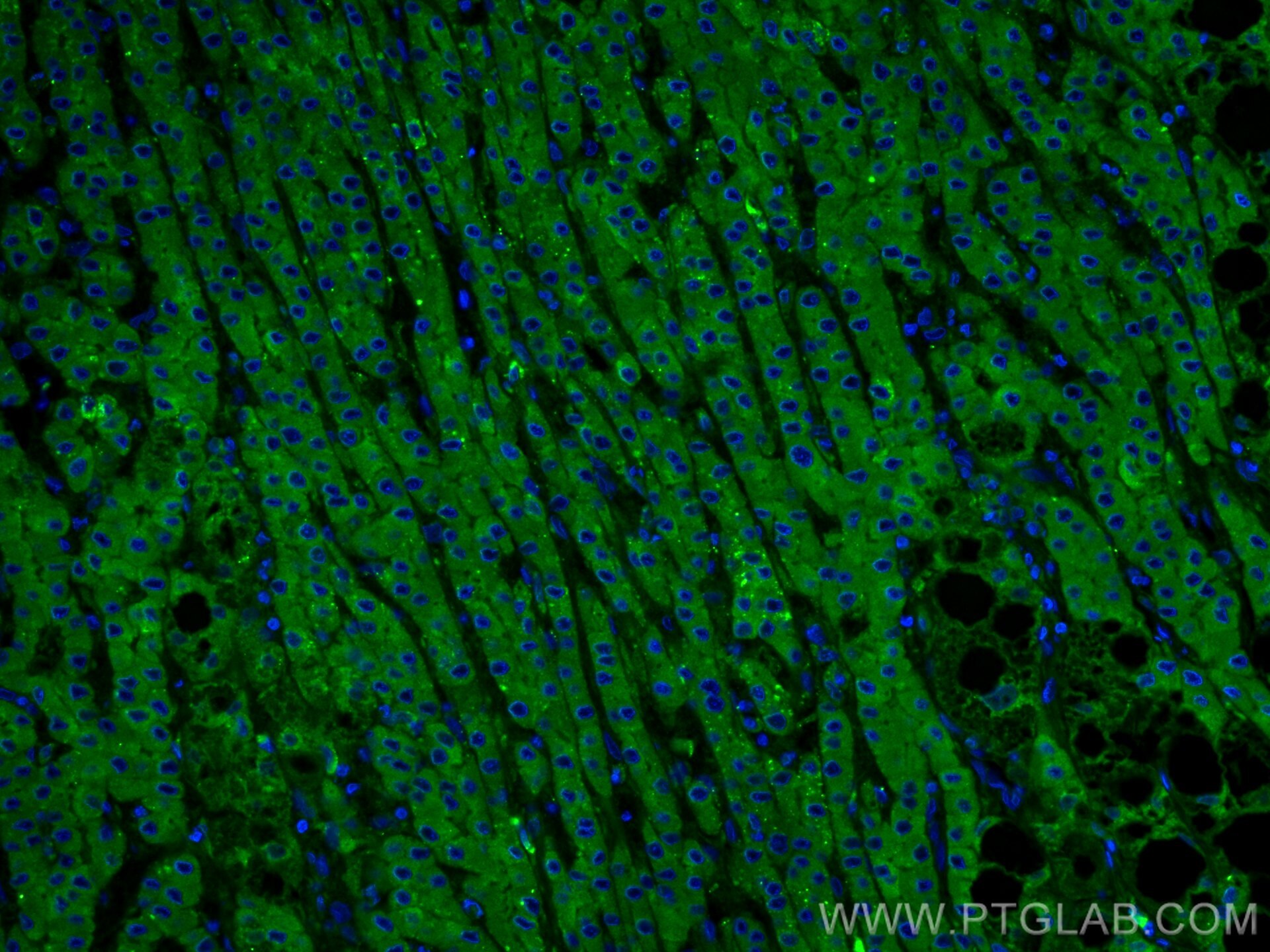
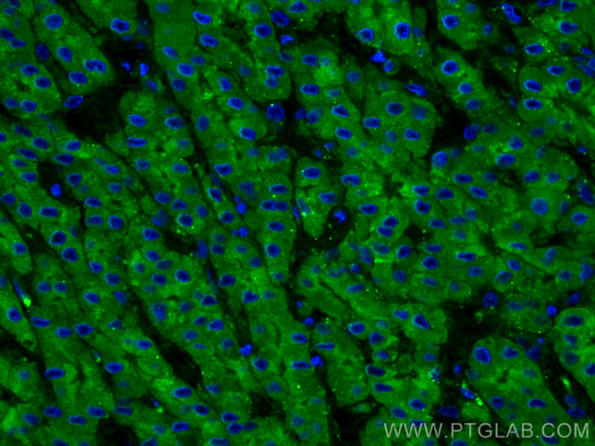
| Positive IF-P detected in | human liver cancer tissue |
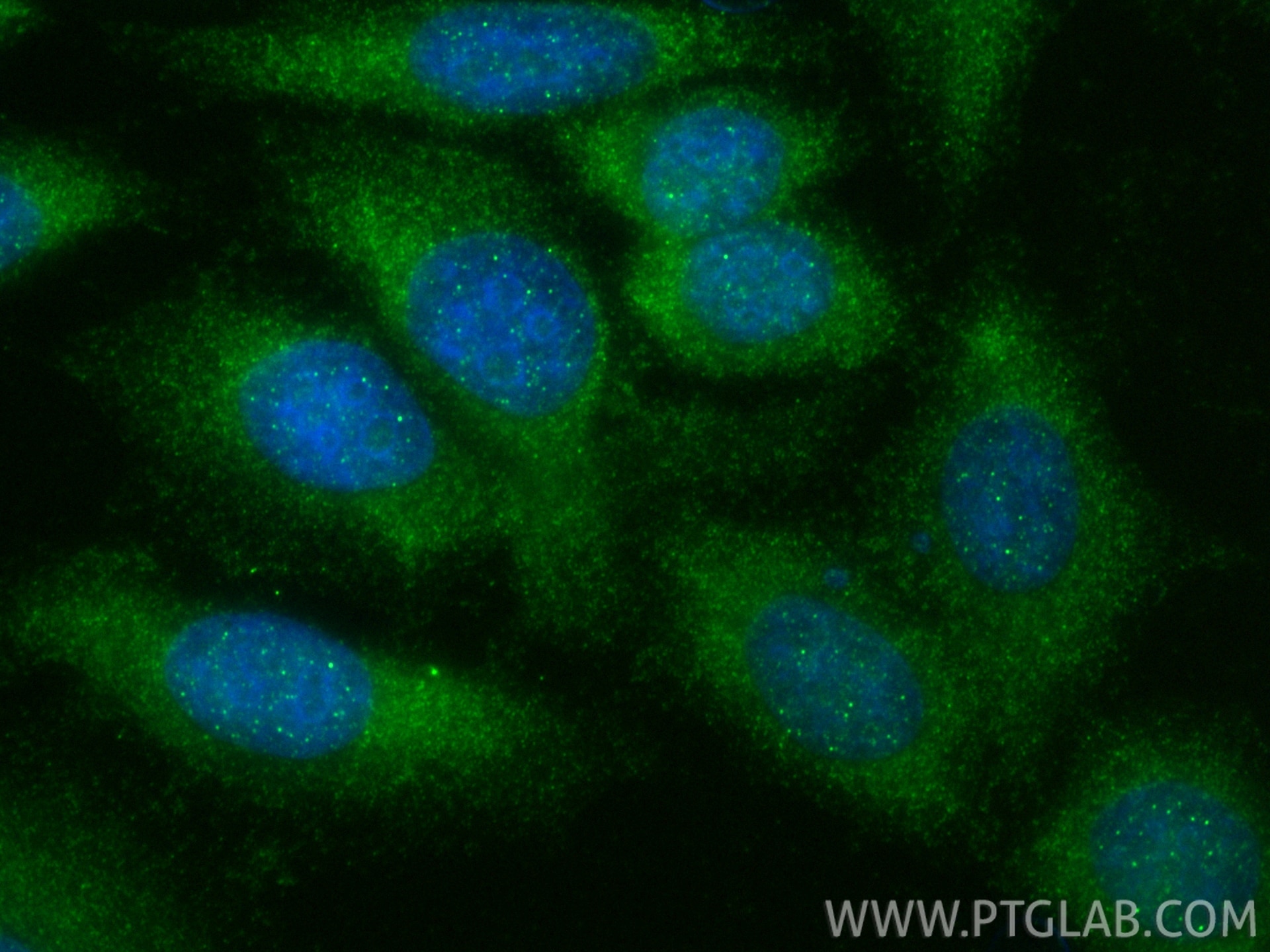
| Positive IF/ICC detected in | HepG2 cells |
Recommended dilution
| Application | Dilution |
|---|---|
| Western Blot (WB) | WB : 1:20000-1:100000 |
| Immunoprecipitation (IP) | IP : 0.5-4.0 ug for 1.0-3.0 mg of total protein lysate |
| Immunohistochemistry (IHC) | IHC : 1:1000-1:4000 |
| Immunofluorescence (IF)-P | IF-P : 1:50-1:500 |
| Immunofluorescence (IF)/ICC | IF/ICC : 1:50-1:500 |
| It is recommended that this reagent should be titrated in each testing system to obtain optimal results. | |
| Sample-dependent, Check data in validation data gallery. | |
Product Information
21337-1-AP targets C3/C3b/C3c in WB, IHC, IF/ICC, IF-P, IP, CoIP, ELISA, Blocking assay applications and shows reactivity with human samples.
| Tested Reactivity | human |
| Cited Reactivity | human, pig, monkey, bovine |
| Host / Isotype | Rabbit / IgG |
| Class | Polyclonal |
| Type | Antibody |
| Immunogen |
CatNo: Ag15537 Product name: Recombinant human C3 protein Source: e coli.-derived, PGEX-4T Tag: GST Domain: 1314-1663 aa of BC150179 Sequence: ESASLLRSEETKENEGFTVTAEGKGQGTLSVVTMYHAKAKDQLTCNKFDLKVTIKPAPETEKRPQDAKNTMILEICTRYRGDQDATMSILDISMMTGFAPDTDDLKQLANGVDRYISKYELDKAFSDRNTLIIYLDKVSHSEDDCLAFKVHQYFNVELIQPGAVKVYAYYNLEESCTRFYHPEKEDGKLNKLCRDELCRCAEENCFIQKSDDKVTLEERLDKACEPGVDYVYKTRLVKVQLSNDFDEYIMAIEQTIKSGSDEVQVGQQRTFISPIKCREALKLEEKKHYLMWGLSSDFWGEKPNLSYIIGKDTWVEHWPEEDECQDEENQKQCQDLGAFTESMVVFGCPN Predict reactive species |
| Full Name | complement component 3 |
| Calculated Molecular Weight | 1663 aa, 187 kDa |
| Observed Molecular Weight | 115 kDa, 70 kDa |
| GenBank Accession Number | BC150179 |
| Gene Symbol | C3 |
| Gene ID (NCBI) | 718 |
| RRID | AB_2878843 |
| Conjugate | Unconjugated |
| Form | Liquid |
| Purification Method | Antigen affinity purification |
| UNIPROT ID | P01024 |
| Storage Buffer | PBS with 0.02% sodium azide and 50% glycerol, pH 7.3. |
| Storage Conditions | Store at -20°C. Stable for one year after shipment. Aliquoting is unnecessary for -20oC storage. 20ul sizes contain 0.1% BSA. |
Background Information
The complement system is an important effector that bridges the innate and adaptive immune systems (PMID: 20010915). The third component of complement, C3, plays a central role in activating the complement system. Its processing by C3 convertase is the central reaction in both classical and alternative complement pathways (PMID: 11414361). Human C3 (190-195 kDa), composed of α and β chains (115-120 and 75 kDa, respectively), is cleaved into C3a and C3b by C3 convertase. C3b is composed of the α' chain and β chain (PMID: 27210597). Factor I cleaves the α′ chain of C3b to 68 kDa and 43 kDa degradation products (iC3b). Inactive iC3b stops convertase amplification but acts as an opsonin via CR3/CR4. Factor I and CR1 further cleave iC3b into C3d, linking innate recognition to B-cell activation via CR2 (PMID: 25395424; 14527961). This antibody raised against 1314-1663 aa of human C3 protein recognizes the C3 α chain (115-120 kDa), C3b α' chain (110-115 kDa), and C3c α' chain fragment 2 (43 kDa).
Protocols
| Product Specific Protocols | |
|---|---|
| IF protocol for C3/C3b/C3c antibody 21337-1-AP | Download protocol |
| IHC protocol for C3/C3b/C3c antibody 21337-1-AP | Download protocol |
| IP protocol for C3/C3b/C3c antibody 21337-1-AP | Download protocol |
| WB protocol for C3/C3b/C3c antibody 21337-1-AP | Download protocol |
| Standard Protocols | |
|---|---|
| Click here to view our Standard Protocols |
Publications
What published studies show
| Species | Application | Title |
|---|---|---|
Nat Cell Biol Autophagy enables microglia to engage amyloid plaques and prevents microglial senescence | ||
Brain Behav Immun PDE4 inhibition alleviates HMGB1/C1q/C3-mediated excessive phagocytic pruning of synapses by microglia and depressive-like behaviors in mice | ||
Neuron C5aR1+ microglia exacerbate neuroinflammation and cerebral edema in acute brain injury | ||
J Autoimmun TIGIT-Fc fusion protein alleviates murine lupus nephritis through the regulation of SPI-B-PAX5-XBP1 axis-mediated B-cell differentiation | ||
Gut Microbes Targeted modulation of intestinal barrier and mucosal immune-related microbiota attenuates IgA nephropathy progression |
Reviews
What customers say
The reviews below have been submitted by verified Proteintech customers who received an incentive for providing their feedback.
Boyan (Verified Customer) (08-27-2025) | good for WB, two bands at ~190 and 130 kd, both of which are C3 forms
|
Marisa (Verified Customer) (07-31-2025) | We used the C3/C3b/C3c antibody in immunofluorescence and obtained a detectable signal, though some background was present. With slight optimization, it worked for visualizing complement activation by confocal microscopy.
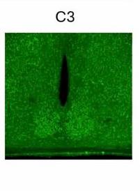 |
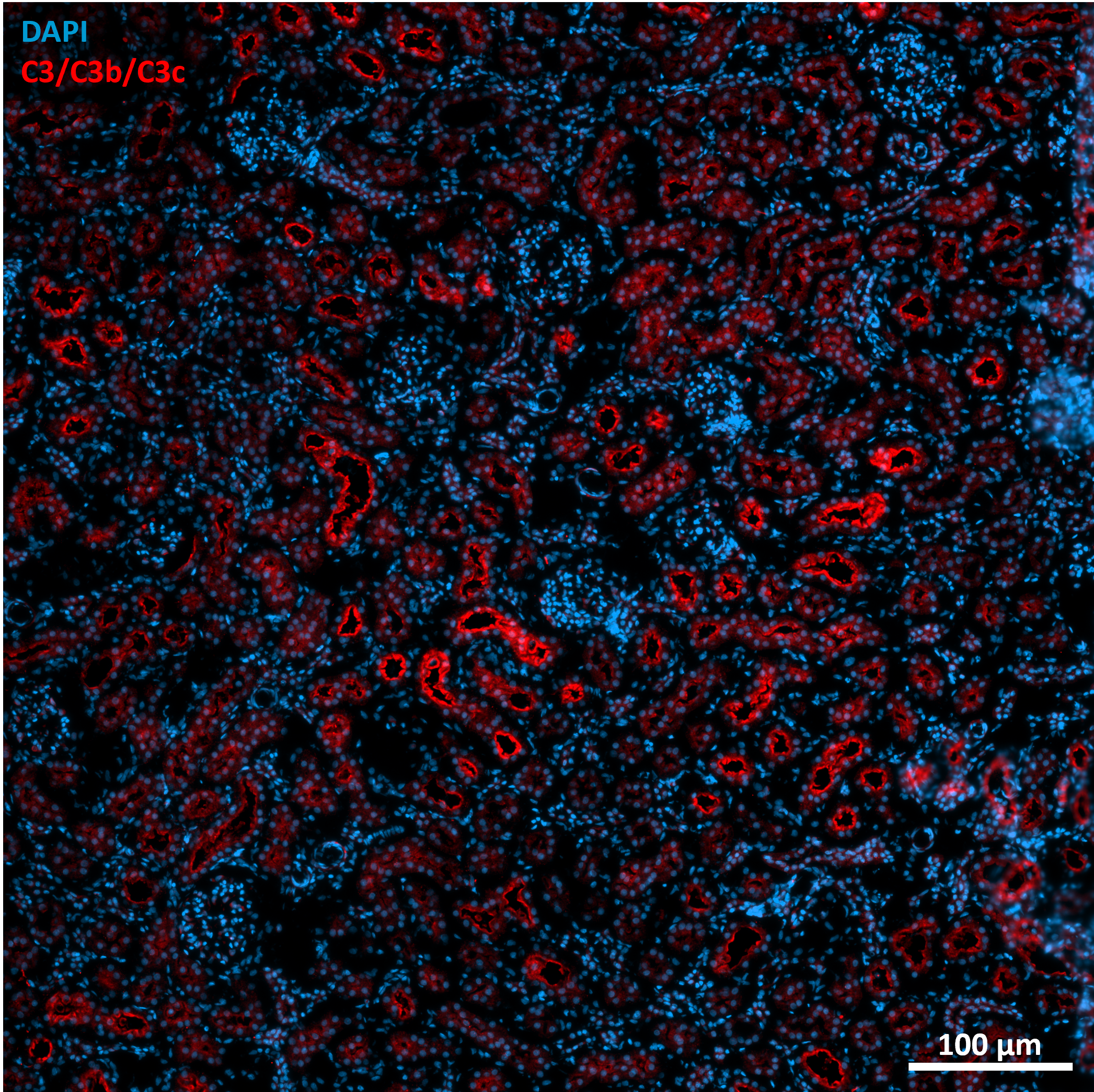
Marion (Verified Customer) (08-01-2024) | LPS induction model Fixative : Paraformaldehyde 4% Permeabilization: 10 min; RT; 0.2% Triton X-100 Blocking: 2 h; RT; 10% Normal donkey serum; 1% BSA Primary antibody: 1 night; 4°C Secondary antibody: ab150064 (Abcam); 1h30; RT Antibodies diluted in blocking solution
 |

