


Tested Applications
| Positive IHC detected in | human breast cancer tissue, human testis tissue Note: suggested antigen retrieval with TE buffer pH 9.0; (*) Alternatively, antigen retrieval may be performed with citrate buffer pH 6.0 |
Recommended dilution
| Application | Dilution |
|---|---|
| Immunohistochemistry (IHC) | IHC : 1:50-1:500 |
| It is recommended that this reagent should be titrated in each testing system to obtain optimal results. | |
| Sample-dependent, Check data in validation data gallery. | |
Published Applications
| IF | See 1 publications below |
Product Information
24214-1-AP targets PPP1R35 in IHC, IF, ELISA applications and shows reactivity with human samples.
| Tested Reactivity | human |
| Cited Reactivity | human |
| Host / Isotype | Rabbit / IgG |
| Class | Polyclonal |
| Type | Antibody |
| Immunogen |
CatNo: Ag18340 Product name: Recombinant human C7orf47 protein Source: e coli.-derived, PET28a Tag: 6*His Domain: 7-253 aa of BC026269 Sequence: ESELKSADGEEAAAVPGPPPEPQVPQLRAPVPEPGLDLSLSPRPDSPQPRHGSPGRRKGRAERRGAARQRRQVRFRLTPPSPVRSEPQPAVPQELEMPVLKSSLALGLELRAAAGSHFDAAKAVEEQLRKSFQIRCGLEESVSEGLNVPRSKRLFRDLVSLQVPEEQVLNAALREKLALLPPQARAPHPKEPPGPGPDMTILCDPETLFYESPHLTLDGLPPLRLQLRPRPSEDTFLMHRTLRRWEA Predict reactive species |
| Full Name | chromosome 7 open reading frame 47 |
| Calculated Molecular Weight | 253 aa, 28 kDa |
| GenBank Accession Number | BC026269 |
| Gene Symbol | C7orf47 |
| Gene ID (NCBI) | 221908 |
| RRID | AB_2879460 |
| Conjugate | Unconjugated |
| Form | Liquid |
| Purification Method | Antigen Affinity purified |
| UNIPROT ID | Q8TAP8 |
| Storage Buffer | PBS with 0.02% sodium azide and 50% glycerol, pH 7.3. |
| Storage Conditions | Store at -20°C. Stable for one year after shipment. Aliquoting is unnecessary for -20oC storage. 20ul sizes contain 0.1% BSA. |
Protocols
| Product Specific Protocols | |
|---|---|
| IHC protocol for PPP1R35 antibody 24214-1-AP | Download protocol |
| Standard Protocols | |
|---|---|
| Click here to view our Standard Protocols |
Reviews
The reviews below have been submitted by verified Proteintech customers who received an incentive for providing their feedback.
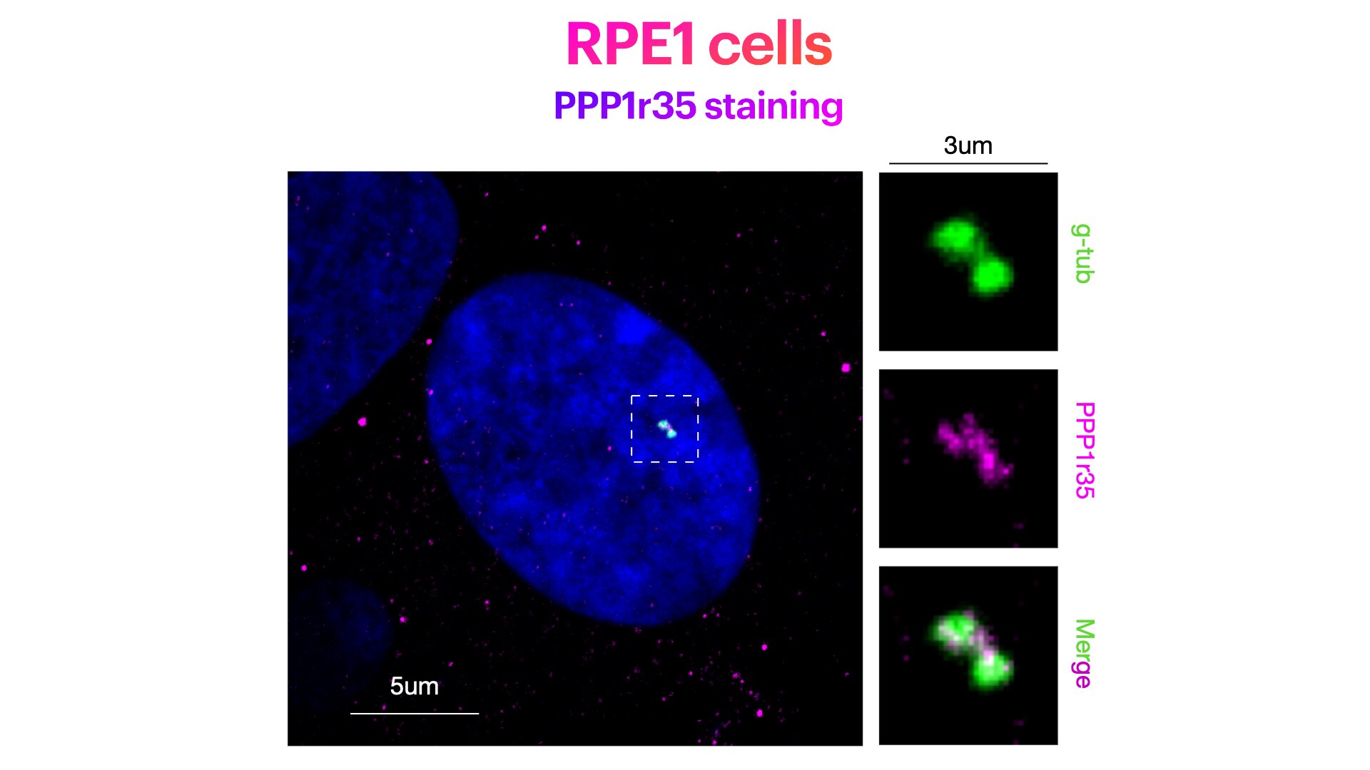
FH Elisa (Verified Customer) (04-24-2023) | Weak centriolar staining. While this antibody works well for Westernblotting, the expected centriolar staining in IF is rather weak. Yet, this antibody works under the following conditions: RPE1 cells stained for Hoechst (DNA marker, in green), PPP1r35 (centriolar marker, in magenta) and g-Tubulin (pericentriolar matrix marker, in green). RPE1 cells were fixed in cold methanol for 10' at -20C. Cells were then rehydrated with PBS for 5'. Membrane permeabilization was then performed with 0.1% Triton + 0.1% Tween +0.01%SDS in PBS for 5'. Cells were finally incubated with blocking buffer (5% BSA+ 0.1% Tween in PBS) for 30' at RT. Primary antibody was diluted in blocking buffer 1:300 and incubated for 1h at room temperature. Alexa-488-Anti-rabbit was used as secondary antibody (1:600 dilution) (1h at room temperature).
 |


