

Tested Applications
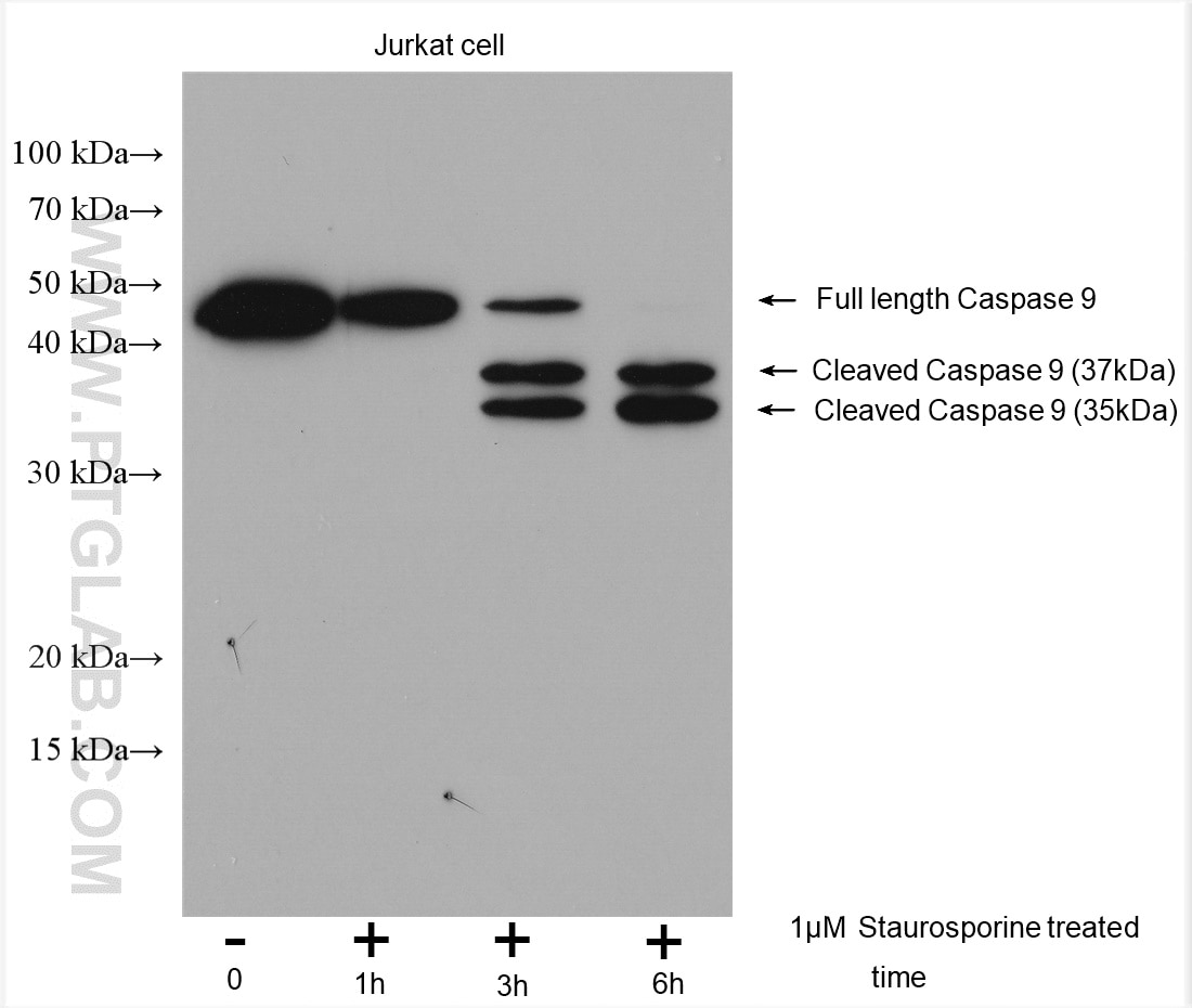
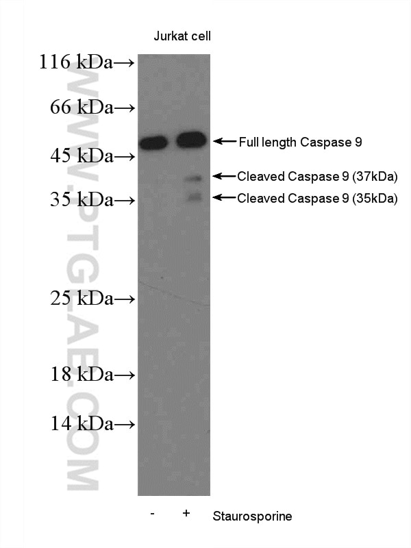
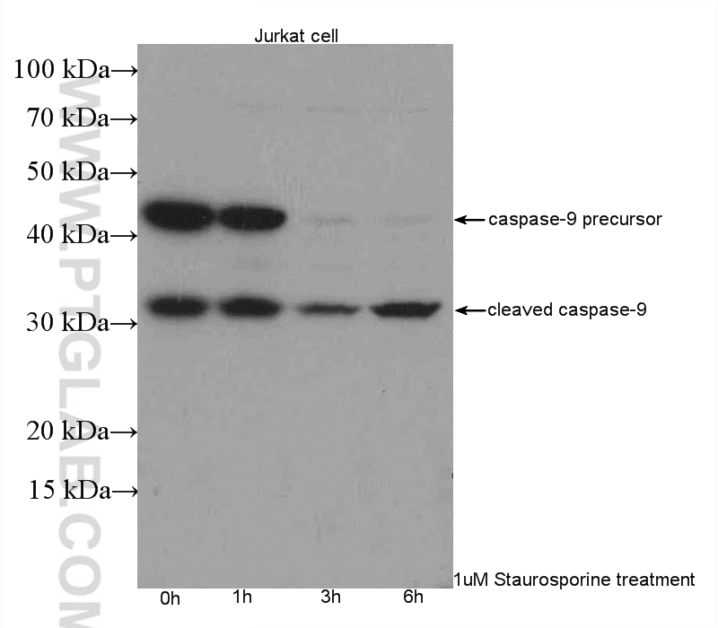
| Positive WB detected in | Jurkat cells, Staurosporine treated Jurkat cells |
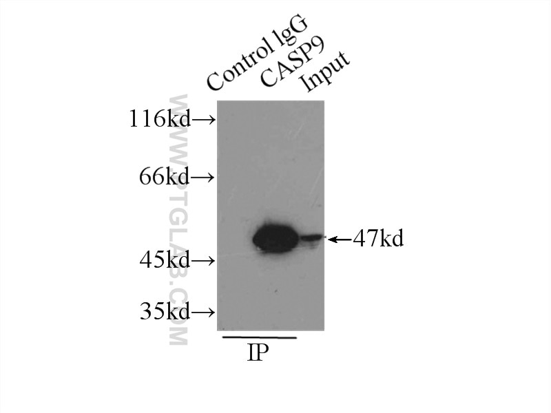
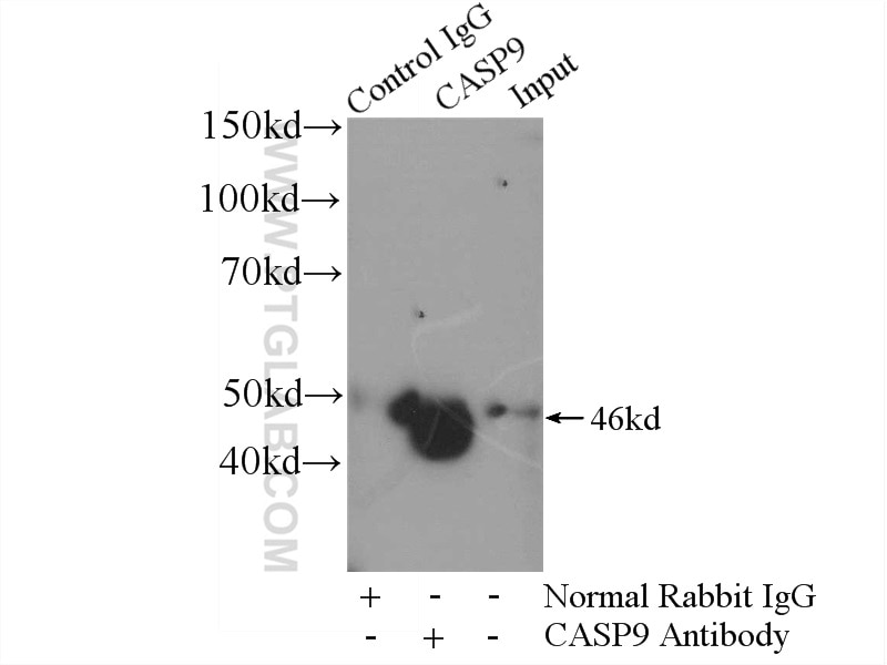
| Positive IP detected in | HeLa cells |
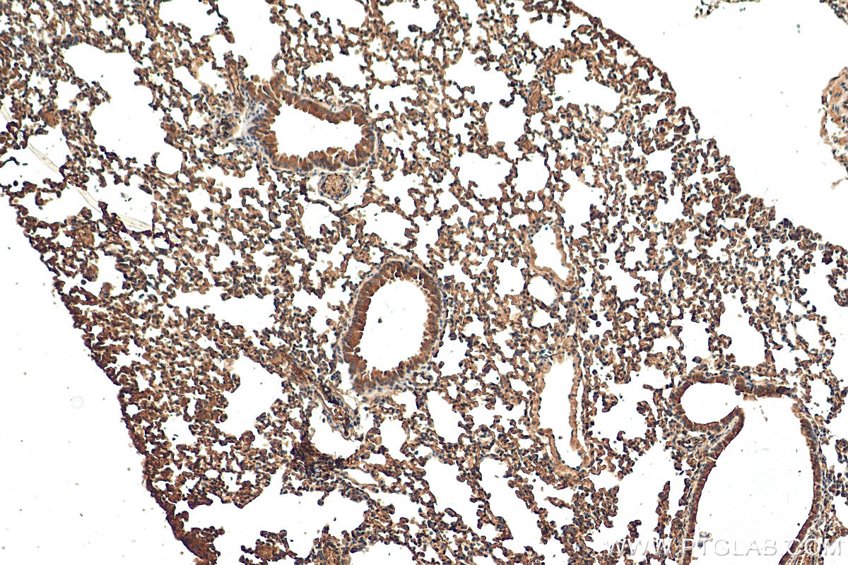
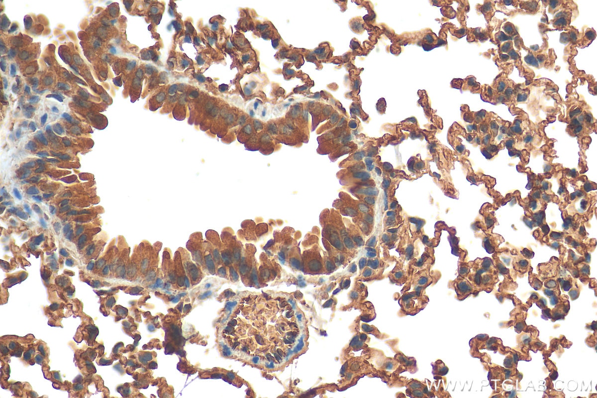
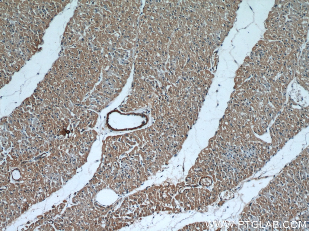
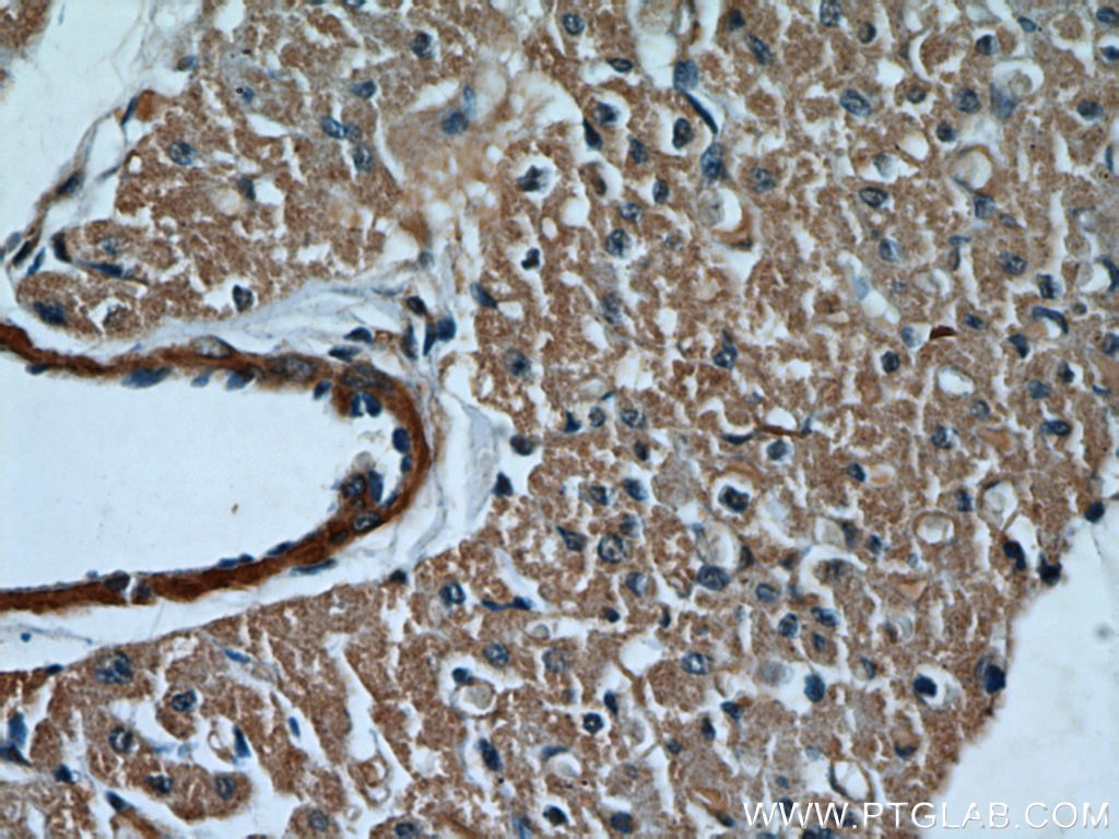
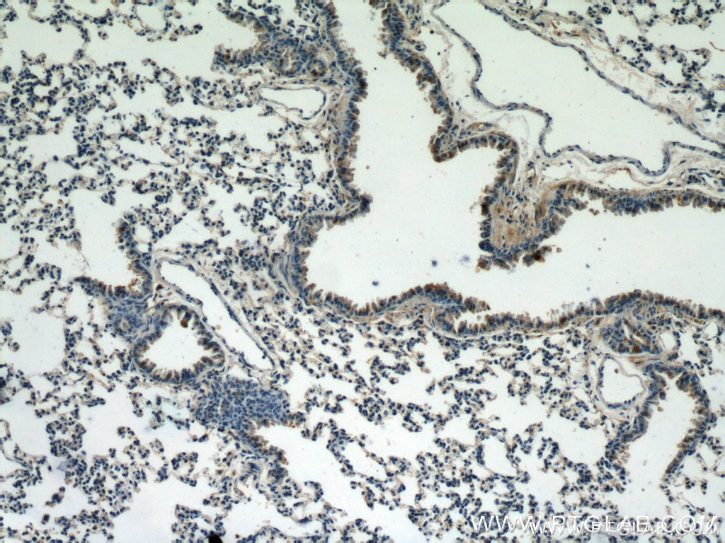
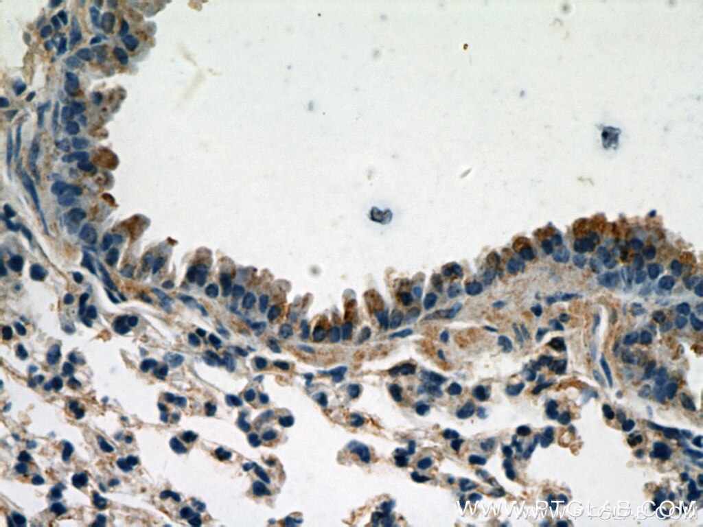
| Positive IHC detected in | mouse lung tissue, human heart tissue Note: suggested antigen retrieval with TE buffer pH 9.0; (*) Alternatively, antigen retrieval may be performed with citrate buffer pH 6.0 |
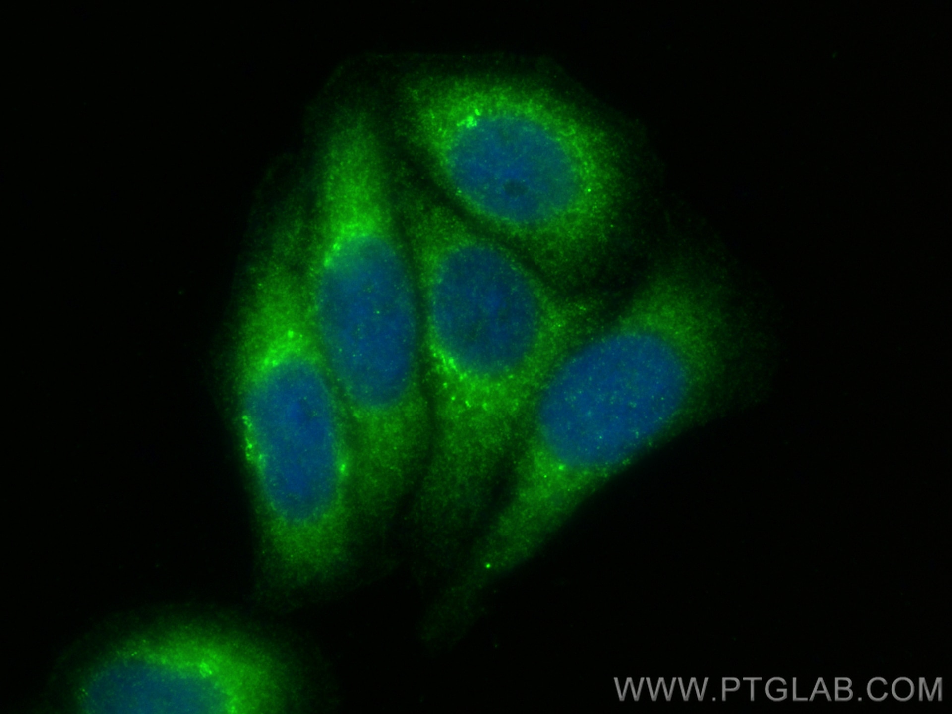
| Positive IF/ICC detected in | HepG2 cells |
Recommended dilution
| Application | Dilution |
|---|---|
| Western Blot (WB) | WB : 1:300-1:1000 |
| Immunoprecipitation (IP) | IP : 0.5-4.0 ug for 1.0-3.0 mg of total protein lysate |
| Immunohistochemistry (IHC) | IHC : 1:50-1:500 |
| Immunofluorescence (IF)/ICC | IF/ICC : 1:200-1:800 |
| It is recommended that this reagent should be titrated in each testing system to obtain optimal results. | |
| Sample-dependent, Check data in validation data gallery. | |
Product Information
10380-1-AP targets Caspase 9/P35 in WB, IHC, IF/ICC, IP, CoIP, RIP, ELISA applications and shows reactivity with human, mouse samples.
| Tested Reactivity | human, mouse |
| Cited Reactivity | human, mouse, rat, rabbit, monkey, chicken, zebrafish, sheep, goat |
| Host / Isotype | Rabbit / IgG |
| Class | Polyclonal |
| Type | Antibody |
| Immunogen |
CatNo: Ag0404 Product name: Recombinant human Caspase 9 protein Source: e coli.-derived, PGEX-4T Tag: GST Domain: 138-389 aa of BC002452 Sequence: DVGALESLRGNADLAYILSMEPCGHCLIINNVNFCRESGLRTRTGSNIDCEKLRRRFSSLHFMVEVKGDLTAKKMVLALLELAQQDHGALDCCVVVILSHGCQASHLQFPGAVYGTDGCPVSVEKIVNIFNGTSCPSLGGKPKLFFIQACGGEQKDHGFEVASTSPEDESPGSNPEPDATPFQEGLRTFDQLDAISSLPTPSDIFVSYSTFPGFVSWRDPKSGSWYVETLDDIFEQWAHSEDLQSLLLRVAN Predict reactive species |
| Full Name | caspase 9, apoptosis-related cysteine peptidase |
| Calculated Molecular Weight | 46 kDa |
| Observed Molecular Weight | 46 kDa, 30-39 kDa |
| GenBank Accession Number | BC002452 |
| Gene Symbol | Caspase 9 |
| Gene ID (NCBI) | 842 |
| RRID | AB_2068632 |
| Conjugate | Unconjugated |
| Form | Liquid |
| Purification Method | Antigen affinity purification |
| UNIPROT ID | P55211 |
| Storage Buffer | PBS with 0.02% sodium azide and 50% glycerol, pH 7.3. |
| Storage Conditions | Store at -20°C. Stable for one year after shipment. Aliquoting is unnecessary for -20oC storage. 20ul sizes contain 0.1% BSA. |
Background Information
Caspase 9, apoptosis-related cysteine protease (CASP9, synonyms: MCH6, APAF3, APAF-3, ICE-LAP6, CASPASE-9c)is a member of the cysteine-aspartic acid protease (caspase) family. Sequential activation of caspases plays a central role in the execution-phase of cell apoptosis. Caspases exist as inactive proenzymes which undergo proteolytic processing at conserved aspartic residues to produce 2 subunits, large and small, that dimerize to form the active enzyme. Caspase 9 is processed by APAF1; this step is thought to be one of the earliest in the caspase activation cascade. 10380-1-AP can recognize the pre- and cleaved- caspase 9. In recent years, the localization of caspase 9 was a focus of interest. Beside its cytoplasmic distribution, a very extensive localization study was done on rat brain tissue, where caspase 9 was found located predominantly in the nucleus and to a lesser extend in the cytoplasm [PMID: 15541731].
Protocols
| Product Specific Protocols | |
|---|---|
| IF protocol for Caspase 9/P35 antibody 10380-1-AP | Download protocol |
| IHC protocol for Caspase 9/P35 antibody 10380-1-AP | Download protocol |
| IP protocol for Caspase 9/P35 antibody 10380-1-AP | Download protocol |
| WB protocol for Caspase 9/P35 antibody 10380-1-AP | Download protocol |
| Standard Protocols | |
|---|---|
| Click here to view our Standard Protocols |
Publications
What published studies show
Able is busy answering your previous question. Please try again later.
| Species | Application | Title |
|---|---|---|
Signal Transduct Target Ther Increased mtDNA copy number promotes cancer progression by enhancing mitochondrial oxidative phosphorylation in microsatellite-stable colorectal cancer. | ||
Mol Cancer hsa_circ_0007919 induces LIG1 transcription by binding to FOXA1/TET1 to enhance the DNA damage response and promote gemcitabine resistance in pancreatic ductal adenocarcinoma | ||
Cancer Discov Cytosolic Phospholipase A2 Determines Intercellular Heterogeneity of Stress Granules and Chemotherapy Response. | ||
Adv Mater Selective Organ-Targeting Hafnium Oxide Nanoparticles with Multienzyme-Mimetic Activities Attenuate Radiation-Induced Tissue Damage | ||
Bioact Mater Silicate ions as soluble form of bioactive ceramics alleviate aortic aneurysm and dissection | ||
Autophagy E-cigarette aerosols induce the hydrolysis of lysosomal glycerophospholipids through PLA2G4A activation initiated by nicotine binding to CHRNA3/α3 nAchr in airway epithelial cells. |
Reviews
What customers say
Users generally find this antibody useful. One reviewer confirmed it works well on both paraffin-embedded adult mouse retina and cryo-embedded postnatal (p7) mouse tissue in immunofluorescence at 1/200 dilution. Another rated it well for detecting apoptosis in mouse heart tissue subjected to ischemia-reperfusion injury by Western blot. However, one Western blot user reported a drawback: a large amount of non-specific (aspecific) bands in the 50-200 kDa range at 1:500 dilution, indicating some background may need optimization. Overall the antibody is considered reliable for its intended apoptosis detection applications.
The reviews below have been submitted by verified Proteintech customers who received an incentive for providing their feedback.
Christoffer (Verified Customer) (03-03-2025) | Works well on adult mouse retina embedded in paraffin as well as cryo embedded postnatal (p7) mouse tissue.
|
Nicola (Verified Customer) (04-11-2023) | Large amount of aspecific band on my western blot in the range 50-200 KDa.
|
WEI (Verified Customer) (01-17-2020) | Good to detect apoptosis in mouse heart subject to IR injury.
|

