Tested Applications
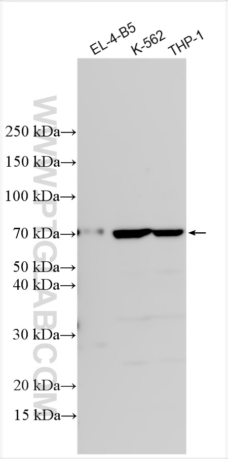
| Positive WB detected in | EL-4-B5 cells, K-562 cells, THP-1 cells |
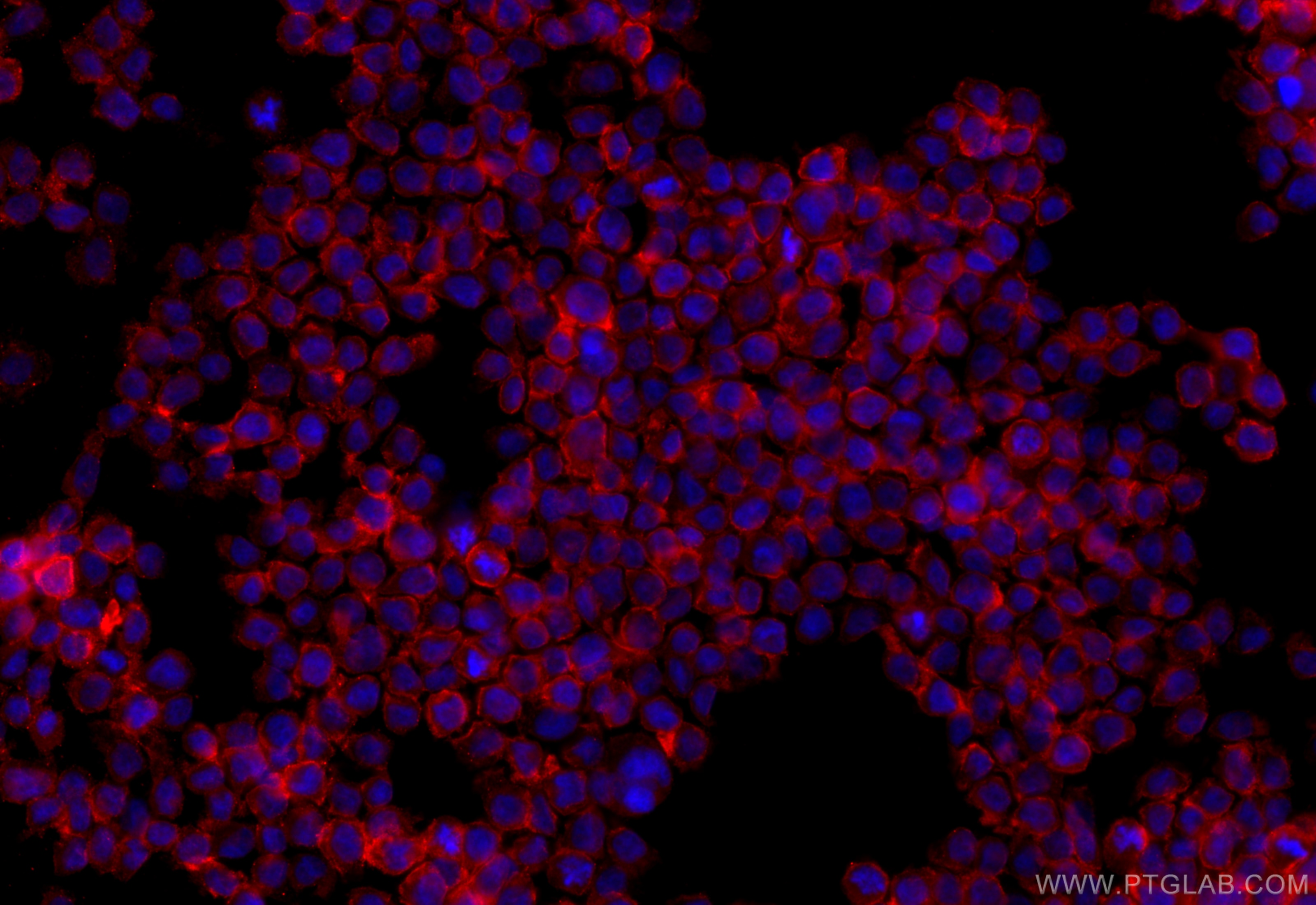
| Positive IF/ICC detected in | THP-1 cells |
Recommended dilution
| Application | Dilution |
|---|---|
| Western Blot (WB) | WB : 1:1000-1:6000 |
| Immunofluorescence (IF)/ICC | IF/ICC : 1:50-1:500 |
| It is recommended that this reagent should be titrated in each testing system to obtain optimal results. | |
| Sample-dependent, Check data in validation data gallery. | |
Published Applications
| IHC | See 5 publications below |
| IF | See 3 publications below |
Product Information
17425-1-AP targets CD33 in WB, IHC, IF/ICC, ELISA applications and shows reactivity with human, mouse samples.
| Tested Reactivity | human, mouse |
| Cited Reactivity | human, mouse |
| Host / Isotype | Rabbit / IgG |
| Class | Polyclonal |
| Type | Antibody |
| Immunogen |
CatNo: Ag11490 Product name: Recombinant human CD33 protein Source: e coli.-derived, PGEX-4T Tag: GST Domain: 49-259 aa of BC028152 Sequence: YYDKNSPVHGYWFREGAIISGDSPVATNKLDQEVQEETQGRFRLLGDPSRNNCSLSIVDARRRDNGSYFFRMERGSTKYSYKSPQLSVHVTDLTHRPKILIPGTLEPGHSKNLTCSVSWACEQGTPPIFSWLSAAPTSLGPRTTHSSVLIITPRPQDHGTNLTCQVKFAGAGVTTERTIQLNVTYVPQNPTTGIFPGDGSGKQETRAGVVH Predict reactive species |
| Full Name | CD33 molecule |
| Calculated Molecular Weight | 364 aa, 40 kDa |
| Observed Molecular Weight | 67 kDa |
| GenBank Accession Number | BC028152 |
| Gene Symbol | CD33 |
| Gene ID (NCBI) | 945 |
| RRID | AB_2228962 |
| Conjugate | Unconjugated |
| Form | Liquid |
| Purification Method | Antigen affinity purification |
| UNIPROT ID | P20138 |
| Storage Buffer | PBS with 0.02% sodium azide and 50% glycerol, pH 7.3. |
| Storage Conditions | Store at -20°C. Stable for one year after shipment. Aliquoting is unnecessary for -20oC storage. 20ul sizes contain 0.1% BSA. |
Background Information
CD33 (also known as sialic acid binding Ig-like lectin 3, or Siglec-3) is a transmembrane receptor expressed on cells of myeloid lineage (https://www.uniprot.org/uniprot/P20138). It belongs to the Siglec family of sialic-acid-binding immunoglobulin-like lectins that are thought to promote cell-cell interactions and regulate the functions of cells in the innate and adaptive immune systems through glycan recognition. CD33 preferentially binds α2-6-linked sialic acids on lactosamine units. It is a member of the immunoglobulin superfamily of proteins and contains two immunoglobulin domains (one IgV and one IgC2 domain) responsible for ligand binding, a transmembrane region, and a cytoplasmic tail that contains two immunoreceptor tyrosine-based inhibitory motifs (ITIMs) that are implicated in the inhibition of cellular activity (PMID:10206955, PMID:10887109).
What is the molecular weight of CD33?
CD33 is a 364 amino acid glycoprotein that has an apparent molecular weight of 67 kDa by SDS-PAGE, with a predicted mass of 40 kDa based on the amino acid sequence. The disparity observed between the predicted and apparent molecular weight is due to the glycosylation of CD33 at five sites.
What is the subcellular localization of CD33?
CD33 is localized to the cell surface, but it can also be detected in the nucleus (PMID: 25613900).
What is the tissue specificity of CD33?
CD33 is expressed by myeloid stem cells (CFU-GEMM, CFU-GM, CFU-G, and E-BFU), myeloblasts and monoblasts, monocytes/macrophages, granulocyte precursors (with expression decreasing with maturation), and mast cells. Mature granulocytes may show a very low level of CD33 expression. It is not expressed on erythrocytes, platelets, B-cells, T-cells, or NK cells. The highest tissue expression of CD33 is observed in the spleen, bone marrow, lymph node, tonsil, and appendix (PMID: 25613900).
What is the function of CD33 in myeloid cells?
CD33 may act as an inhibitory receptor on myeloid cells to affect their function. Upon ligand binding, it becomes phosphorylated at two tyrosine residues (Y340 and Y358) within the ITIMs by Src family kinases. Phospho-Y340 has been shown to recruit the SH2-domain containing protein-tyrosine phosphatases SHP-1 and SHP-2 to the receptor following sialic acid binding, which then inhibits phosphotyrosine-mediated signal transduction events (PMID:10206955, PMID:10887109). It has been shown that CD33 exerts an inhibitory effect on tyrosine phosphorylation and Ca2+ mobilization when co-engaged with the activating FcgammaRI receptor and therefore negatively regulates FcgammaRI signal transduction (PMID:10556798).
What is the CD33 role in leukemia?
CD33 is expressed by more than 90% of cases of acute myeloid leukemia (AML) and in virtually all cases of chronic myeloid leukemia (CML). It has been validated as a target for antigen-specific immunotherapy; gemtuzumab ozogamicin (Mylotarg) is an antibody-drug conjugate that was developed for the treatment of patients with acute myeloid leukemia. The drug is a recombinant humanized anti-CD33 monoclonal antibody covalently attached to the cytotoxic antitumor antibiotic calicheamicin (N-acetyl-γ-calicheamicin) via a bifunctional linker (4-(4-acetylphenoxy)butanoic acid). It is prescribed for the treatment of adults with newly diagnosed CD33-positive AML, or patients over the age of 2 with CD33-positive AML whose disease returned or did not respond to previous treatment (PMID:29021230). A number of single nucleotide polymorphisms (SNPs) have been identified in AML patients that influence the effectiveness of gemtuzumab ozogamicin immunotherapy. SNP rs12459419 C>T in the splice enhancer region present in exon2 regulates the expression of an alternatively spliced CD33 isoform lacking exon2 (D2-CD33) that deletes the IgV domain, which is the antibody-binding site for gemtuzumab ozogamicin, and patients with this SNP are refractory to immunotherapy (PMID:28644774).
Protocols
| Product Specific Protocols | |
|---|---|
| IF protocol for CD33 antibody 17425-1-AP | Download protocol |
| WB protocol for CD33 antibody 17425-1-AP | Download protocol |
| Standard Protocols | |
|---|---|
| Click here to view our Standard Protocols |
Publications
| Species | Application | Title |
|---|---|---|
Int Immunopharmacol Targeting upregulation of the immunosuppressive activity of MDSCs with indirubin as a novel strategy to alleviate psoriasis | ||
Front Genet Transmembrane Protein 170B is a Prognostic Biomarker and Associated With Immune Infiltrates in Pancreatic Adenocarcinoma. | ||
PLoS One Myeloid derived suppressor cells contribute to the malignant progression of oral squamous cell carcinoma. | ||
Med Oncol CD33?/p-STAT1? double-positive cell as a prognostic factor for stage IIIa gastric cancer. | ||
Aging (Albany NY) Bridging the gap between clear cell renal cell carcinoma and cutaneous melanoma: the role of SCARB1 in dysregulated cholesterol metabolism | ||
Mol Ther Optimized CAR-T therapy based on spatiotemporal changes and chemotactic mechanisms of MDSCs induced by hypofractionated radiotherapy |
Reviews
The reviews below have been submitted by verified Proteintech customers who received an incentive for providing their feedback.
FH Emma (Verified Customer) (11-29-2021) | We struggled to get this antibody working by IF on FFPE tissue. We tried a 1:50 dilution and Tris-EDTA antigen retrieval.
|


