

Tested Applications
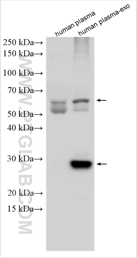
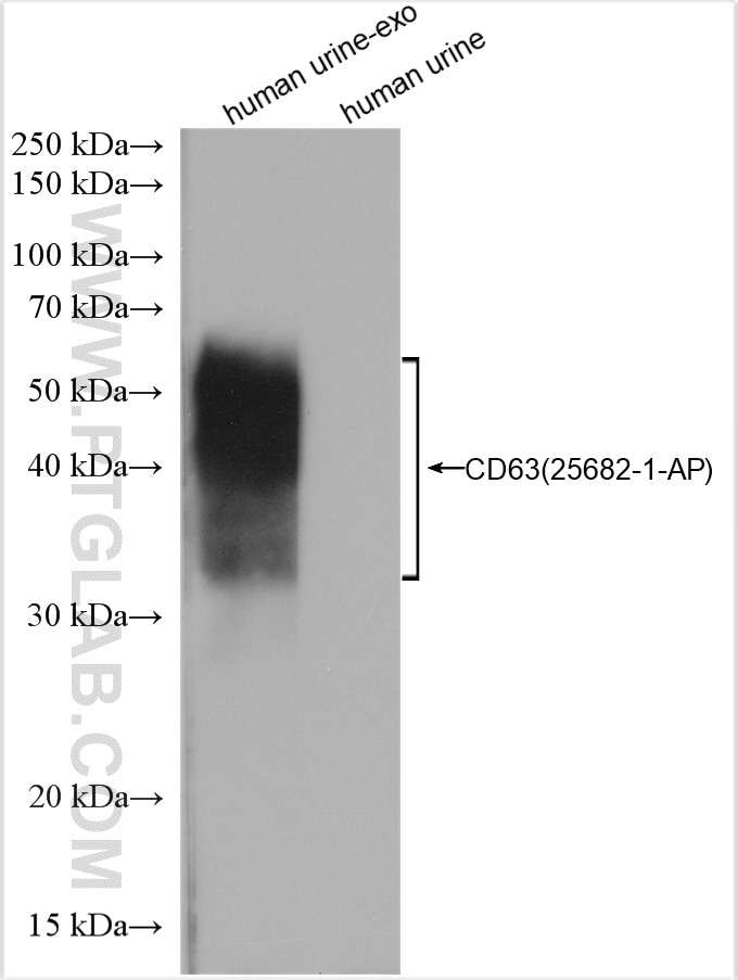
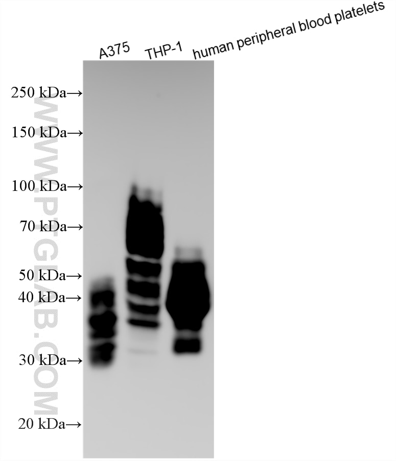
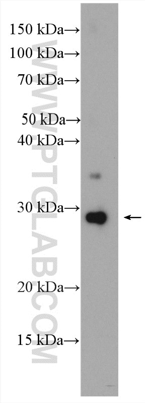
| Positive WB detected in | human urine exosomes tissue, A375 cells, human serum exosomes, THP-1 cells, human peripheral blood platelets |
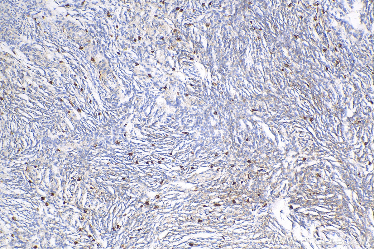
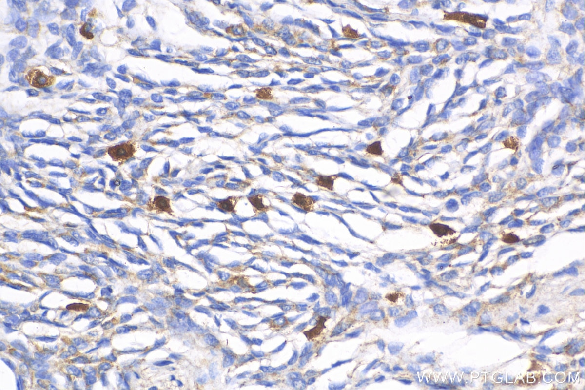
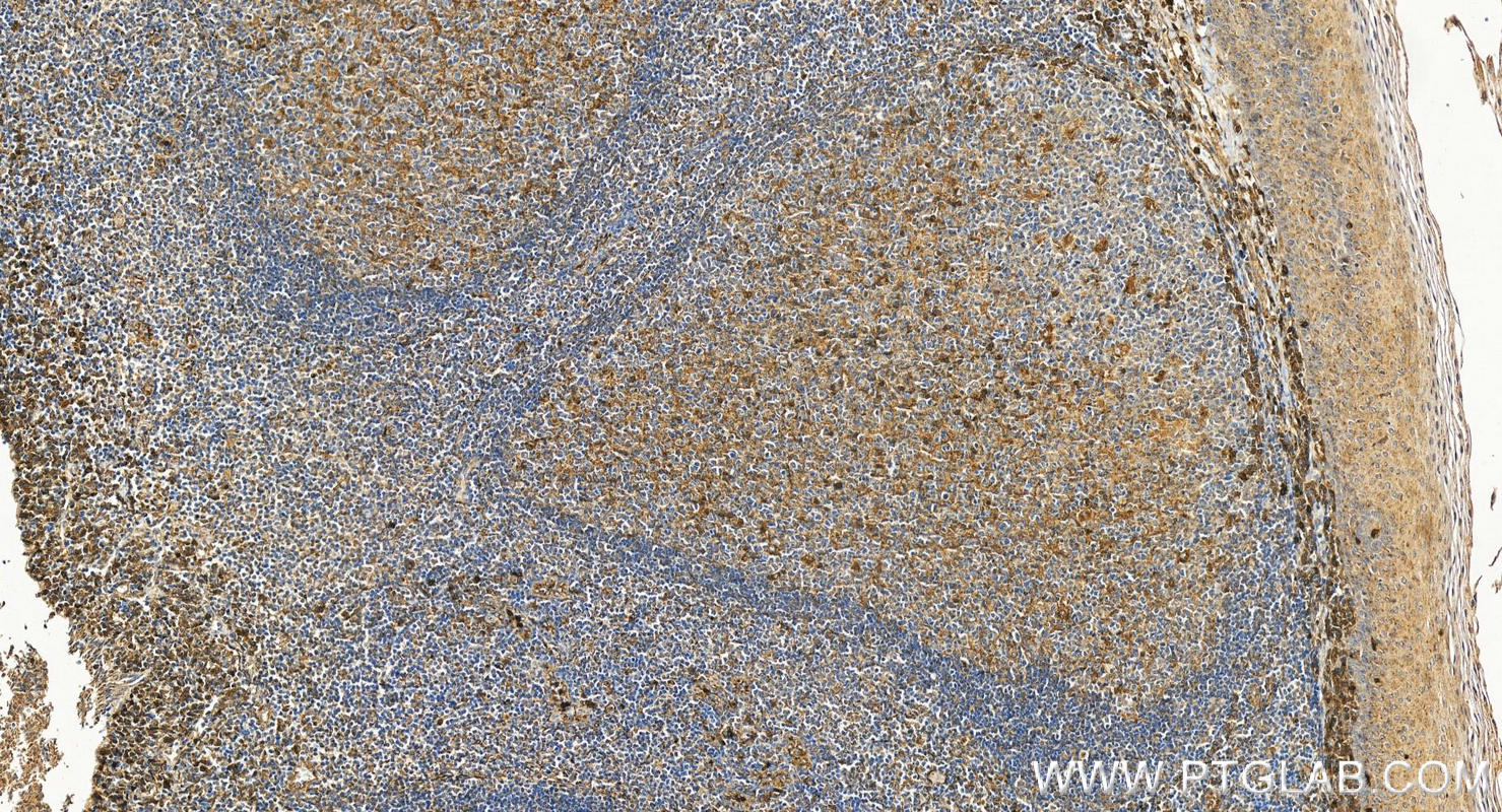
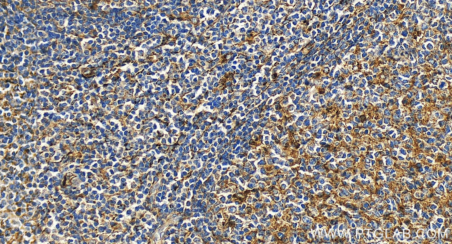
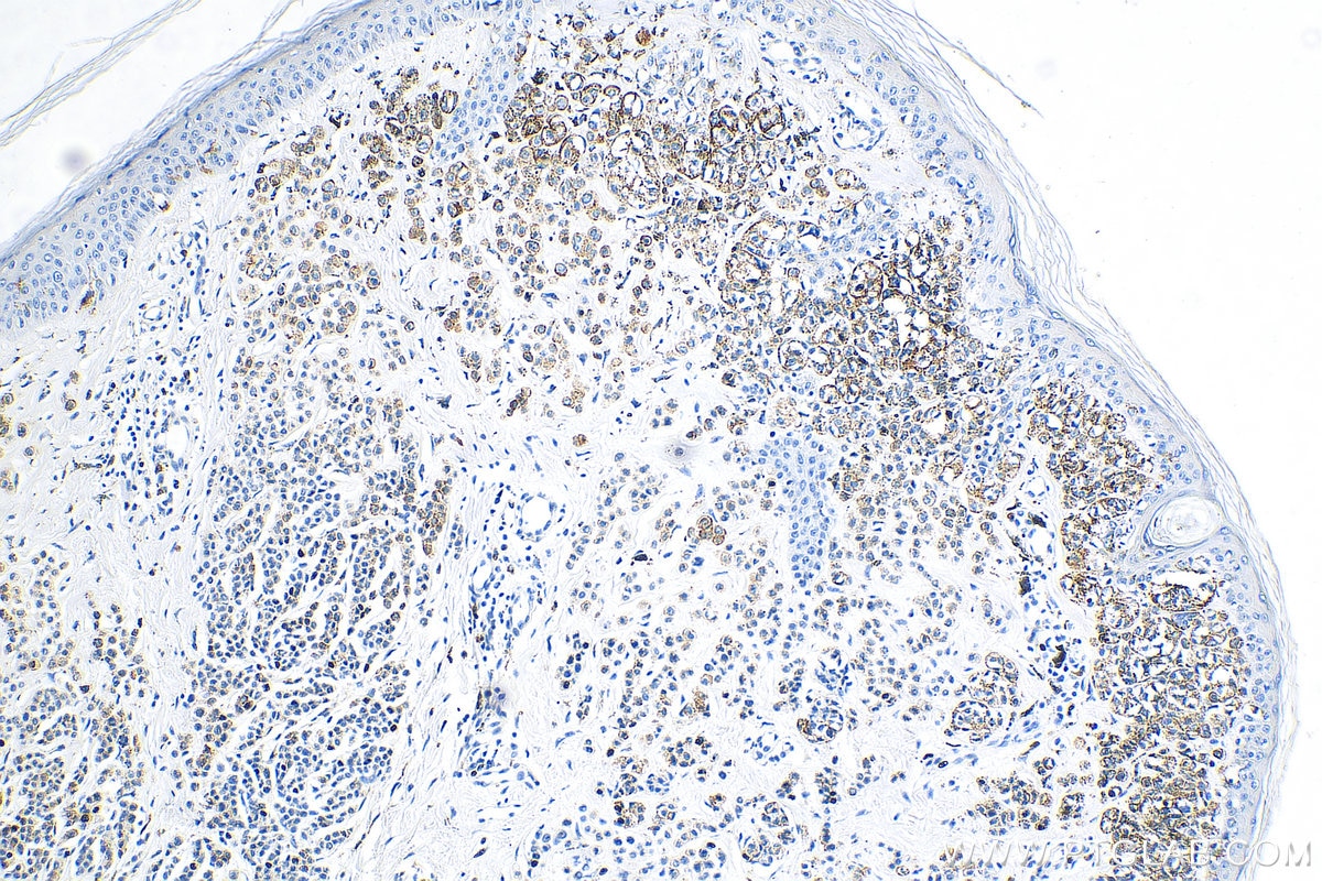
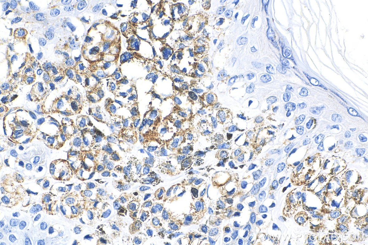
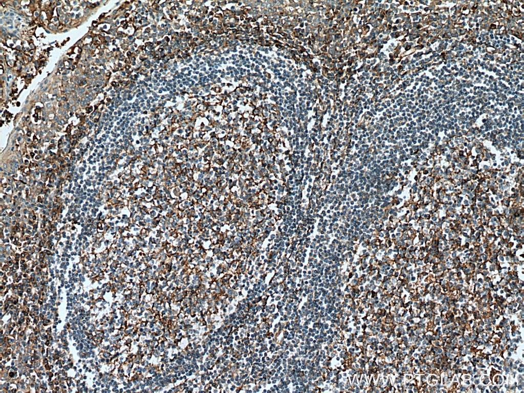
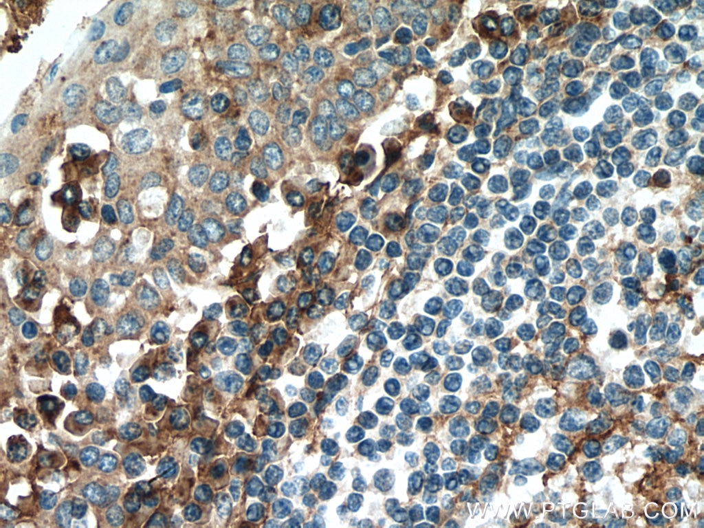
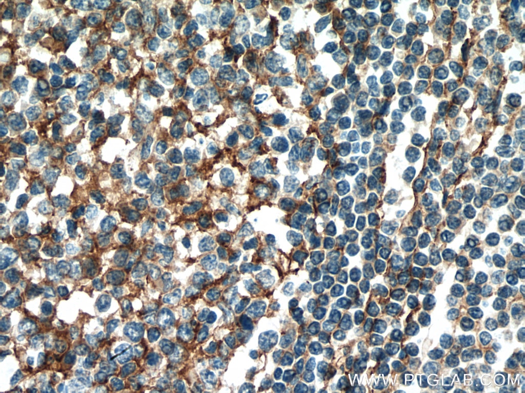
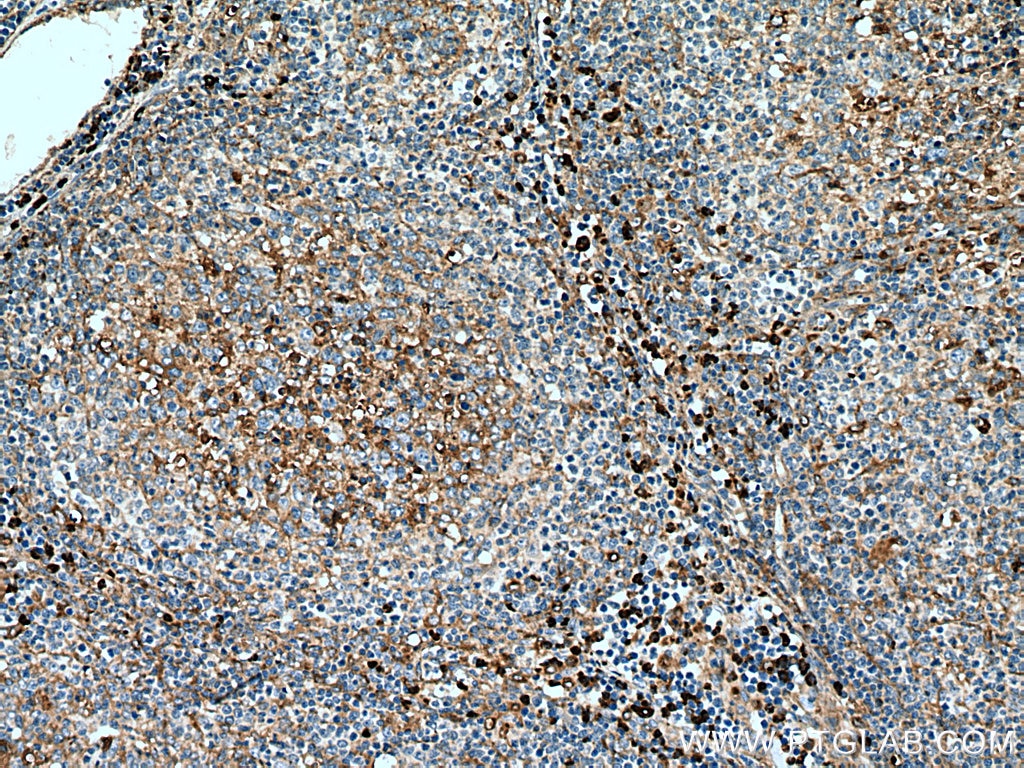
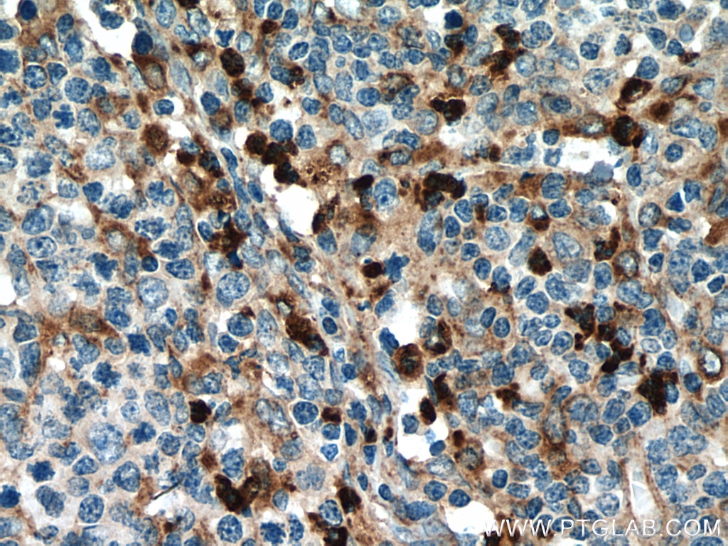
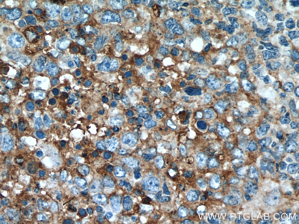
| Positive IHC detected in | human malignant melanoma tissue, human lymphoma tissue, human tonsillitis tissue Note: suggested antigen retrieval with TE buffer pH 9.0; (*) Alternatively, antigen retrieval may be performed with citrate buffer pH 6.0 |
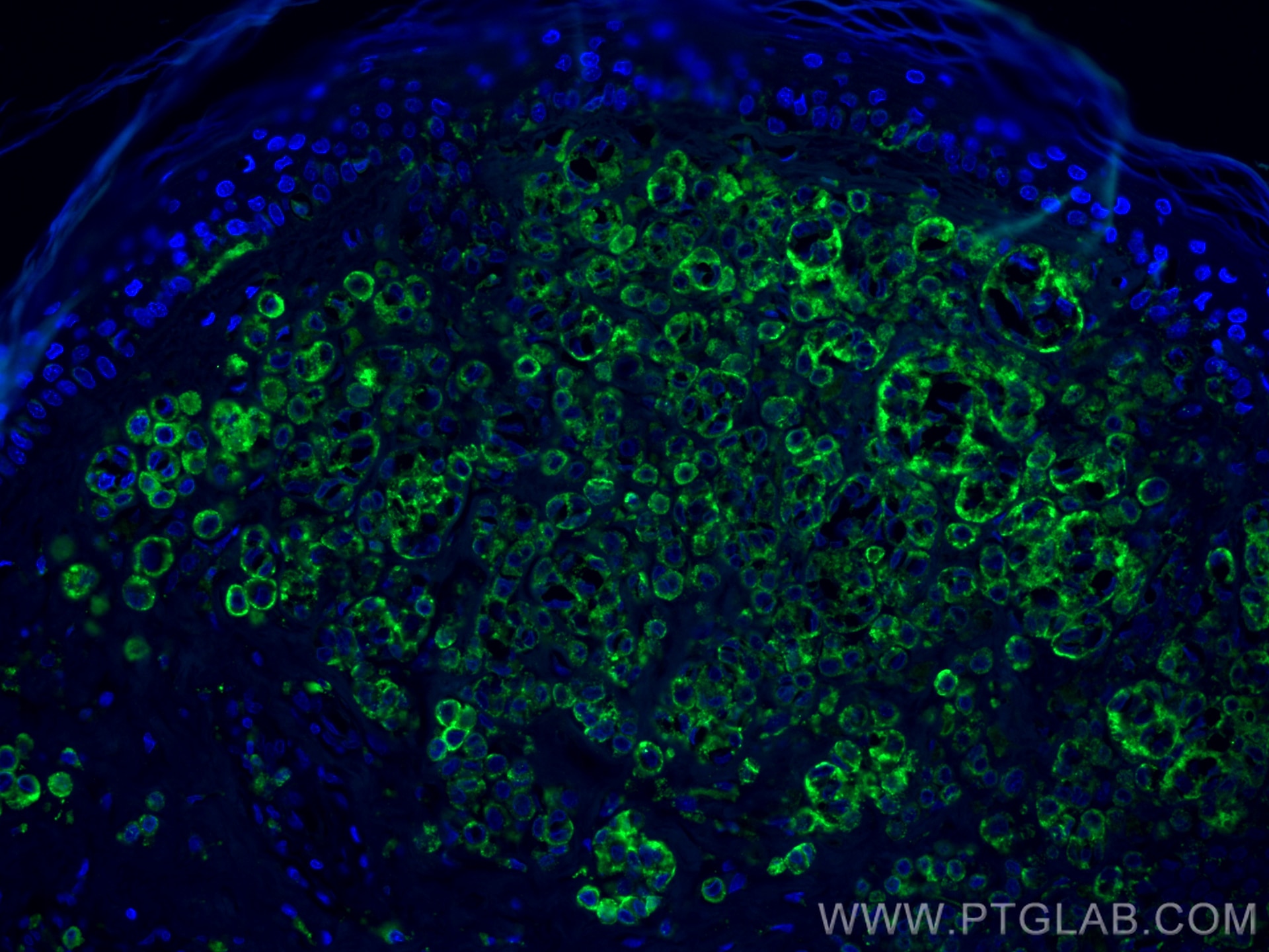
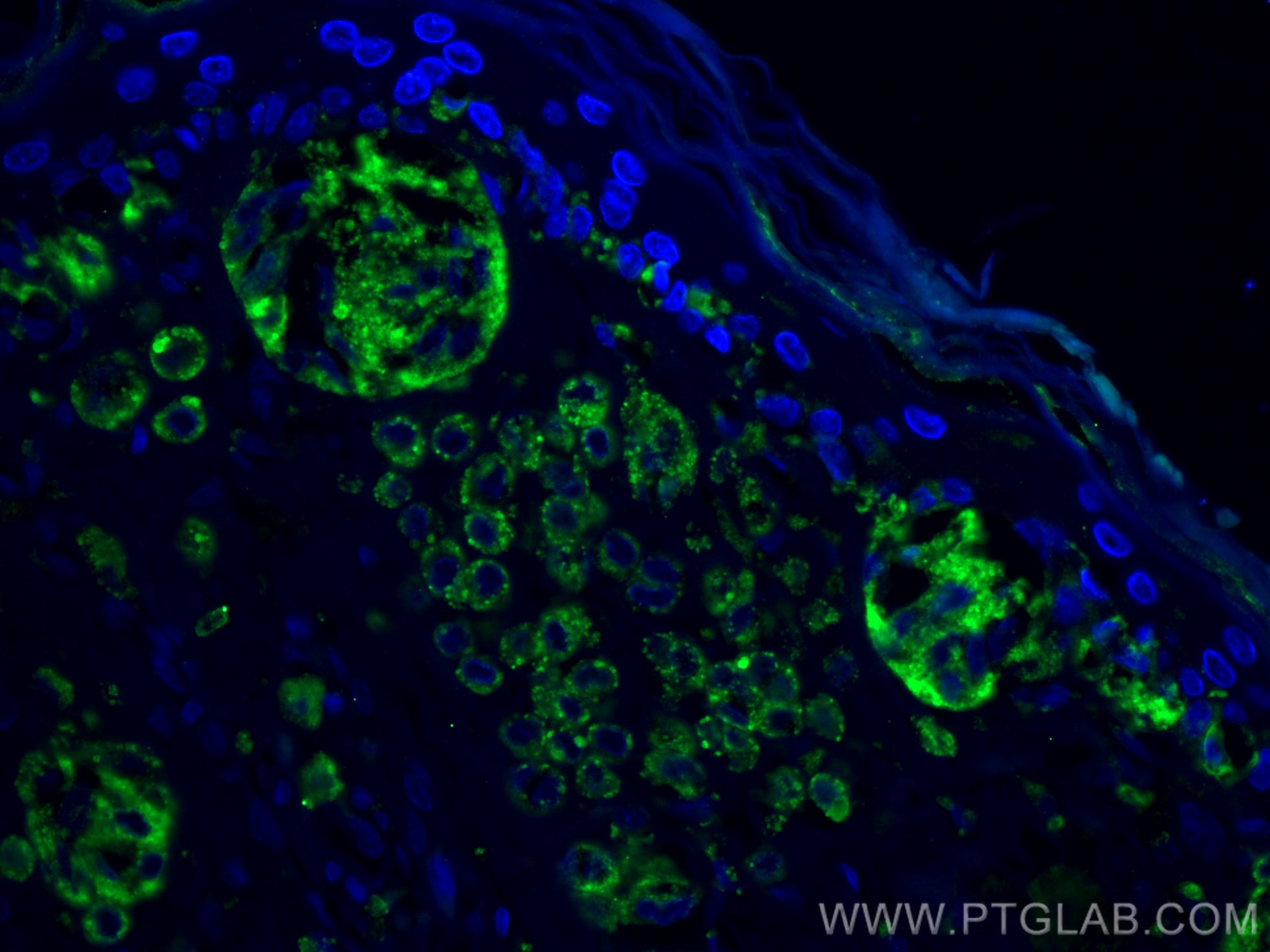
| Positive IF-P detected in | human malignant melanoma tissue |
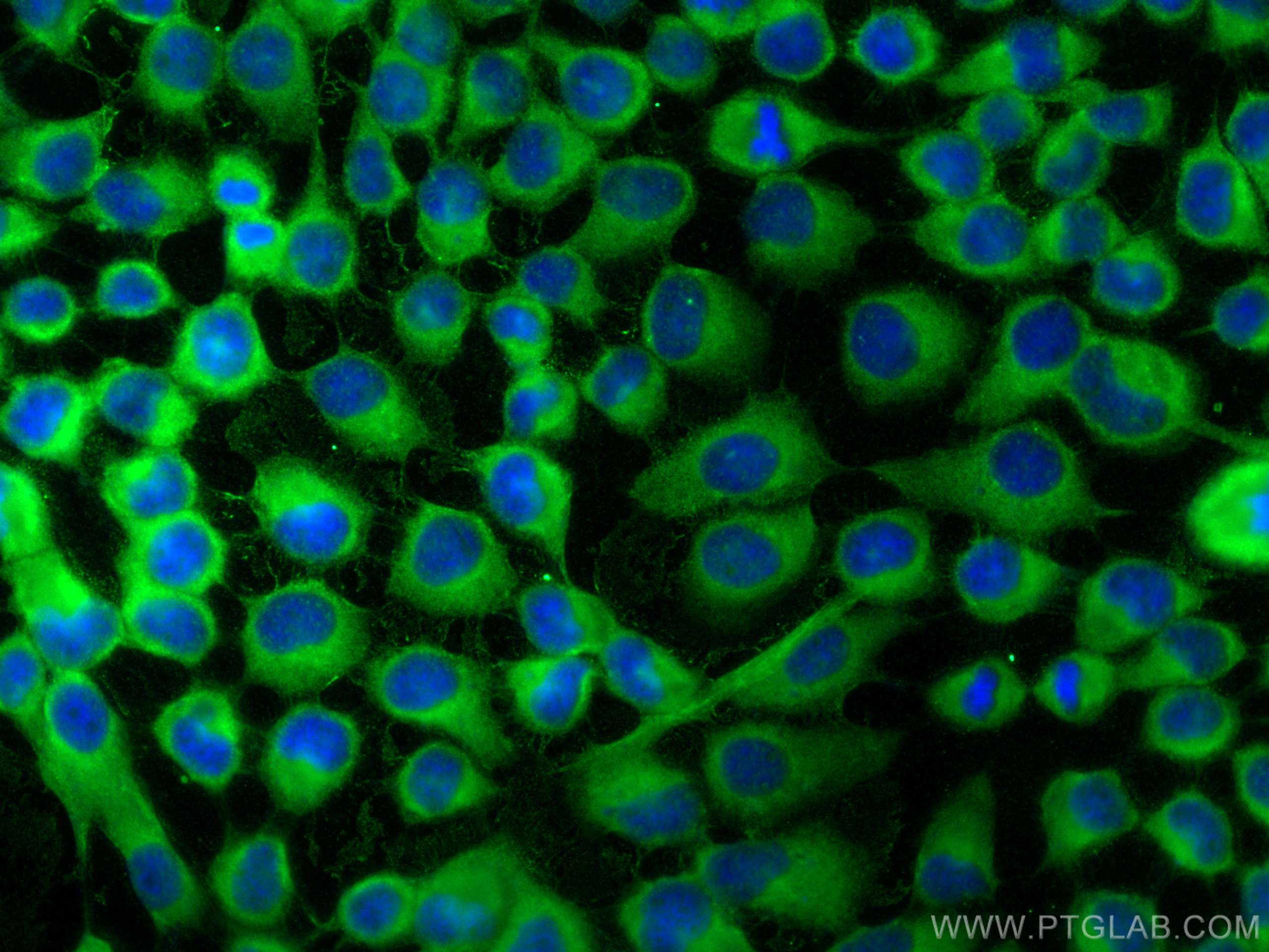
| Positive IF/ICC detected in | A431 cells |
Recommended dilution
| Application | Dilution |
|---|---|
| Western Blot (WB) | WB : 1:500-1:40000 |
| Immunohistochemistry (IHC) | IHC : 1:400-1:1600 |
| Immunofluorescence (IF)-P | IF-P : 1:50-1:500 |
| Immunofluorescence (IF)/ICC | IF/ICC : 1:200-1:800 |
| It is recommended that this reagent should be titrated in each testing system to obtain optimal results. | |
| Sample-dependent, Check data in validation data gallery. | |
Product Information
25682-1-AP targets CD63 in WB, IHC, IF/ICC, IF-P, IP, ChIP, ELISA applications and shows reactivity with human samples.
| Tested Reactivity | human |
| Cited Reactivity | human, pig, canine, monkey, bovine, hamster, goat |
| Host / Isotype | Rabbit / IgG |
| Class | Polyclonal |
| Type | Antibody |
| Immunogen |
CatNo: Ag19690 Product name: Recombinant human CD63 protein Source: e coli.-derived, PET28a Tag: 6*His Domain: 104-209 aa of BC002349 Sequence: GYVFRDKVMSEFNNNFRQQMENYPKNNHTASILDRMQADFKCCGAANYTDWEKIPSMSKNRVPDSCCINVTVGCGINFNEKAIHKEGCVEKIGGWLRKNVLVVAAA Predict reactive species |
| Full Name | CD63 molecule |
| Calculated Molecular Weight | 26 kDa |
| Observed Molecular Weight | 30-60 kDa |
| GenBank Accession Number | BC002349 |
| Gene Symbol | CD63 |
| Gene ID (NCBI) | 967 |
| RRID | AB_2783831 |
| Conjugate | Unconjugated |
| Form | Liquid |
| Purification Method | Antigen affinity purification |
| UNIPROT ID | P08962 |
| Storage Buffer | PBS with 0.02% sodium azide and 50% glycerol, pH 7.3. |
| Storage Conditions | Store at -20°C. Stable for one year after shipment. Aliquoting is unnecessary for -20oC storage. 20ul sizes contain 0.1% BSA. |
Background Information
CD63 is a 30-60 kDa lysosomal membrane protein that belongs to the tetraspanin family. This protein plays many important roles in immuno-physiological functions. It mediates signal transduction events that play a role in the regulation of cell development, activation, growth, and motility. CD63 is expressed on activated platelets, thus it may function as a blood platelet activation marker. CD63 is a lysosomal membrane glycoprotein that is translocated to plasma membrane after platelet activation. The CD63 tetraspanin is highly expressed in the early stages of melanoma and decreases in advanced lesions, suggesting it as a possible suppressor of tumor progression. Deficiency of this protein is associated with Hermansky-Pudlak syndrome.
Protocols
| Product Specific Protocols | |
|---|---|
| IF protocol for CD63 antibody 25682-1-AP | Download protocol |
| IHC protocol for CD63 antibody 25682-1-AP | Download protocol |
| WB protocol for CD63 antibody 25682-1-AP | Download protocol |
| Standard Protocols | |
|---|---|
| Click here to view our Standard Protocols |
Publications
What published studies show
The CD63 Polyclonal antibody (Cat# 25682-1-AP) has 645 total citations. Citations appear in high-impact journals including Nature Communications, Cell Metabolism, Nature Cancer, Nature Aging, Cell Research, Advanced Materials, ACS Nano, Theranostics, Journal of Extracellular Vesicles, Autophagy, Biomaterials, Advanced Science, Oncogene, and Cancer Letters, among others. Research fields span extracellular vesicle/exosome biology, oncology, tissue engineering, wound healing, neurodegeneration, diabetes, cardiovascular disease, and immunology.
| Species | Application | Title |
|---|---|---|
Signal Transduct Target Ther Human adipose and umbilical cord mesenchymal stem cell-derived extracellular vesicles mitigate photoaging via TIMP1/Notch1 | ||
J Hematol Oncol Exosomes released by hepatocarcinoma cells endow adipocytes with tumor-promoting properties. | ||
Cell Metab Hepatic sphingomyelin phosphodiesterase 3 promotes steatohepatitis by disrupting membrane sphingolipid metabolism | ||
Cell Metab Nondigestible stachyose binds membranous HSP90β on small intestinal epithelium to regulate the exosomal miRNAs: A new function and mechanism | ||
Cell Metab Arachidonic acid triggers spermidine synthase secretion from primary tumor to induce skeletal muscle weakness upon irradiation | ||
Cell Metab Serotonin-licensed macrophages potentiate chemoresistance via inositol metabolic crosstalk in ovarian cancer. |
Reviews
What customers say
Users report strong performance across multiple applications. Western blot results were positive in cancer cells, SH-SY5Y lysates, and exosomes, with one reviewer noting excellent signal even at low dilution and impressive results compared to other markers. One WB user observed smearing between 30–60 kDa in ARPE-19 cells. Immunohistochemistry on tonsil tissue using a Leica Bond Max (ER2, 95°C, 30 min) yielded good results. Immunofluorescence on HeLa cells and Lamp1-expressing stem cells was also successful. Overall, 4 out of 5 reviewers gave 5 stars, reflecting high satisfaction with specificity and sensitivity.
The reviews below have been submitted by verified Proteintech customers who received an incentive for providing their feedback.
Sonam (Verified Customer) (11-17-2025) | Works well
|
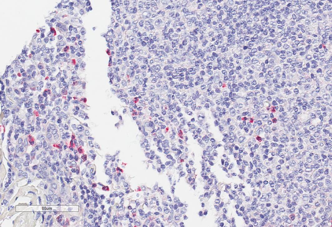
Federica (Verified Customer) (11-30-2023) | I used a Leica Bond Max on a healthy Tonsil tissue. Antigen Retrival ER2 95C for 30min
 |
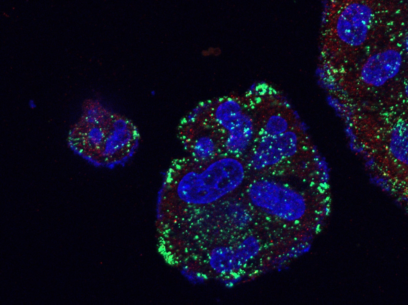
Nydia (Verified Customer) (06-11-2022) | The HeLa cells as well as Lamp1-expressing stem cells used in the immunostaining were transfected or alternatively transfected by splitting the cells later on the glass coverslips. In order to increase cell spreading and adhesion, the coverslips used were acid washed and treated with fibronectin. Afterwards, the cells were fixed with 4% PFA, permeabilized with 0.2% Triton X-100 in phosphate- buffered saline (PBS) for 10 minutes and for 1 hour the cells were blocked with 5% BSA in PBS. Then overnight, at 4 degrees Celsius, the primary antibodies were added followed by a wash with PBS 3 times.
 |
Guorong (Verified Customer) (03-22-2022) | Smears between 30 and 60 kDa was detected
 |
David (Verified Customer) (03-30-2020) | Used for both SH-SY5Y lysate and exosomes/ extracellular vesicles. Very good signal in lysates (could be used at much higher dilution). Also good signal in exosomes, which are notoriously difficult to assess by immunoblot. Very impressive results compared to other markers.
|

