

Tested Applications
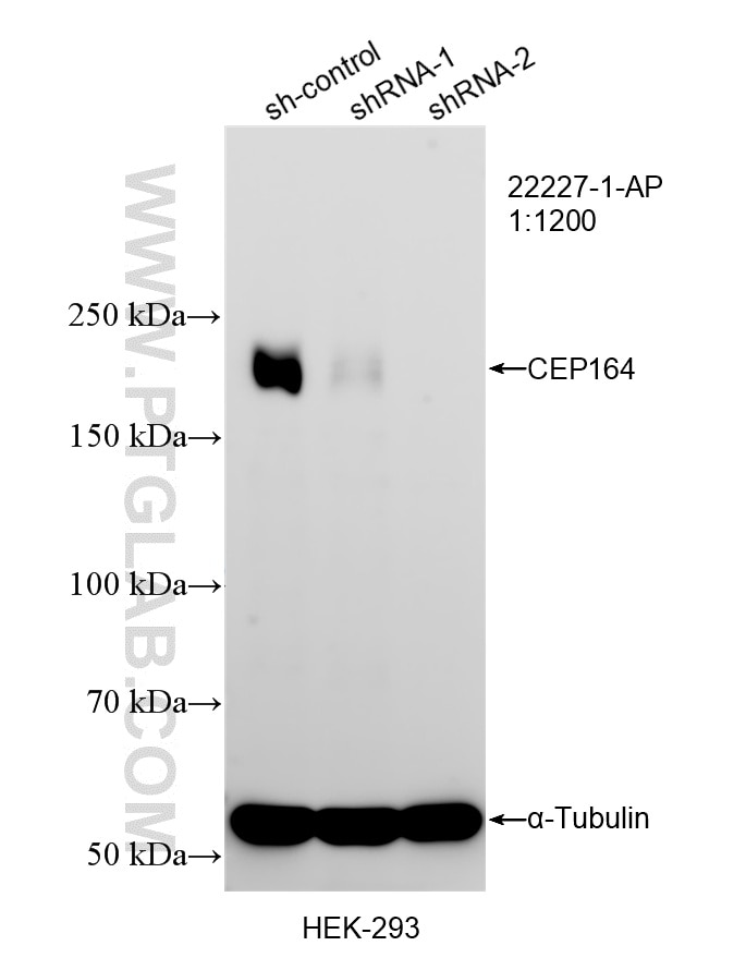
| Positive WB detected in | HEK-293 cells |
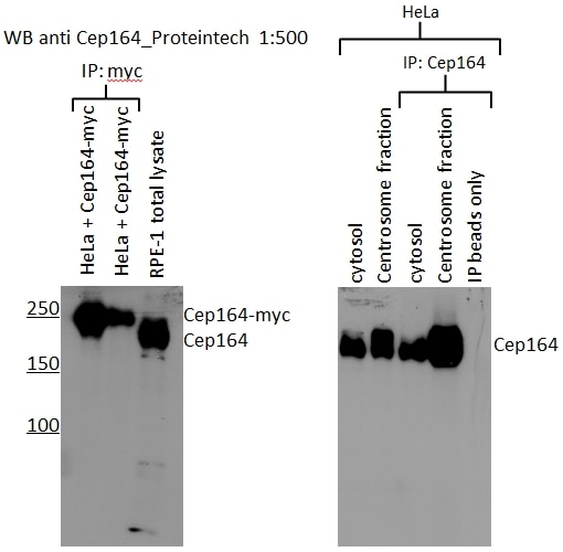
| Positive IP detected in | RPE1 cells |
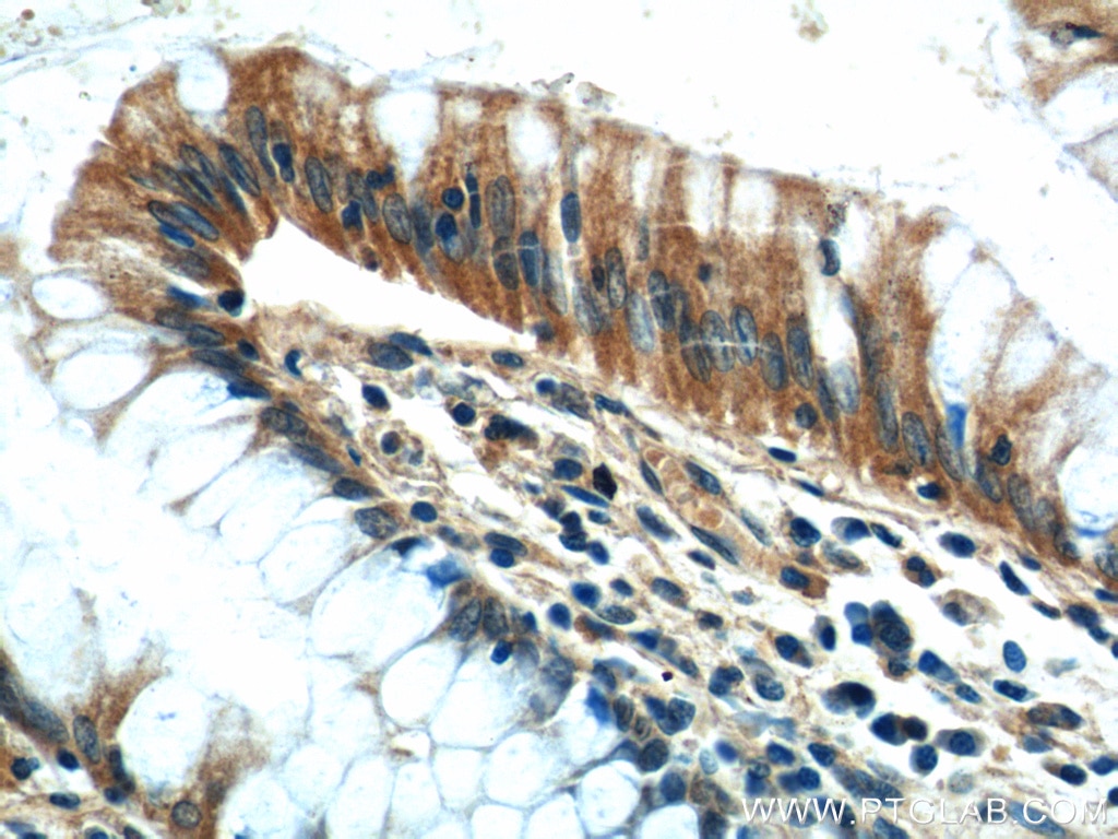
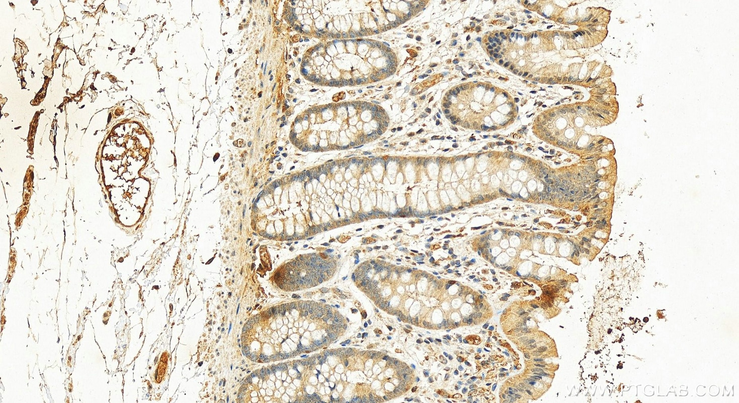
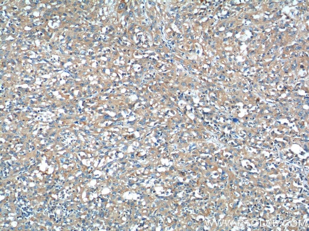
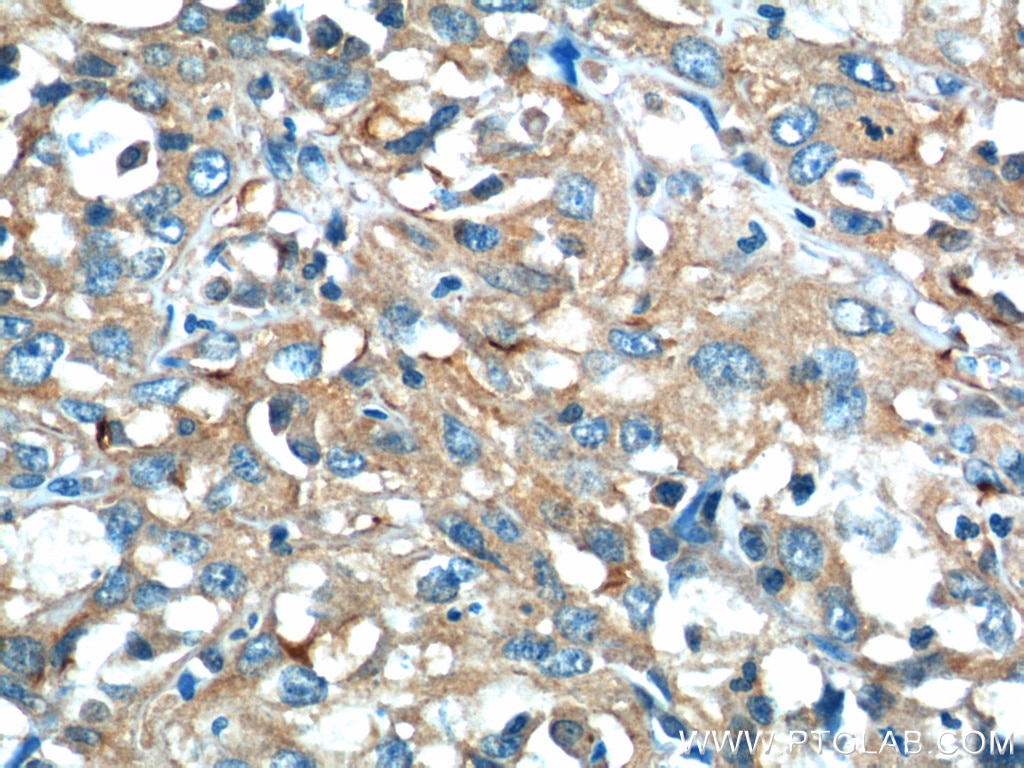
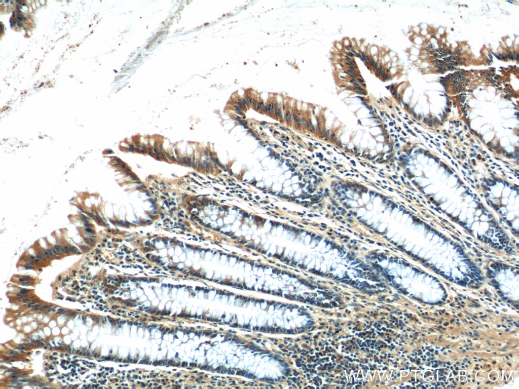
| Positive IHC detected in | human colon tissue, human cervical cancer tissue Note: suggested antigen retrieval with TE buffer pH 9.0; (*) Alternatively, antigen retrieval may be performed with citrate buffer pH 6.0 |
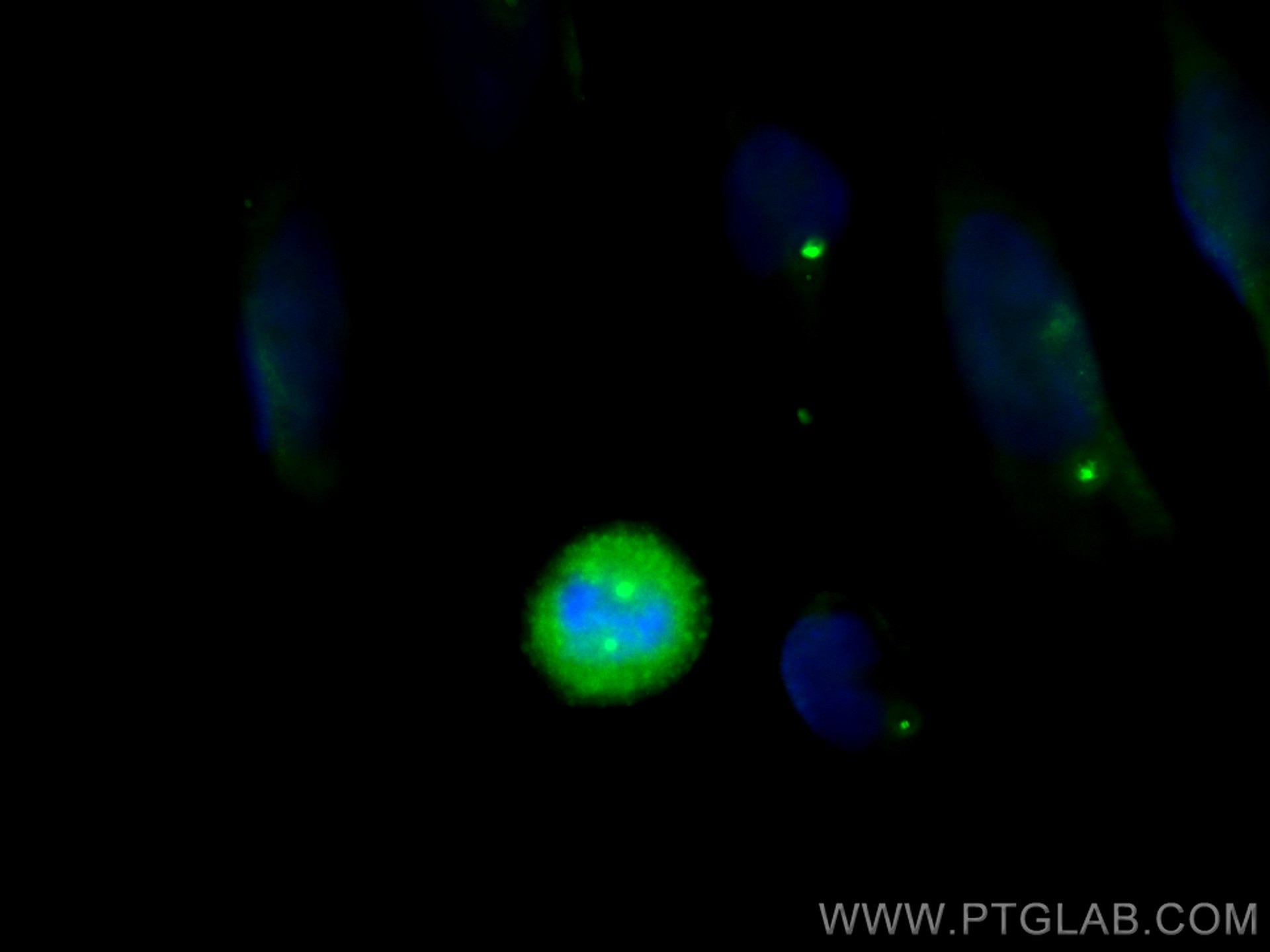
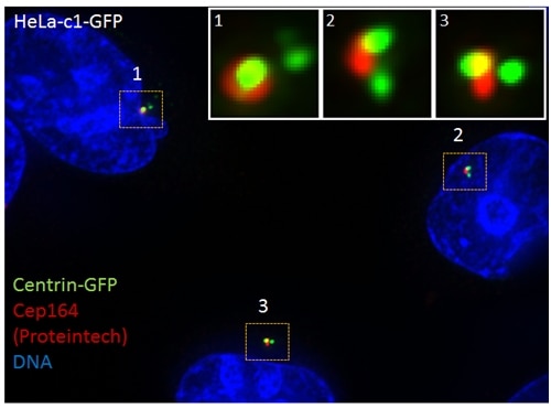
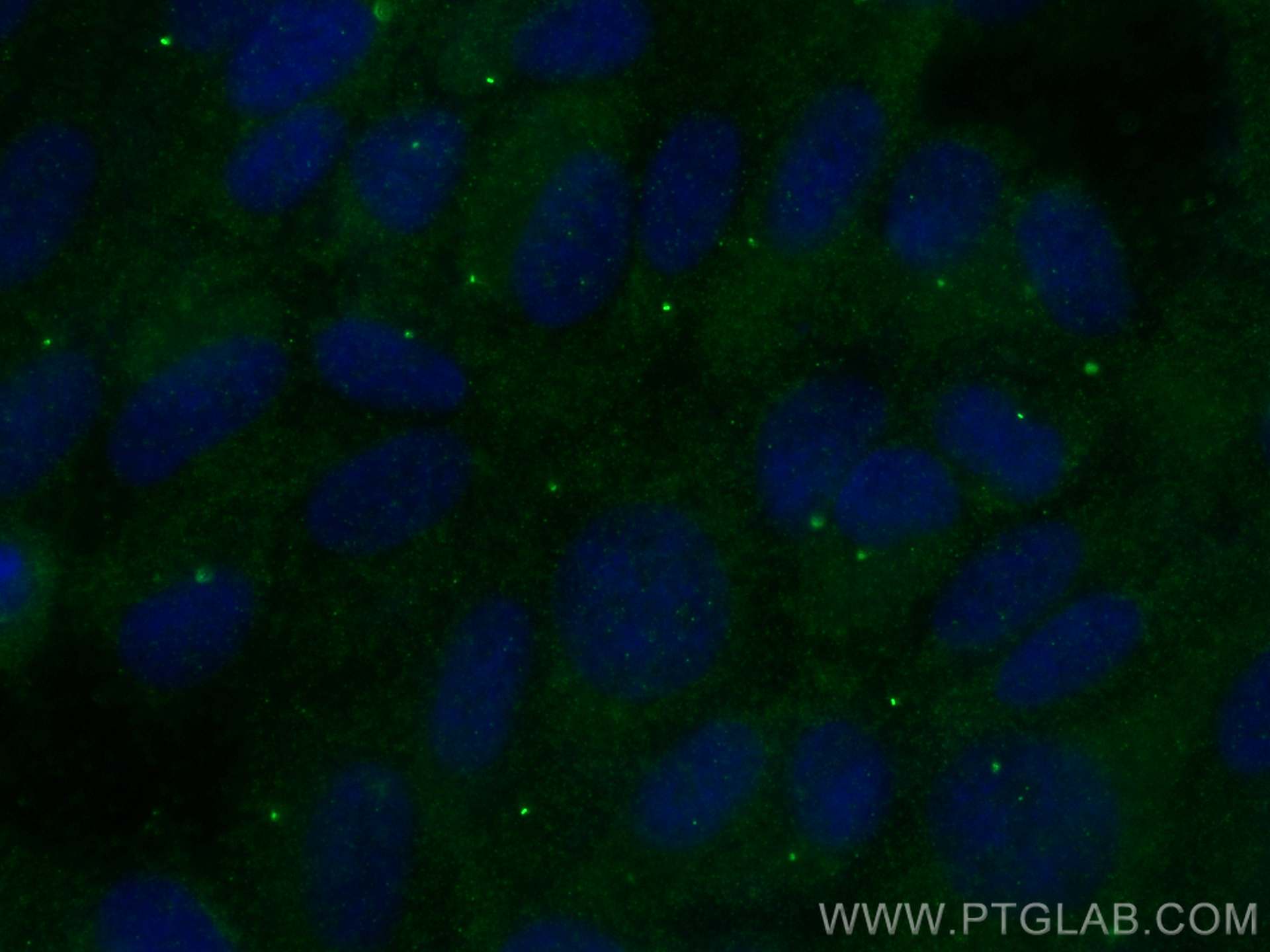
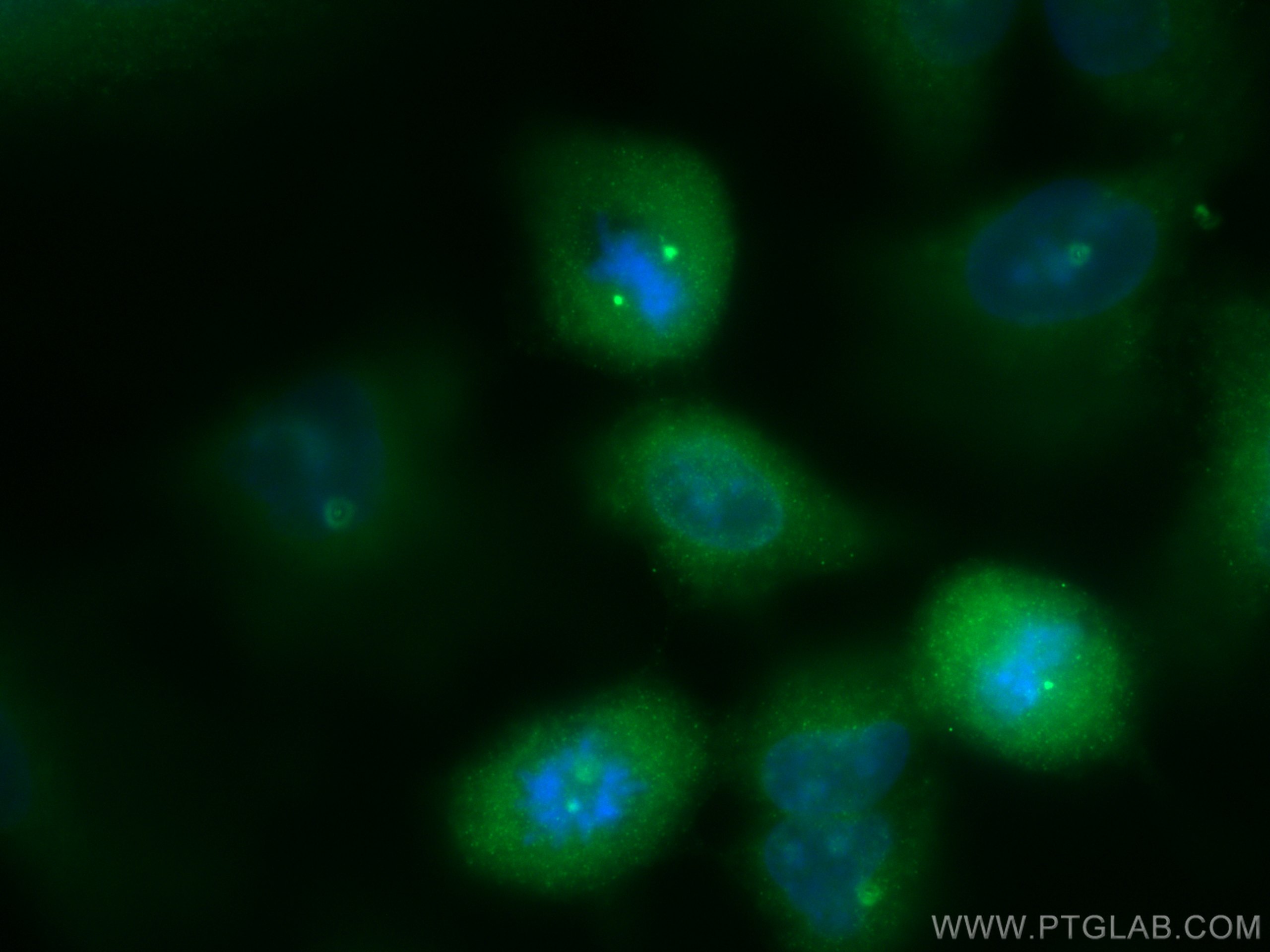
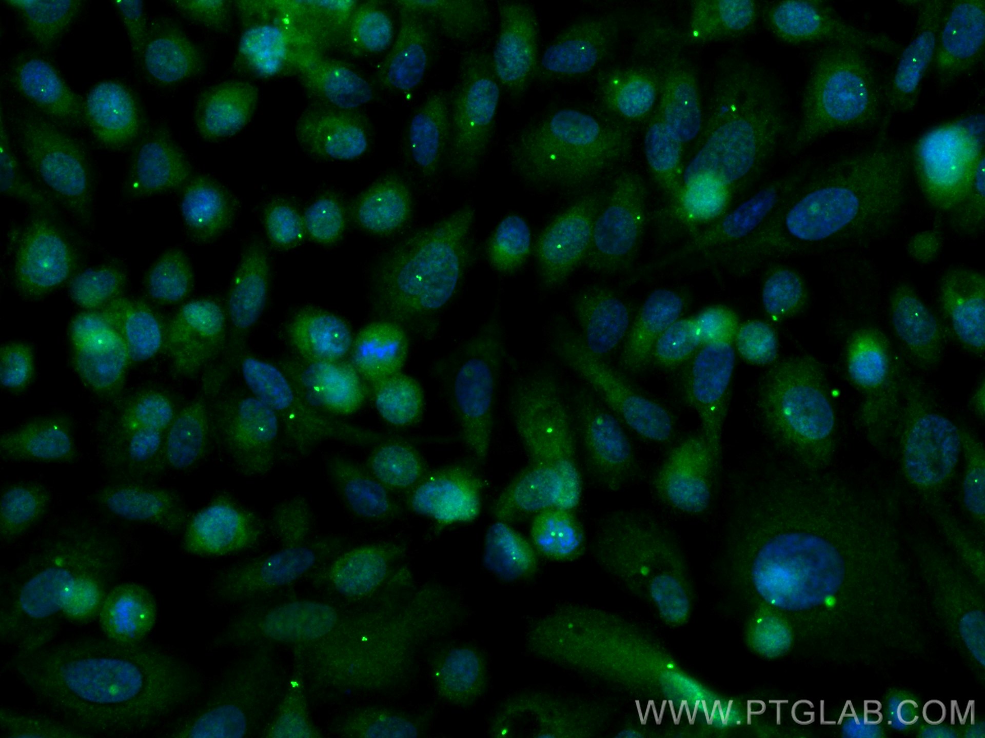
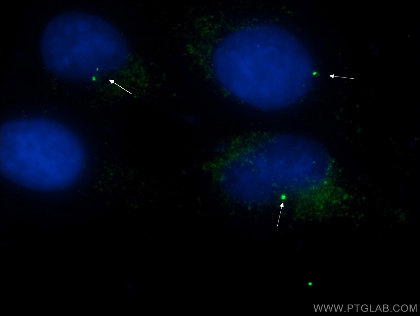
| Positive IF/ICC detected in | HeLa cells, A549 cells, PC-3 cells, hTERT-RPE1 cells, MDCK cells |
Recommended dilution
| Application | Dilution |
|---|---|
| Western Blot (WB) | WB : 1:200-1:1000 |
| Immunoprecipitation (IP) | IP : 0.5-4.0 ug for 1.0-3.0 mg of total protein lysate |
| Immunohistochemistry (IHC) | IHC : 1:100-1:1000 |
| Immunofluorescence (IF)/ICC | IF/ICC : 1:200-1:800 |
| It is recommended that this reagent should be titrated in each testing system to obtain optimal results. | |
| Sample-dependent, Check data in validation data gallery. | |
Product Information
22227-1-AP targets CEP164 in WB, IHC, IF/ICC, IP, ELISA applications and shows reactivity with human, canine samples.
| Tested Reactivity | human, canine |
| Cited Reactivity | human, mouse, xenopus |
| Host / Isotype | Rabbit / IgG |
| Class | Polyclonal |
| Type | Antibody |
| Immunogen |
CatNo: Ag17570 Product name: Recombinant human CEP164 protein Source: e coli.-derived, PGEX-4T Tag: GST Domain: 1-112 aa of BC000602 Sequence: MAGRPLRIGDQLVLEEDYDETYIPSEQEILEFAREIGIDPIKEPELMWLAREGIVAPLPGEWKPCQDITGDIYYFNFANGQSMWDHPCDEHYRSLVIQERAKLSTSGAIKKK Predict reactive species |
| Full Name | centrosomal protein 164kDa |
| Calculated Molecular Weight | 1460 aa, 164 kDa |
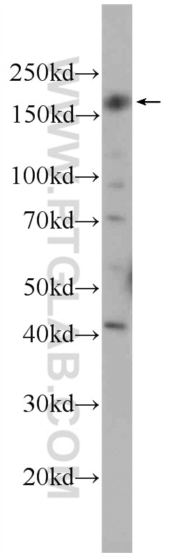
| Observed Molecular Weight | 164 kDa |
| GenBank Accession Number | BC000602 |
| Gene Symbol | CEP164 |
| Gene ID (NCBI) | 22897 |
| RRID | AB_2651175 |
| Conjugate | Unconjugated |
| Form | Liquid |
| Purification Method | Antigen affinity purification |
| UNIPROT ID | Q9UPV0 |
| Storage Buffer | PBS with 0.02% sodium azide and 50% glycerol, pH 7.3. |
| Storage Conditions | Store at -20°C. Stable for one year after shipment. Aliquoting is unnecessary for -20oC storage. 20ul sizes contain 0.1% BSA. |
Background Information
CEP164, also called KIAA1052 or NPHP15, is a 1460 amino acid protein containing 1 WW domain. CEP164 localizes in the microtubule organizing center and is expressed in several cell lines. CEP164 plays a role in microtubule organization and/or maintenance for the formation of primary cilia, a microtubule-based structure that protrudes from the surface of epithelial cells. CEP164 plays a critical role in the G2/M checkpoint and nuclear divisions. The expression of CEP164 is normally limited to the mother centriole, and CEP164 can be used as a useful marker for mother centriole.
Protocols
| Product Specific Protocols | |
|---|---|
| IF protocol for CEP164 antibody 22227-1-AP | Download protocol |
| IHC protocol for CEP164 antibody 22227-1-AP | Download protocol |
| WB protocol for CEP164 antibody 22227-1-AP | Download protocol |
| Standard Protocols | |
|---|---|
| Click here to view our Standard Protocols |
Publications
What published studies show
Able is busy answering your previous question. Please try again later.
| Species | Application | Title |
|---|---|---|
Cell SARS-CoV-2 replication in airway epithelia requires motile cilia and microvillar reprogramming | ||
Cell Time-series reconstruction of the molecular architecture of human centriole assembly | ||
Cell Unraveling cellular complexity with transient adapters in highly multiplexed super-resolution imaging | ||
Cell Res NudCL2 is an autophagy receptor that mediates selective autophagic degradation of CP110 at mother centrioles to promote ciliogenesis. | ||
Nat Commun Spatiotemporal H2O2 flashes coordinate actin cytoskeletal remodeling and regulate cell migration and wound healing |
Reviews
What customers say
The three reviews for 22227-1-AP (anti-CEP164) are all highly positive (5/5). Users consistently praise it for identifying the distal appendage of the primary cilium and mother centriole, calling it a "fabulous" antibody that "works so well." It performs excellently in immunofluorescence at dilutions of 1:300 to 1:2000 across human cell lines including H9, RPE1, HEK293, and HeLa Kyoto, and also works well in Western blot (1:1000). Detailed protocols with methanol fixation and blocking are shared, confirming reliable, clear staining results.
The reviews below have been submitted by verified Proteintech customers who received an incentive for providing their feedback.
Ludovic (Verified Customer) (02-27-2024) | A fabulous antibody to identify the distal appendage of the primary cilium. Never seen an antibody that works so well!
|
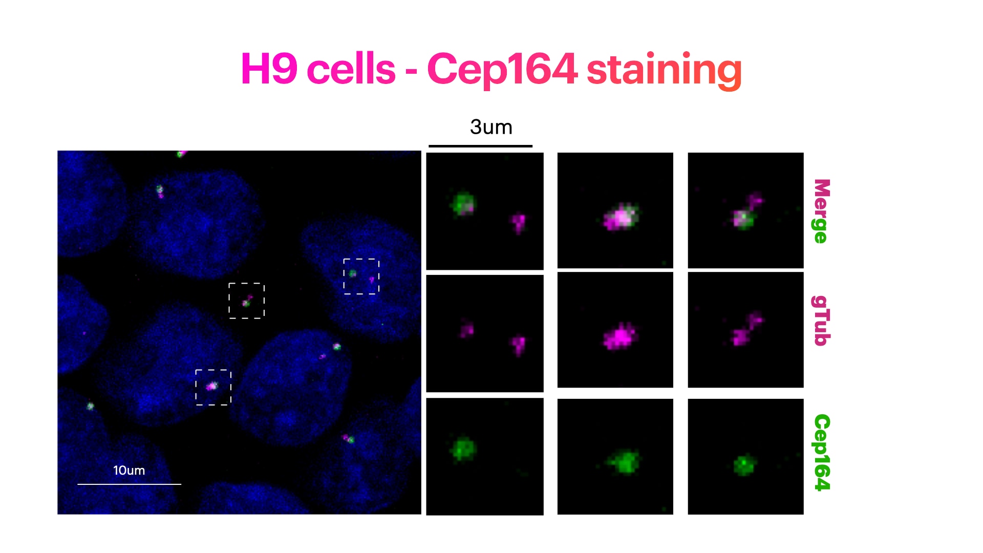
Elisa (Verified Customer) (04-04-2023) | H9 cells stained for Hoechst (DNA marker, in green), Cep164 (mother centriole distal appendage marker, in green) and g-Tubulin (pericentriolar matrix marker, in magenta). H9 cells were fixed in cold methanol for 10' at -20C. Cells were then rehydrated with PBS for 5'. Membrane permeabilization was then performed with 0.1% Triton + 0.1% Tween +0.01%SDS in PBS for 5'. Cells were finally incubated with blocking buffer (5% BSA+ 0.1% Tween in PBS) for 30' at RT. Primary antibody was diluted in blocking buffer 1:300 and incubated for 1h at room temperature. Alexa-488-Anti-rabbit was used as secondary antibody (1:600 dilution) (1h at room temperature).
 |
Pierrick (Verified Customer) (10-24-2019) | Great antibody for immunofluorescence on human cells.Works well in WB
|

