

Tested Applications
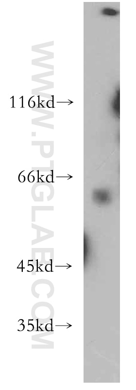
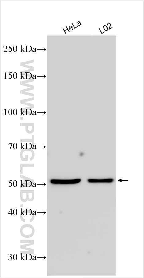
| Positive WB detected in | HeLa cells, human brain tissue, L02 cells |
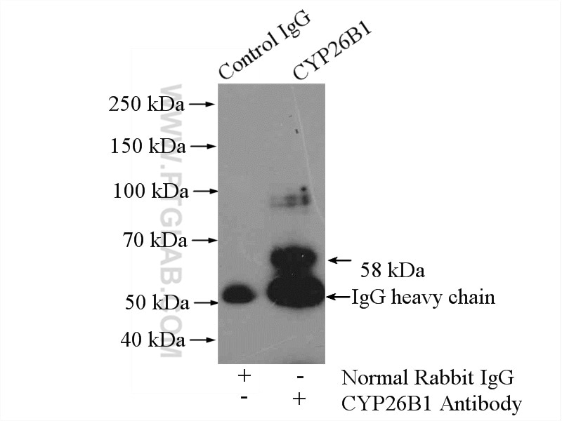
| Positive IP detected in | L02 cells |
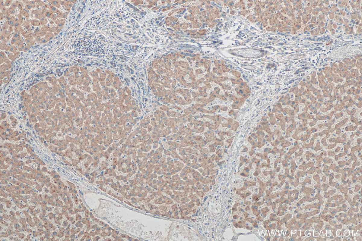
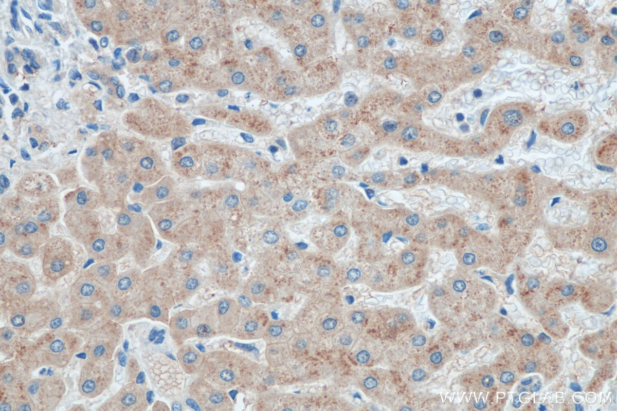
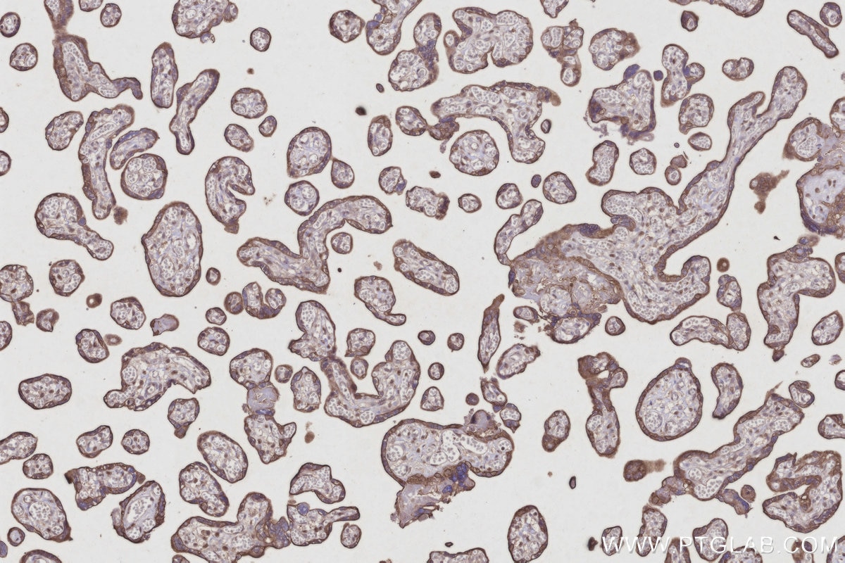
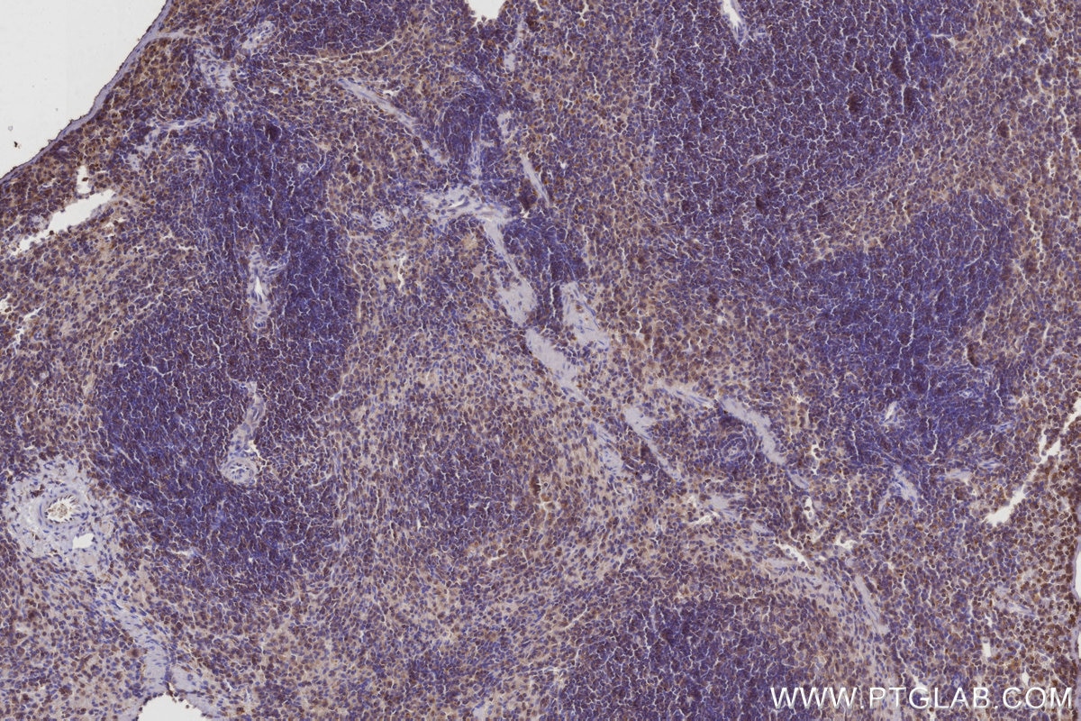
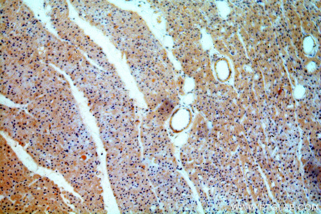
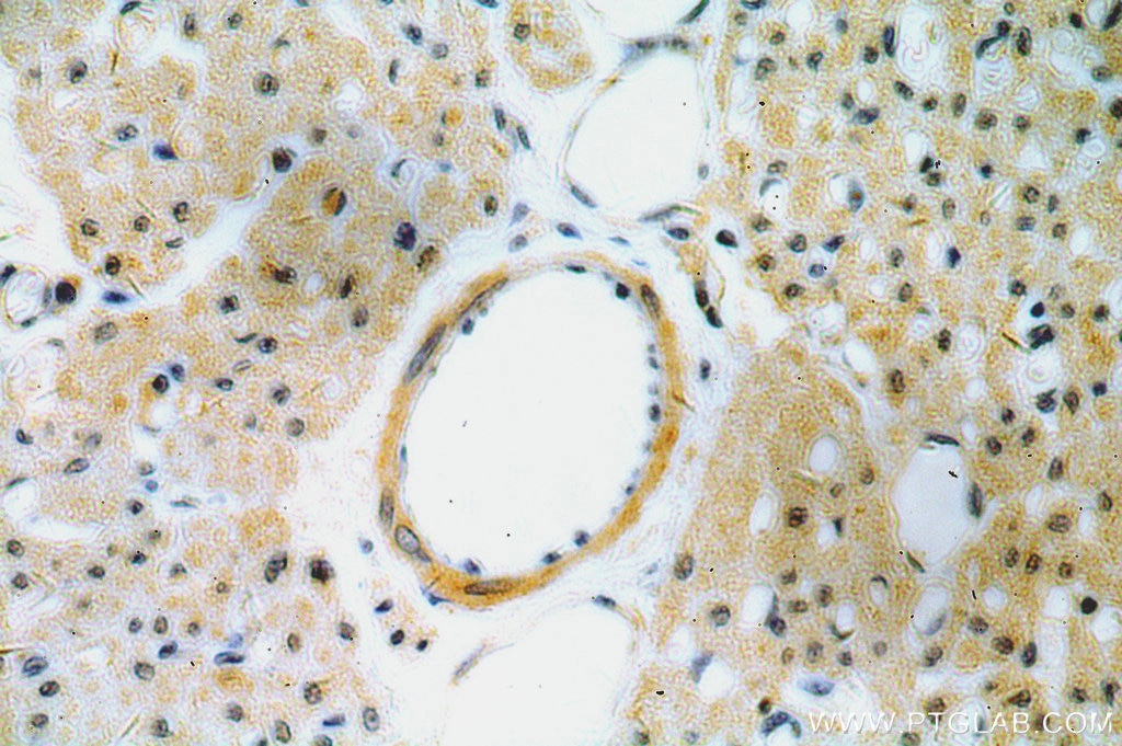
| Positive IHC detected in | human hepatocirrhosis tissue, human heart tissue, mouse spleen tissue, human placenta tissue Note: suggested antigen retrieval with TE buffer pH 9.0; (*) Alternatively, antigen retrieval may be performed with citrate buffer pH 6.0 |
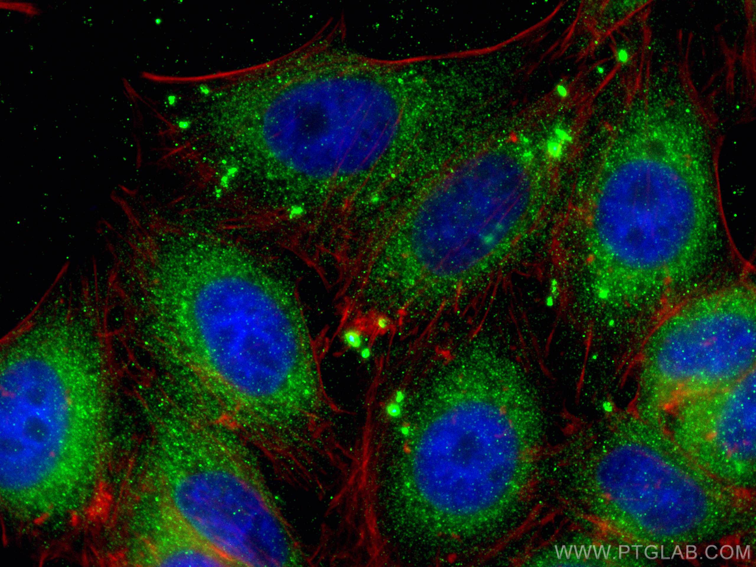
| Positive IF/ICC detected in | HepG2 cells |
Recommended dilution
| Application | Dilution |
|---|---|
| Western Blot (WB) | WB : 1:1000-1:4000 |
| Immunoprecipitation (IP) | IP : 0.5-4.0 ug for 1.0-3.0 mg of total protein lysate |
| Immunohistochemistry (IHC) | IHC : 1:50-1:500 |
| Immunofluorescence (IF)/ICC | IF/ICC : 1:50-1:500 |
| It is recommended that this reagent should be titrated in each testing system to obtain optimal results. | |
| Sample-dependent, Check data in validation data gallery. | |
Product Information
21555-1-AP targets CYP26B1 in WB, IHC, IF/ICC, IP, ELISA applications and shows reactivity with human samples.
| Tested Reactivity | human |
| Cited Reactivity | human, mouse, rat |
| Host / Isotype | Rabbit / IgG |
| Class | Polyclonal |
| Type | Antibody |
| Immunogen |
CatNo: Ag15368 Product name: Recombinant human CYP26B1 protein Source: e coli.-derived, PGEX-4T Tag: GST Domain: 104-300 aa of BC069443 Sequence: KILMGEHHLVSTEWPRSTRMLLGPNTVSNSIGDIHRNKRKVFSKIFSHEALESYLPKIQLVIQDTLRAWSSHPEAINVYQEAQKLTFRMAIRVLLGFSIPEEDLGHLFEVYQQFVDNVFSLPVDLPFSGYRRGIQARQILQKGLEKAIREKLQCTQGKDYLDALDLLIESSKEHGKEMTMQELKDGTLELIFAAYAT Predict reactive species |
| Full Name | cytochrome P450, family 26, subfamily B, polypeptide 1 |
| Calculated Molecular Weight | 512 aa, 58 kDa |
| Observed Molecular Weight | 49-58 kDa |
| GenBank Accession Number | BC069443 |
| Gene Symbol | CYP26B1 |
| Gene ID (NCBI) | 56603 |
| RRID | AB_10733471 |
| Conjugate | Unconjugated |
| Form | Liquid |
| Purification Method | Antigen affinity purification |
| UNIPROT ID | Q9NR63 |
| Storage Buffer | PBS with 0.02% sodium azide and 50% glycerol, pH 7.3. |
| Storage Conditions | Store at -20°C. Stable for one year after shipment. Aliquoting is unnecessary for -20oC storage. 20ul sizes contain 0.1% BSA. |
Background Information
CYP26B1(Cytochrome P450 26B1) is involved in the metabolism of retinoic acid (RA), rendering this classical morphogen inactive through oxidation.It has 3 isoforms produced by alternative splicing.Cyp26b1 deficiency induces pronounced apoptosis in the developing limb and delayed chondrocyte maturation(PMID:15030763).Defects in CYP26B1 are the cause of radiohumeral fusions with other skeletal and craniofacial anomalies (RHFCA).
Protocols
| Product Specific Protocols | |
|---|---|
| IF protocol for CYP26B1 antibody 21555-1-AP | Download protocol |
| IHC protocol for CYP26B1 antibody 21555-1-AP | Download protocol |
| IP protocol for CYP26B1 antibody 21555-1-AP | Download protocol |
| WB protocol for CYP26B1 antibody 21555-1-AP | Download protocol |
| Standard Protocols | |
|---|---|
| Click here to view our Standard Protocols |
Publications
What published studies show
Able is busy answering your previous question. Please try again later.
| Species | Application | Title |
|---|---|---|
Nat Genet Exome-wide analyses identify low-frequency variant in CYP26B1 and additional coding variants associated with esophageal squamous cell carcinoma.
| ||
Cell Stem Cell Multi-omic analysis reveals retinoic acid molecular drivers for dermal fibrosis and regenerative repair in the skin. | ||
Cell Death Dis Single-cell RNA-seq and pathological phenotype reveal the functional atlas and precise roles of Sox30 in testicular cell development and differentiation | ||
Mater Today Bio Self-assembled nanoparticles from natural herbs alleviate osteoarthritis by targeting retinol metabolism and inhibiting chondrocyte ferroptosis. | ||
Hepatol Int Pathological transcriptional retention of Cyp26b1 in perirenal adipose-derived stem cells contributes to MASLD progression. | ||
Antioxid Redox Signal Sulfur Dioxide Alleviates Aortic Dissection Through Inhibiting Vascular Smooth Muscle Cell Phenotype Switch, Migration, and Proliferation via miR-184-3p/Cyp26b1 Axis |
Reviews
What customers say
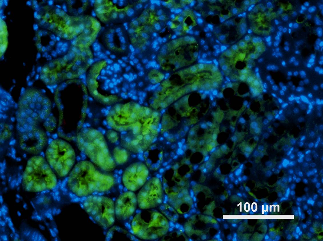
The single review for this product rates it 4 out of 5 stars. The reviewer used the antibody at a 1:200 dilution for immunofluorescence on mouse kidney tissue and reported that it successfully stained a subset of proximal tubules, indicating clear and specific signal in the intended tissue context.
The reviews below have been submitted by verified Proteintech customers who received an incentive for providing their feedback.
Daniel (Verified Customer) (02-06-2020) | The antibody stains a subset of proximal tubulues.
 |

