



Tested Applications
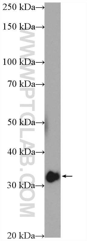
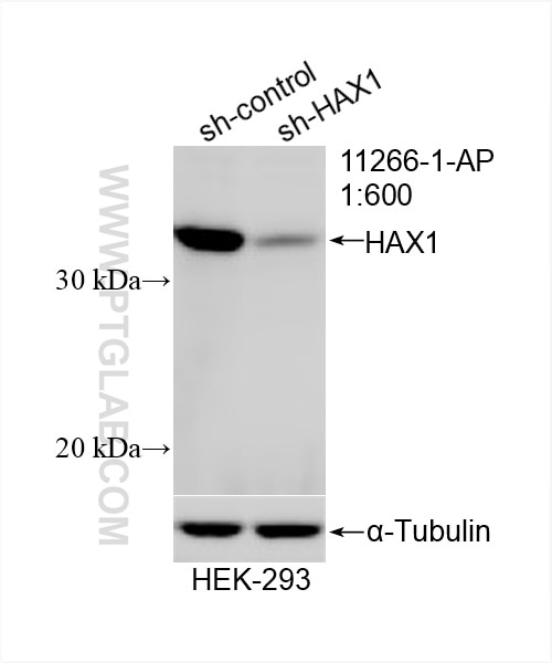
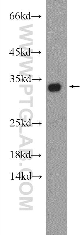
| Positive WB detected in | HeLa cells, HEK-293 cells |
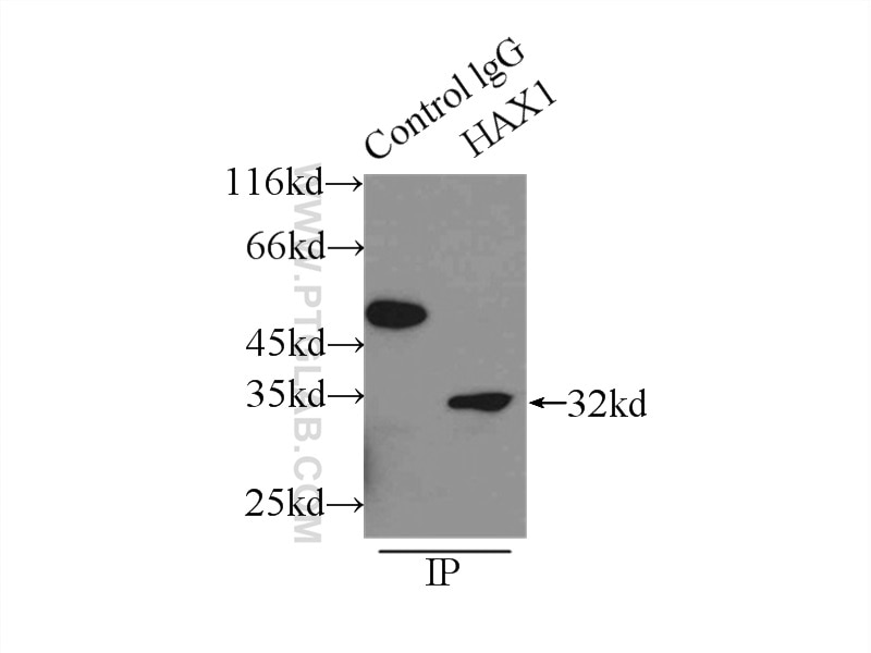
| Positive IP detected in | mouse brain tissue |
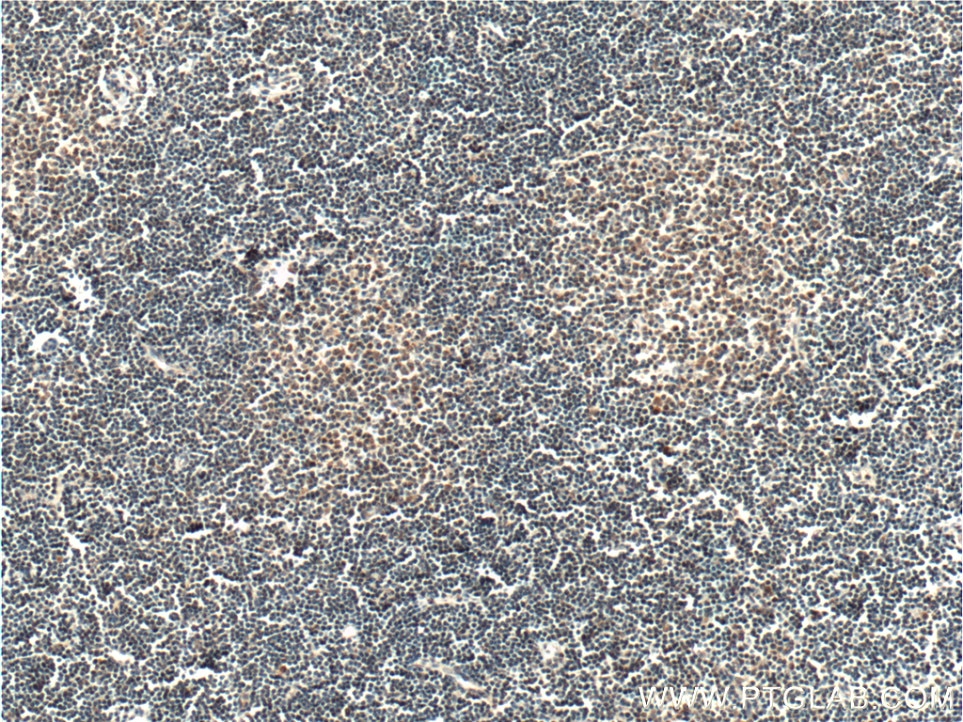
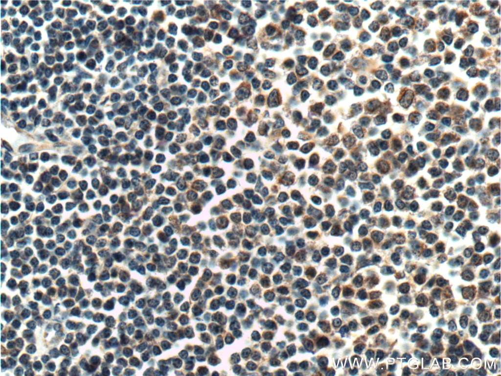
| Positive IHC detected in | human lymphoma tissue, mouse brain tissue Note: suggested antigen retrieval with TE buffer pH 9.0; (*) Alternatively, antigen retrieval may be performed with citrate buffer pH 6.0 |
| Positive IF/ICC detected in | HeLa cells |
Recommended dilution
| Application | Dilution |
|---|---|
| Western Blot (WB) | WB : 1:500-1:2000 |
| Immunoprecipitation (IP) | IP : 0.5-4.0 ug for 1.0-3.0 mg of total protein lysate |
| Immunohistochemistry (IHC) | IHC : 1:50-1:500 |
| Immunofluorescence (IF)/ICC | IF/ICC : 1:50-1:500 |
| It is recommended that this reagent should be titrated in each testing system to obtain optimal results. | |
| Sample-dependent, Check data in validation data gallery. | |
Product Information
11266-1-AP targets HAX1 in WB, IHC, IF/ICC, IP, CoIP, RIP, ELISA applications and shows reactivity with human, mouse samples.
| Tested Reactivity | human, mouse |
| Cited Reactivity | human, mouse, pig, monkey, hamster |
| Host / Isotype | Rabbit / IgG |
| Class | Polyclonal |
| Type | Antibody |
| Immunogen |
CatNo: Ag1792 Product name: Recombinant human HAX1 protein Source: e coli.-derived, PGEX-4T Tag: GST Domain: 1-279 aa of BC014314 Sequence: MSLFDLFRGFFGFPGPRSHRDPFFGGMTRDEDDDEEEEEEGGSWGRGNPRFHSPQHPPEEFGFGFSFSPGGGIRFHDNFGFDDLVRDFNSIFSDMGAWTLPSHPPELPGPESETPGERLREGQTLRDSMLKYPDSHQPRIFGGVLESDARSESPQPAPDWGSQRPFHRFDDVWPMDPHPRTREDNDLDSQVSQEGLGPVLQPQPKSYFKSISVTKITKPDGIVEERRTVVDSEGRTETTVTRHEADSSPRGDPESPRPPALDDAFSILDLFLGRWFRSR Predict reactive species |
| Full Name | HCLS1 associated protein X-1 |
| Calculated Molecular Weight | 32 kDa |
| Observed Molecular Weight | 32 kDa |
| GenBank Accession Number | BC014314 |
| Gene Symbol | HAX1 |
| Gene ID (NCBI) | 10456 |
| RRID | AB_2263720 |
| Conjugate | Unconjugated |
| Form | Liquid |
| Purification Method | Antigen affinity purification |
| UNIPROT ID | O00165 |
| Storage Buffer | PBS with 0.02% sodium azide and 50% glycerol, pH 7.3. |
| Storage Conditions | Store at -20°C. Stable for one year after shipment. Aliquoting is unnecessary for -20oC storage. 20ul sizes contain 0.1% BSA. |
Background Information
HAX1, also named as HS1BP1, is a 32 kDa protein that is ubiquitously expressed. Hax1 has been demonstrated to be a negative regulator of apoptosis in many immune cell types. Furthermore, Hax1 has been shown to have additional roles in regulating cell motility and adhesion, and is overexpressed in many types of cancer. Kristina and Cavnar demonstrate in their paper validation of two research antibodies including Proteintech's anti-Hax1 (11266-1-AP). They have evidence that the rabbit anti-Hax1 (11266-1-AP, Proteintech Group Inc.) results in a more robust and consistent detection of Hax1.
Protocols
| Product Specific Protocols | |
|---|---|
| IF protocol for HAX1 antibody 11266-1-AP | Download protocol |
| IHC protocol for HAX1 antibody 11266-1-AP | Download protocol |
| IP protocol for HAX1 antibody 11266-1-AP | Download protocol |
| WB protocol for HAX1 antibody 11266-1-AP | Download protocol |
| Standard Protocols | |
|---|---|
| Click here to view our Standard Protocols |
Publications
What published studies show
Able is busy answering your previous question. Please try again later.
| Species | Application | Title |
|---|---|---|
Nat Commun Tumour initiated purinergic signalling promotes cardiomyocyte RBFOX1 degradation and cardiotoxicity from DNA damaging anticancer agents | ||
Nat Commun Cerebellar Kv3.3 potassium channels activate TANK-binding kinase 1 to regulate trafficking of the cell survival protein Hax-1. | ||
Nat Commun The EIF3H-HAX1 axis increases RAF-MEK-ERK signaling activity to promote colorectal cancer progression
| ||
Nat Commun Interactome profiling of Crimean-Congo hemorrhagic fever virus glycoproteins
| ||
Mol Cell The IMiD target CRBN determines HSP90 activity toward transmembrane proteins essential in multiple myeloma. |
Reviews
What customers say
The single review for this product is a 5-star rating from an immunofluorescence application. The reviewer used a 1:150 dilution to stain mouse primary lymphocytes and reported strong, clean staining that localized to the mitochondria, consistent with published literature. Overall, the feedback is positive, highlighting reliable performance for IF on primary cells.
The reviews below have been submitted by verified Proteintech customers who received an incentive for providing their feedback.
Sara (Verified Customer) (10-06-2024) | It works well for immunofluorescence. I stained mouse primary lymphocytes with it and gave a nice staining, it seems to localise to the mitochondria as described in the literature.
 |


