

Tested Applications
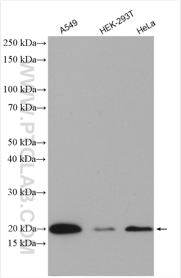
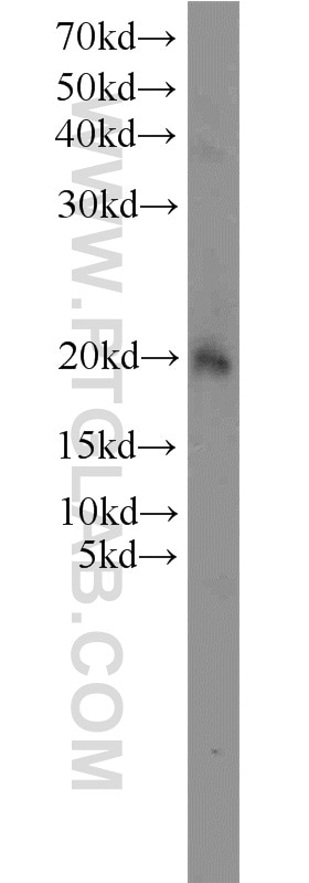
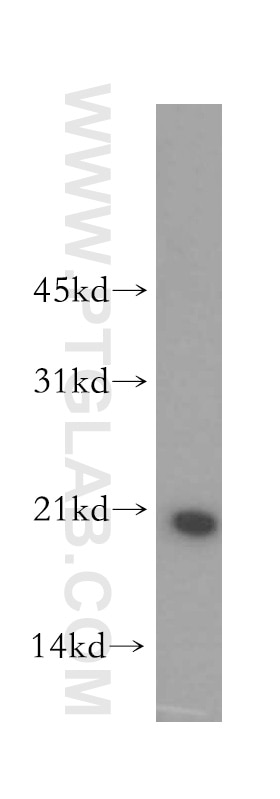
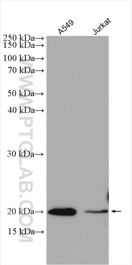
| Positive WB detected in | A549 cells, human kidney tissue, mouse thymus tissue, Jurkat cells, HeLa cells, HEK-293T cells |
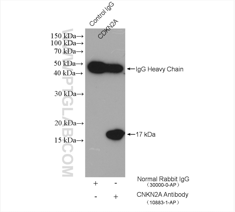
| Positive IP detected in | HeLa cells |
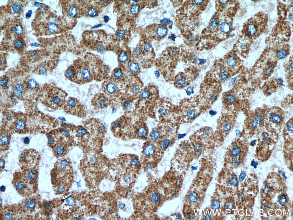
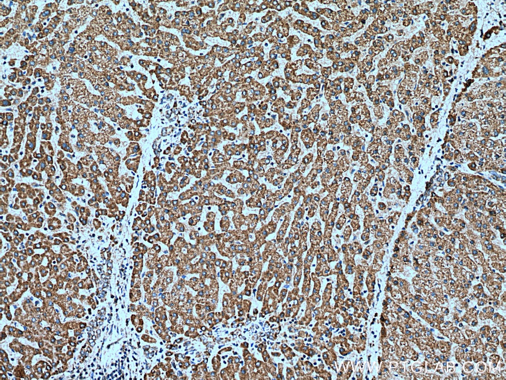
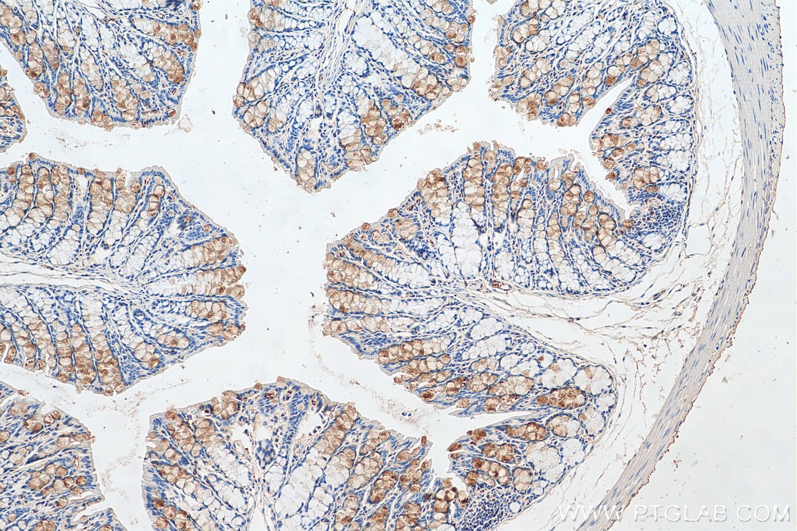
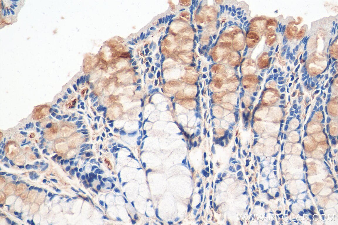
| Positive IHC detected in | human liver tissue, mouse colon tissue Note: suggested antigen retrieval with TE buffer pH 9.0; (*) Alternatively, antigen retrieval may be performed with citrate buffer pH 6.0 |
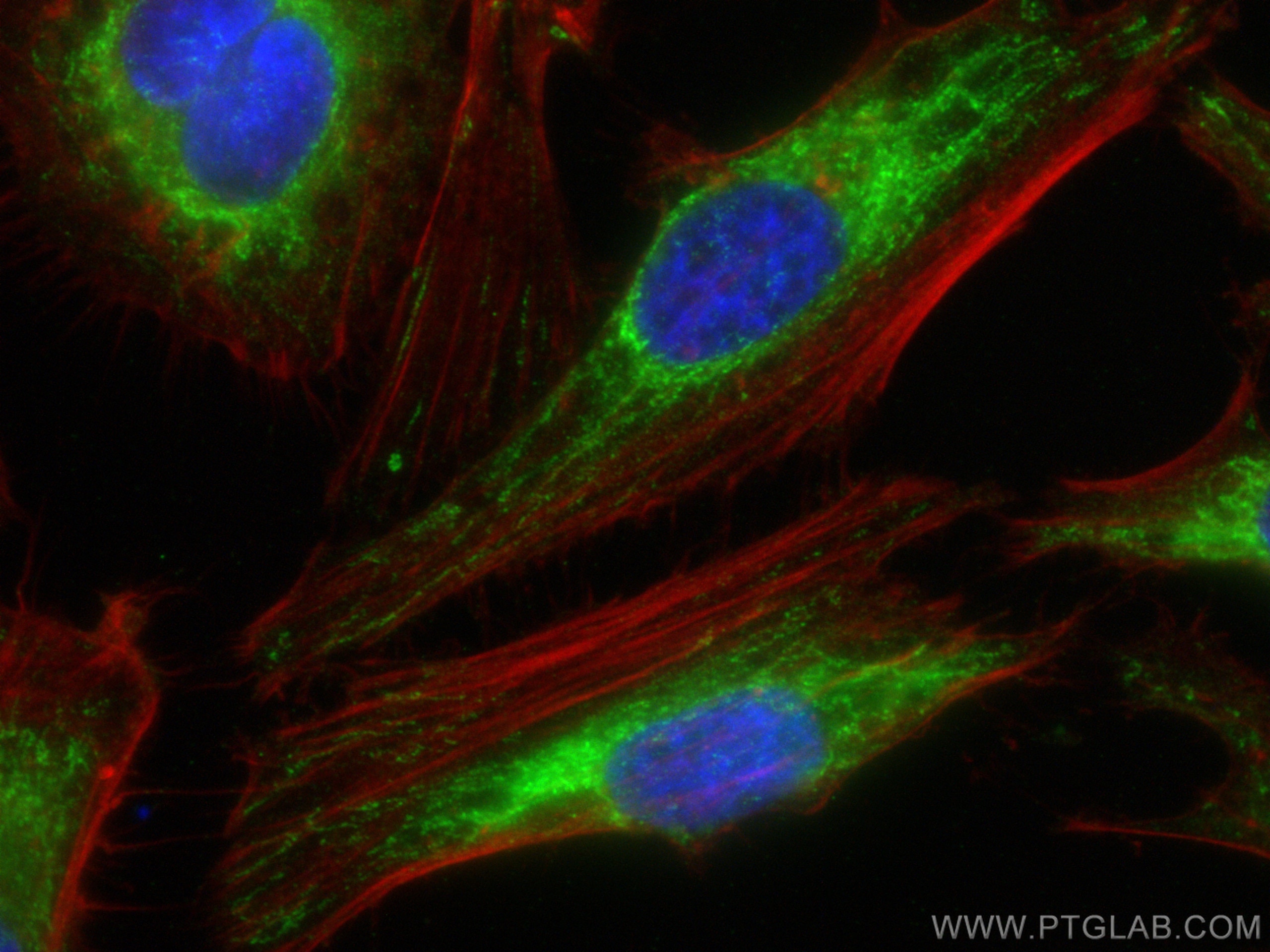
| Positive IF/ICC detected in | HeLa cells |
Recommended dilution
| Application | Dilution |
|---|---|
| Western Blot (WB) | WB : 1:500-1:2000 |
| Immunoprecipitation (IP) | IP : 0.5-4.0 ug for 1.0-3.0 mg of total protein lysate |
| Immunohistochemistry (IHC) | IHC : 1:50-1:500 |
| Immunofluorescence (IF)/ICC | IF/ICC : 1:50-1:500 |
| It is recommended that this reagent should be titrated in each testing system to obtain optimal results. | |
| Sample-dependent, Check data in validation data gallery. | |
Product Information
15638-1-AP targets OPA3 in WB, IHC, IF/ICC, IP, ELISA applications and shows reactivity with human, mouse, rat samples.
| Tested Reactivity | human, mouse, rat |
| Cited Reactivity | human, mouse, rat, bovine |
| Host / Isotype | Rabbit / IgG |
| Class | Polyclonal |
| Type | Antibody |
| Immunogen |
CatNo: Ag8103 Product name: Recombinant human OPA3 protein Source: e coli.-derived, PGEX-4T Tag: GST Domain: 24-179 aa of BC005059 Sequence: ANRIKEAARRSEFFKTYICLPPAQLYHWVEMRTKMRIMGFRGTVIKPLNEEAAAELGAELLGEATIFIVGGGCLVLEYWRHQAQQRHKEEEQRAAWNALRDEVGHLALALEALQAQVQAAPPQGALEELRTELQEVRAQLCNPGRSASHAVPASKK Predict reactive species |
| Full Name | optic atrophy 3 (autosomal recessive, with chorea and spastic paraplegia) |
| Calculated Molecular Weight | 179 aa, 20 kDa |
| Observed Molecular Weight | 20 kDa |
| GenBank Accession Number | BC005059 |
| Gene Symbol | OPA3 |
| Gene ID (NCBI) | 80207 |
| RRID | AB_2158168 |
| Conjugate | Unconjugated |
| Form | Liquid |
| Purification Method | Antigen affinity purification |
| UNIPROT ID | Q9H6K4 |
| Storage Buffer | PBS with 0.02% sodium azide and 50% glycerol, pH 7.3. |
| Storage Conditions | Store at -20°C. Stable for one year after shipment. Aliquoting is unnecessary for -20oC storage. 20ul sizes contain 0.1% BSA. |
Background Information
The OPA3 cDNA encodes a deduced 179-amino acid protein. Northern blot analysis demonstrated a primary transcript of approximately 5.0 kb that was ubiquitously expressed, most prominently in skeletal muscle and kidney. Mutations in this gene have been shown to result in 3-methylglutaconic aciduria type III and autosomal dominant optic atrophy and cataract.
Protocols
| Product Specific Protocols | |
|---|---|
| IF protocol for OPA3 antibody 15638-1-AP | Download protocol |
| IHC protocol for OPA3 antibody 15638-1-AP | Download protocol |
| IP protocol for OPA3 antibody 15638-1-AP | Download protocol |
| WB protocol for OPA3 antibody 15638-1-AP | Download protocol |
| Standard Protocols | |
|---|---|
| Click here to view our Standard Protocols |
Publications
What published studies show
The OPA3 Polyclonal antibody (Cat# 15638-1-AP) has 7 citations published in journals including Invest Ophthalmol Vis Sci, Cancers (Basel), Cell Signal, Genomics, Hum Mol Genet, Bioengineered, and In Vitro Cell Dev Biol Anim. Research fields span mitochondrial biology, ophthalmology, oncology (pancreatic, hepatocellular, colorectal cancer), cardiotoxicity, ferroptosis, Costeff syndrome, and the cGAS-STING signaling pathway.
| Species | Application | Title |
|---|---|---|
Invest Ophthalmol Vis Sci Mitochondrial localization and ocular expression of mutant Opa3 in a mouse model of 3-methylglutaconicaciduria type III. | ||
Cancers (Basel) Oncogenic K-ras Induces Mitochondrial OPA3 Expression to Promote Energy Metabolism in Pancreatic Cancer Cells. | ||
Cell Signal Hydrogen sulfide alleviates mitochondrial damage and ferroptosis by regulating OPA3-NFS1 axis in doxorubicin-induced cardiotoxicity | ||
Genomics A nonsense mutation in the optic atrophy 3 gene (OPA3) causes dilated cardiomyopathy in Red Holstein cattle. | ||
Hum Mol Genet Disrupted mitochondrial function in the Opa3L122P mouse model for Costeff Syndrome impairs skeletal integrity. | ||
Bioengineered MYB proto-oncogene like 2 promotes hepatocellular carcinoma growth and glycolysis via binding to the Optic atrophy 3 promoter and activating its expression.
|

