


Tested Applications
| Positive WB detected in | HEK-293 cells, human liver tissue, human testis tissue, HeLa cells, Jurkat cells, U-251 cells |
| Positive IP detected in | HeLa cells, HEK-293 cells |
| Positive IHC detected in | human prostate cancer tissue Note: suggested antigen retrieval with TE buffer pH 9.0; (*) Alternatively, antigen retrieval may be performed with citrate buffer pH 6.0 |
| Positive IF/ICC detected in | MCF-7 cells |
Recommended dilution
| Application | Dilution |
|---|---|
| Western Blot (WB) | WB : 1:500-1:2000 |
| Immunoprecipitation (IP) | IP : 0.5-4.0 ug for 1.0-3.0 mg of total protein lysate |
| Immunohistochemistry (IHC) | IHC : 1:50-1:500 |
| Immunofluorescence (IF)/ICC | IF/ICC : 1:50-1:500 |
| It is recommended that this reagent should be titrated in each testing system to obtain optimal results. | |
| Sample-dependent, Check data in validation data gallery. | |
Published Applications
| WB | See 6 publications below |
| IHC | See 1 publications below |
| IF | See 1 publications below |
Product Information
15611-1-AP targets OXSR1 in WB, IHC, IF/ICC, IP, ELISA applications and shows reactivity with human, mouse, rat samples.
| Tested Reactivity | human, mouse, rat |
| Cited Reactivity | human, rat |
| Host / Isotype | Rabbit / IgG |
| Class | Polyclonal |
| Type | Antibody |
| Immunogen |
CatNo: Ag8002 Product name: Recombinant human OXSR1 protein Source: e coli.-derived, PGEX-4T Tag: GST Domain: 1-288 aa of BC008726 Sequence: MSEDSSALPWSINRDDYELQEVIGSGATAVVQAAYCAPKKEKVAIKRINLEKCQTSMDELLKEIQAMSQCHHPNIVSYYTSFVVKDELWLVMKLLSGGSVLDIIKHIVAKGEHKSGVLDESTIATILREVLEGLEYLHKNGQIHRDVKAGNILLGEDGSVQIADFGVSAFLATGGDITRNKVRKTFVGTPCWMAPEVMEQVRGYDFKADIWSFGITAIELATGAAPYHKYPPMKVLMLTLQNDPPSLETGVQDKEMLKKYGKSFRKMISLCLQKDPEKRPTAAELLRH Predict reactive species |
| Full Name | oxidative-stress responsive 1 |
| Calculated Molecular Weight | 58 kDa |
| Observed Molecular Weight | 58 kDa |
| GenBank Accession Number | BC008726 |
| Gene Symbol | OXSR1 |
| Gene ID (NCBI) | 9943 |
| RRID | AB_2299030 |
| Conjugate | Unconjugated |
| Form | Liquid |
| Purification Method | Antigen affinity purification |
| UNIPROT ID | O95747 |
| Storage Buffer | PBS with 0.02% sodium azide and 50% glycerol, pH 7.3. |
| Storage Conditions | Store at -20°C. Stable for one year after shipment. Aliquoting is unnecessary for -20oC storage. 20ul sizes contain 0.1% BSA. |
Background Information
Oxidative-stress responsive 1(OXSR1) is also named as KIAA1101, OSR1 and belongs to the STE Ser/Thr protein kinase family. It contains an N-terminal Ste20-like ser/thr kinase domain and 2 C-terminal regions, which has a putative caspase-3 cleavage site at the end. OXSR1's interaction with WNK1 is required for NKCC function, and it modulates the G protein sensitivity of PAK by phosphorylation of PAK1.Western blot analysis detected Oxsr1 at an apparent molecular mass of 58 kD in all mouse tissues examined except thymus. Cell fractionation and immunofluorescence analysis of HeLa cells showed that OXSR1 was distributed throughout the cell and OXSR1 could phosphorylate a test substrate and itself(PMID:14707132).
Protocols
| Product Specific Protocols | |
|---|---|
| IF protocol for OXSR1 antibody 15611-1-AP | Download protocol |
| IHC protocol for OXSR1 antibody 15611-1-AP | Download protocol |
| IP protocol for OXSR1 antibody 15611-1-AP | Download protocol |
| WB protocol for OXSR1 antibody 15611-1-AP | Download protocol |
| Standard Protocols | |
|---|---|
| Click here to view our Standard Protocols |
Publications
| Species | Application | Title |
|---|---|---|
Bioengineered Upregulation of Oxidative stress-responsive 1(OXSR1) Predicts Poor Prognosis and Promotes Hepatocellular Carcinoma Progression. | ||
Adv Sci (Weinh) Modulation of Cerebrospinal Fluid Dysregulation via a SPAK and OSR1 Targeted Framework Nucleic Acid in Hydrocephalus | ||
Heliyon Sevoflurane-induced regulation of NKCC1/KCC2 phosphorylation through activation of Spak/OSR1 kinase and cognitive impairment in ischemia-reperfusion injury in rats | ||
Exp Mol Pathol Proteomic analysis of the effects of Dictyophora polysaccharide on arsenic-induced hepatotoxicity in rats | ||
Nat Commun Thermal proteome profiling reveals fructose-1,6-bisphosphate as a phosphate donor to activate phosphoglycerate mutase 1 |
Reviews
The reviews below have been submitted by verified Proteintech customers who received an incentive for providing their feedback.
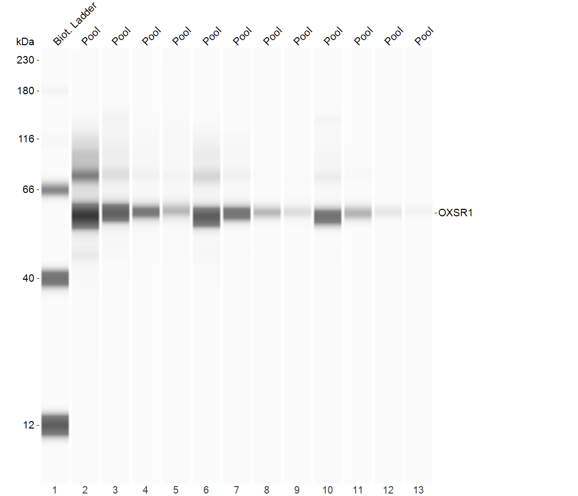
FH Jennifer (Verified Customer) (03-23-2022) | Image provided from optimisation plate for OXSR1 for rat brain lysates. Analysed using Simple Western Analysis (WES) by Protein Simple. Finalised antibody and protein concentration for samples produced a single band with no non-specific binding.
 |


