
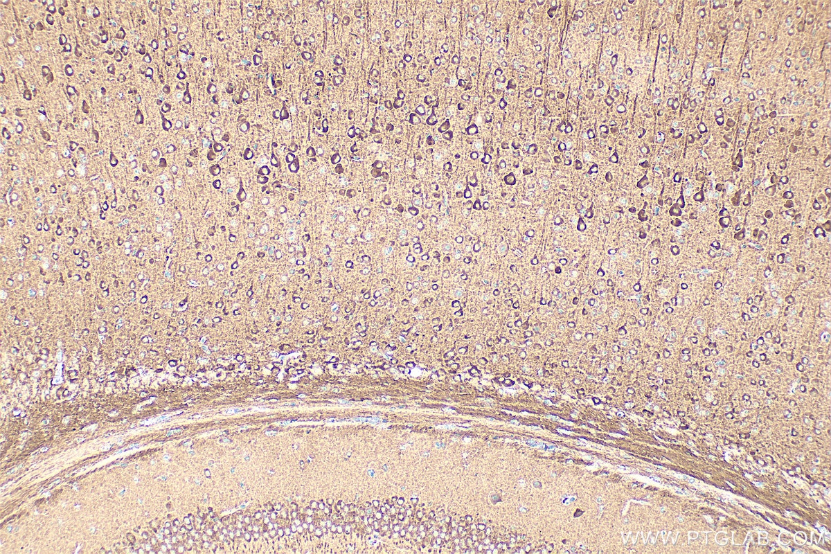

Tested Applications
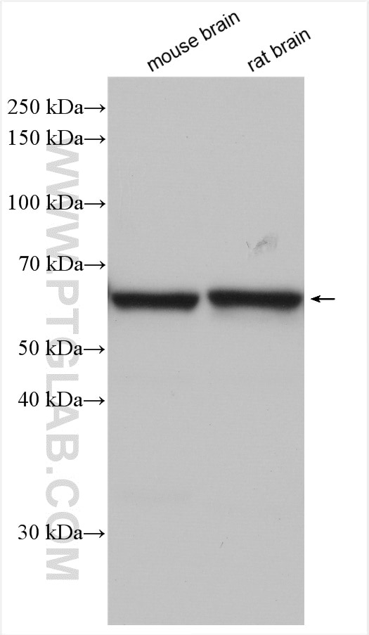
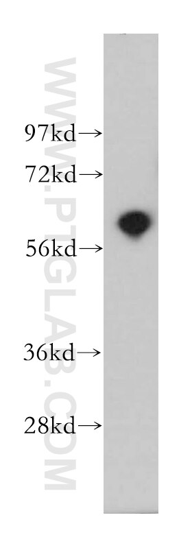
| Positive WB detected in | mouse brain tissue, human brain tissue, rat brain tissue |
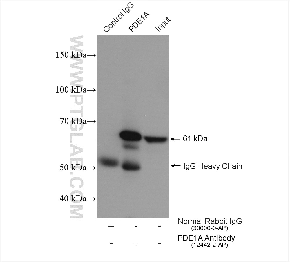
| Positive IP detected in | mouse brain tissue |
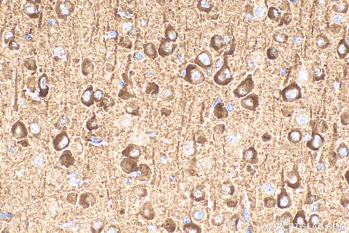
| Positive IHC detected in | mouse brain tissue Note: suggested antigen retrieval with TE buffer pH 9.0; (*) Alternatively, antigen retrieval may be performed with citrate buffer pH 6.0 |
Recommended dilution
| Application | Dilution |
|---|---|
| Western Blot (WB) | WB : 1:1000-1:6000 |
| Immunoprecipitation (IP) | IP : 0.5-4.0 ug for 1.0-3.0 mg of total protein lysate |
| Immunohistochemistry (IHC) | IHC : 1:200-1:800 |
| It is recommended that this reagent should be titrated in each testing system to obtain optimal results. | |
| Sample-dependent, Check data in validation data gallery. | |
Product Information
12442-2-AP targets PDE1A in WB, IHC, IP, ELISA applications and shows reactivity with human, mouse, rat samples.
| Tested Reactivity | human, mouse, rat |
| Cited Reactivity | human, mouse |
| Host / Isotype | Rabbit / IgG |
| Class | Polyclonal |
| Type | Antibody |
| Immunogen |
CatNo: Ag3157 Product name: Recombinant human PDE1A protein Source: e coli.-derived, PGEX-4T Tag: GST Domain: 185-545 aa of BC022480 Sequence: TRYDLINRFKIPVSCLITFAEALEVGYSKYKNPYHNLIHAADVTQTVHYIMLHTGIMHWLTELEILAMVFAAAIHDYEHTGTTNNFHIQTRSDVAILYNDRSVLENHHVSAAYRLMQEEEMNILINLSKDDWRDLRNLVIEMVLSTDMSGHFQQIKNIRNSLQQPEGIDRAKTMSLILHAADISHPAKSWKLHYRWTMALMEEFFLQGDKEAELGLPFSPLCDRKSTMVAQSQIGFIDFIVEPTFSLLTDSTEKIVIPLIEEASKAETSSYVASSSTTIVGLHIADALRRSNTKGSMSDGSYSPDYSLAAVDLKSFKNNLVDIIQQNKERWKELAAQGESDLHKNSEDLVNAEEKHDETHS Predict reactive species |
| Full Name | phosphodiesterase 1A, calmodulin-dependent |
| Calculated Molecular Weight | 545 aa, 61 kDa |
| Observed Molecular Weight | 61 kDa |
| GenBank Accession Number | BC022480 |
| Gene Symbol | PDE1A |
| Gene ID (NCBI) | 5136 |
| RRID | AB_2162893 |
| Conjugate | Unconjugated |
| Form | Liquid |
| Purification Method | Antigen affinity purification |
| UNIPROT ID | P54750 |
| Storage Buffer | PBS with 0.02% sodium azide and 50% glycerol, pH 7.3. |
| Storage Conditions | Store at -20°C. Stable for one year after shipment. Aliquoting is unnecessary for -20oC storage. 20ul sizes contain 0.1% BSA. |
Background Information
PDE1A(Calcium/calmodulin-dependent 3',5'-cyclic nucleotide phosphodiesterase 1A) is also named as Cam-PDE 1A, 61 kDa Cam-PDE, hCam-1 and belongs to the cyclic nucleotide phosphodiesterase (PDEs) family. The PDEs play a role in signal transduction by regulating intracellular cyclic nucleotide concentrations through hydrolysis of cAMP and/or cGMP to their respective nucleoside 5-prime monophosphates. This protein has 9 isoforms produced by alternative splicing with the molecular weight from 57 kDa to 63 kDa. There is a report showing the major 67-kD PDE1A isoform identified in human sperm associates tightly with calmodulin and is not activated by Ca(2+)/calmodulin(PMID:12135876).
Protocols
| Product Specific Protocols | |
|---|---|
| IHC protocol for PDE1A antibody 12442-2-AP | Download protocol |
| IP protocol for PDE1A antibody 12442-2-AP | Download protocol |
| WB protocol for PDE1A antibody 12442-2-AP | Download protocol |
| Standard Protocols | |
|---|---|
| Click here to view our Standard Protocols |
Publications
What published studies show
The PDE1A antibody (Cat# 12442-2-AP) has 8 citations across journals including Mol Cancer, Mol Ther, Cell Rep, Biol Reprod, Front Genet, PLoS One, and eLife. Research fields span breast cancer senescence and drug resistance, renal fibrosis, striatal development, ovarian follicle biology, Crohn's disease and enteric smooth muscle pathology, Pde1a knockout mouse characterization, and non-small cell lung cancer progression. Applications include WB and IHC in human and mouse samples.
| Species | Application | Title |
|---|---|---|
Mol Cancer Therapy-induced senescence is a transient drug resistance mechanism in breast cancer | ||
Mol Ther Phosphodiesterase 1A overexpression contributes to the progression of renal fibrosis. | ||
Cell Rep Single-Cell Analysis of Foxp1-Driven Mechanisms Essential for Striatal Development. | ||
Biol Reprod Follicle-stimulating hormone and luteinizing hormone increase Ca2+ in the granulosa cells of mouse ovarian follicles†. | ||
Biol Reprod Luteinizing Hormone Causes Phosphorylation and Activation of the cGMP Phosphodiesterase PDE5 in Rat Ovarian Follicles, Contributing, Together with PDE1 Activity, to the Resumption of Meiosis. | ||
Front Genet Transcriptome profiling of intact bowel wall reveals that PDE1A and SEMA3D are possible markers with roles in enteric smooth muscle apoptosis, proliferative disorders, and dysautonomia in Crohn's disease |

