


Tested Applications
| Positive WB detected in | Jurkat cells, HEK-293 cells, human kidney tissue, human liver tissue, K-562 cells |
| Positive IP detected in | Jurkat cells |
| Positive IHC detected in | mouse kidney tissue, human kidney tissue, human stomach tissue Note: suggested antigen retrieval with TE buffer pH 9.0; (*) Alternatively, antigen retrieval may be performed with citrate buffer pH 6.0 |
Recommended dilution
| Application | Dilution |
|---|---|
| Western Blot (WB) | WB : 1:1000-1:8000 |
| Immunoprecipitation (IP) | IP : 0.5-4.0 ug for 1.0-3.0 mg of total protein lysate |
| Immunohistochemistry (IHC) | IHC : 1:50-1:500 |
| It is recommended that this reagent should be titrated in each testing system to obtain optimal results. | |
| Sample-dependent, Check data in validation data gallery. | |
Published Applications
| KD/KO | See 5 publications below |
| WB | See 38 publications below |
| IHC | See 2 publications below |
| IF | See 6 publications below |
| IP | See 2 publications below |
| CoIP | See 2 publications below |
Product Information
12704-1-AP targets SNAP29 in WB, IHC, IF, IP, CoIP, ELISA applications and shows reactivity with human, mouse, rat samples.
| Tested Reactivity | human, mouse, rat |
| Cited Reactivity | human, mouse, rat |
| Host / Isotype | Rabbit / IgG |
| Class | Polyclonal |
| Type | Antibody |
| Immunogen |
CatNo: Ag3382 Product name: Recombinant human SNAP29 protein Source: e coli.-derived, PGEX-4T Tag: GST Domain: 1-258 aa of BC009715 Sequence: MSAYPKSYNPFDDDGEDEGARPAPWRDARDLPDGPDAPADRQQYLRQEVLRRAEATAASTSRSLALMYESEKVGVASSEELARQRGVLERTEKMVDKMDQDLKISQKHINSIKSVFGGLVNYFKSKPVETPPEQNGTLTSQPNNRLKEAISTSKEQEAKYQASHPNLRKLDDTDPVPRGAGSAMSTDAYPKNPHLRAYHQKIDSNLDELSMGLGRLKDIALGMQTEIEEQDDILDRLTTKVDKLDVNIKSTERKVRQL Predict reactive species |
| Full Name | synaptosomal-associated protein, 29kDa |
| Calculated Molecular Weight | 258 aa, 29 kDa |
| Observed Molecular Weight | 29 kDa |
| GenBank Accession Number | BC009715 |
| Gene Symbol | SNAP29 |
| Gene ID (NCBI) | 9342 |
| RRID | AB_2192340 |
| Conjugate | Unconjugated |
| Form | Liquid |
| Purification Method | Antigen affinity purification |
| UNIPROT ID | O95721 |
| Storage Buffer | PBS with 0.02% sodium azide and 50% glycerol, pH 7.3. |
| Storage Conditions | Store at -20°C. Stable for one year after shipment. Aliquoting is unnecessary for -20oC storage. 20ul sizes contain 0.1% BSA. |
Background Information
SNAREs, soluble N-ethylmaleimide-sensitive factor-attachment protein receptors, are essential proteins for the fusion of cellular membranes. SNAREs localized on opposing membranes assemble to form a trans-SNARE complex, an extended, parallel four alpha-helical bundle that drives membrane fusion. SNAP29 is a SNARE involved in autophagy through the direct control of autophagosome membrane fusion with the lysosome membrane. SNAP29 also plays a role in ciliogenesis by regulating membrane fusions.
Protocols
| Product Specific Protocols | |
|---|---|
| IHC protocol for SNAP29 antibody 12704-1-AP | Download protocol |
| IP protocol for SNAP29 antibody 12704-1-AP | Download protocol |
| WB protocol for SNAP29 antibody 12704-1-AP | Download protocol |
| Standard Protocols | |
|---|---|
| Click here to view our Standard Protocols |
Publications
| Species | Application | Title |
|---|---|---|
Nat Cell Biol Early steps in primary cilium assembly require EHD1/EHD3-dependent ciliary vesicle formation.
| ||
Nat Cell Biol Early steps in primary cilium assembly require EHD1/EHD3-dependent ciliary vesicle formation.
| ||
Autophagy SDC1-dependent TGM2 determines radiosensitivity in glioblastoma by coordinating EPG5-mediated fusion of autophagosomes with lysosomes | ||
Nat Commun Kansl1 haploinsufficiency impairs autophagosome-lysosome fusion and links autophagic dysfunction with Koolen-de Vries syndrome in mice. | ||
Autophagy The ORF7a protein of SARS-CoV-2 initiates autophagy and limits autophagosome-lysosome fusion via degradation of SNAP29 to promote virus replication. |
Reviews
The reviews below have been submitted by verified Proteintech customers who received an incentive for providing their feedback.
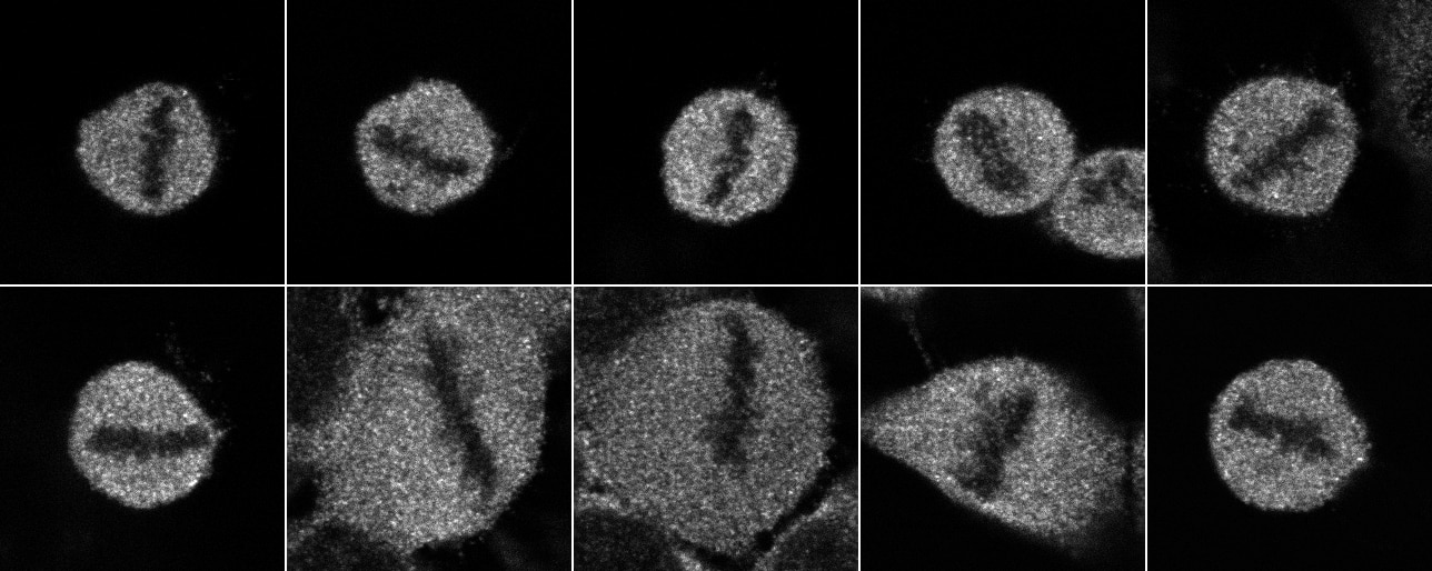
FH Simone (Verified Customer) (03-02-2023) | I used the antibody one time for a westernblot analysis of cells (Stable HeLa cell line expressing sec61b-GFP) which I transfected with siRNA targeting SNAP29 on the one hand and scrambled siRNA on the other hand. I observed a probably specific signal at around 30 kDa, indicated by a strong reduction in the sample from the cells in which I down regulated the protein. I observed a strong unspecific signal at around 40 kDa and weaker unspecific signals at around 70 kDa. I also used the antibody for immunofluorescence one time. I transfected the cells as for the western blot and fixed them with PLP on coverslips and incubated the coverslips overnight with the antibody at 4°C. On the next day I stained the coverslips using an anti rabbit antibody, coupled with Alexa 568 fluorophore. I imaged mainly mitotic cells (see picture attached). I observed a broad staining of the hole cells, leaving out the chromosomal area. I also imaged a few interphase cells, but also observed a rather broad signal. In some interphase cells, especially at the edge of some kind of vesicles I observed a stronger, probably specific staining.
 |


