

"Lamin A/C Antibodies" Comparison
View side-by-side comparison of Lamin A/C antibodies from other vendors to find the one that best suits your research needs.

Tested Applications
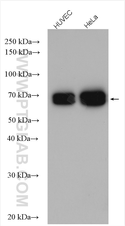
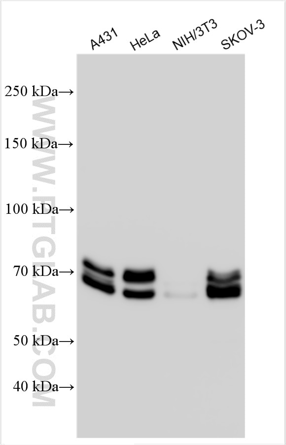
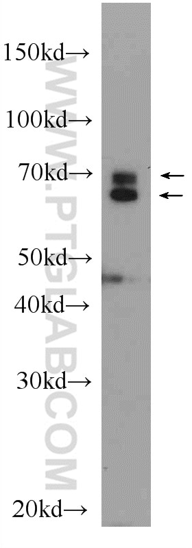
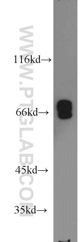
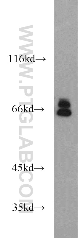
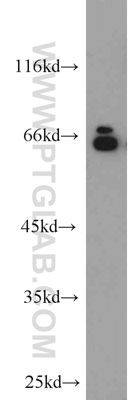
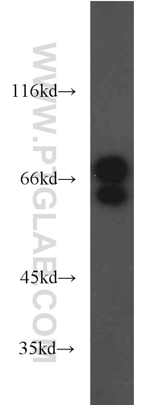
| Positive WB detected in | A431 cells, HEK-293 cells, HUVEC cells, mouse ovary tissue, SKOV-3 cells, HeLa cells, A375 cells, C6 cells, NIH/3T3 cells |
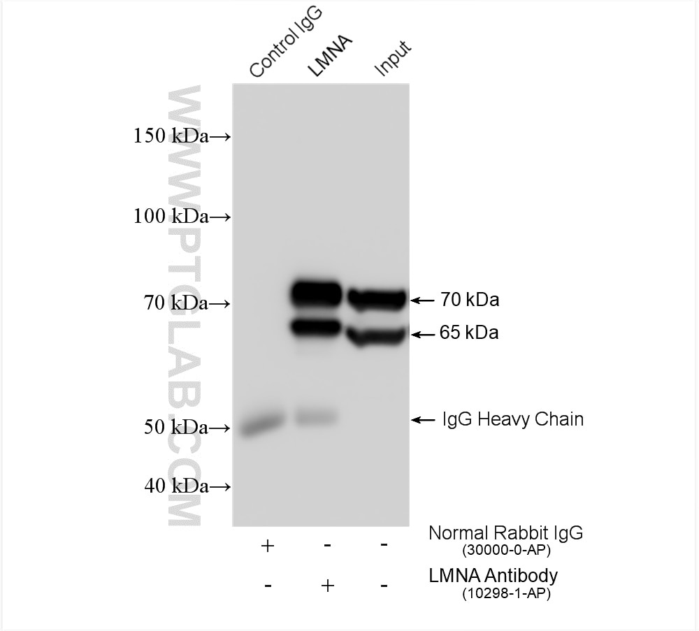
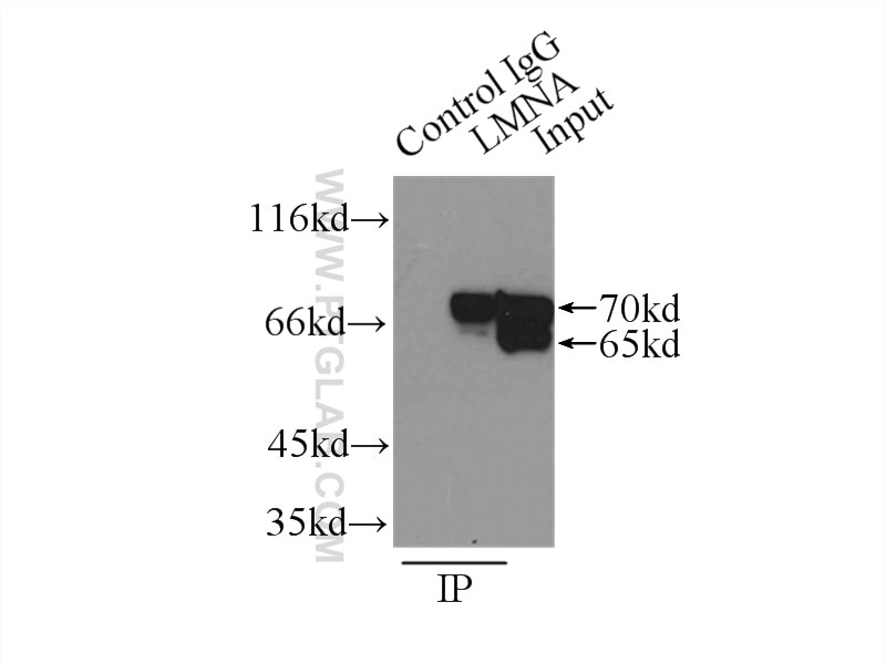
| Positive IP detected in | HeLa cells, A375 cells |
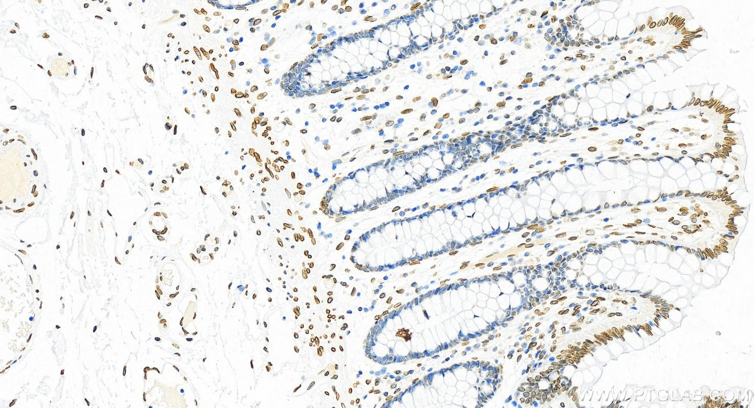
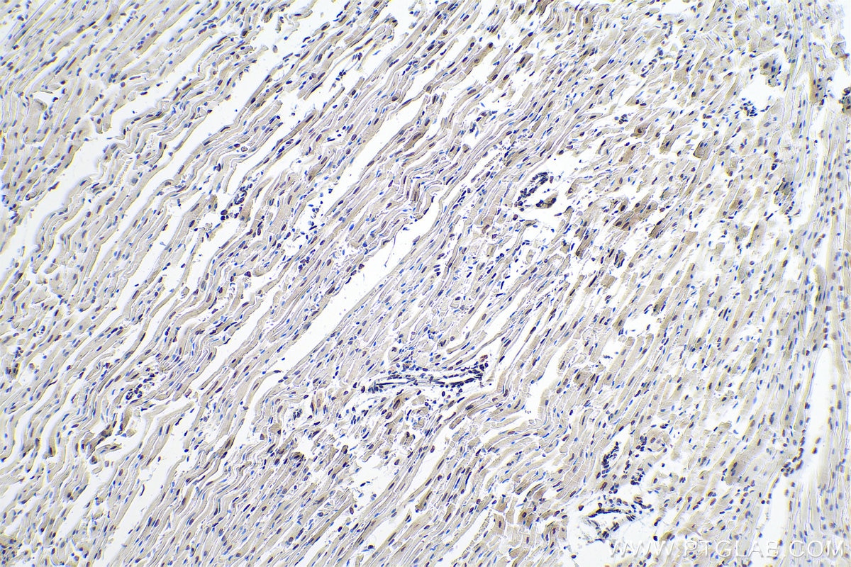
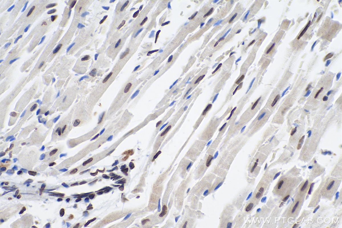
| Positive IHC detected in | mouse heart tissue, human normal colon Note: suggested antigen retrieval with TE buffer pH 9.0; (*) Alternatively, antigen retrieval may be performed with citrate buffer pH 6.0 |
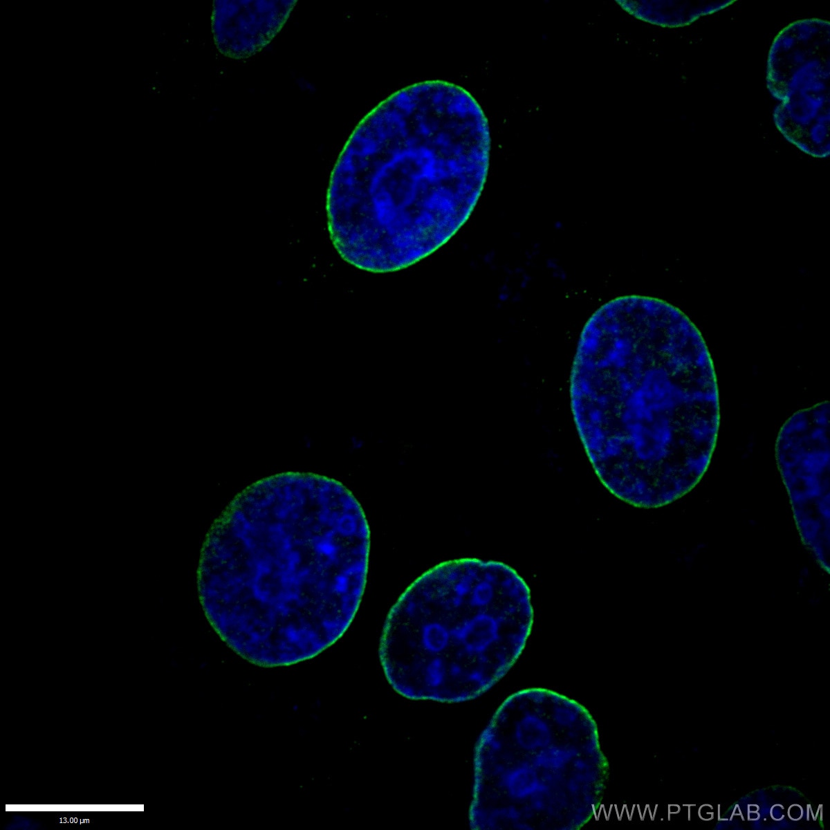
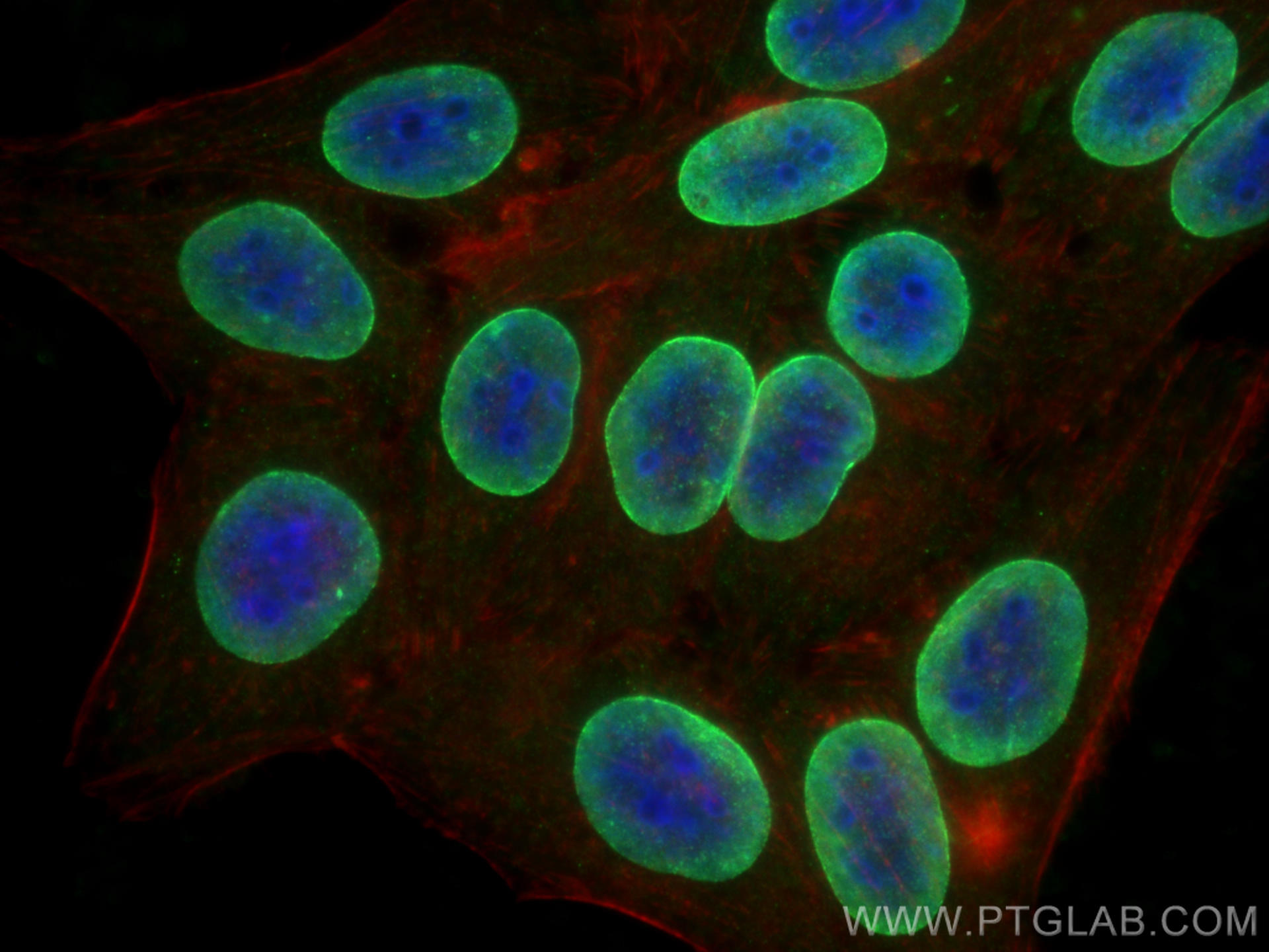
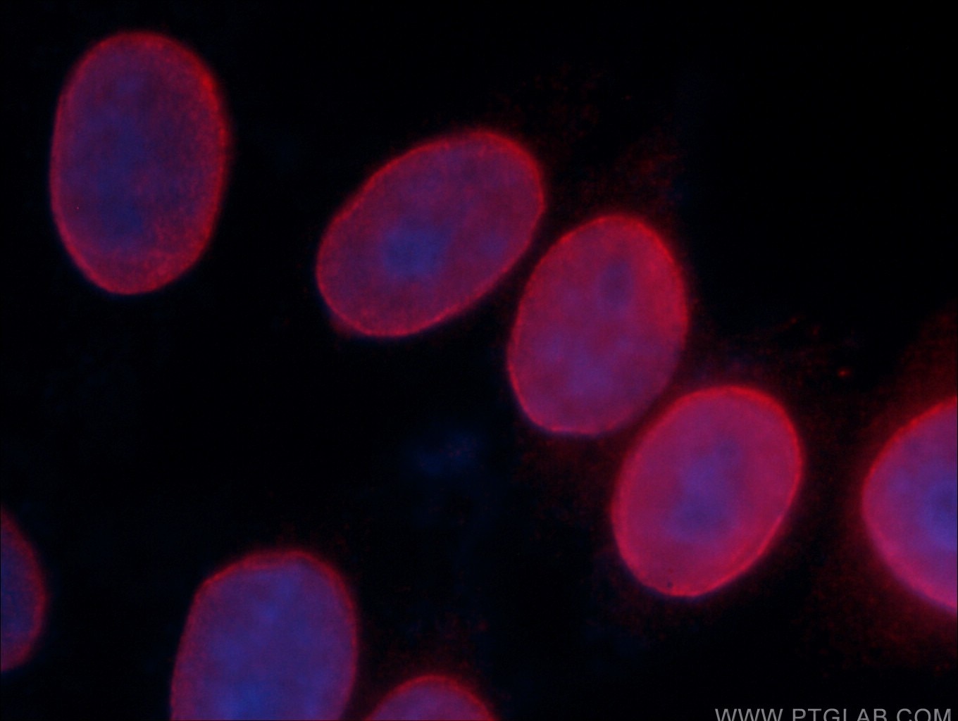
| Positive IF/ICC detected in | HepG2 cells, HeLa cells |
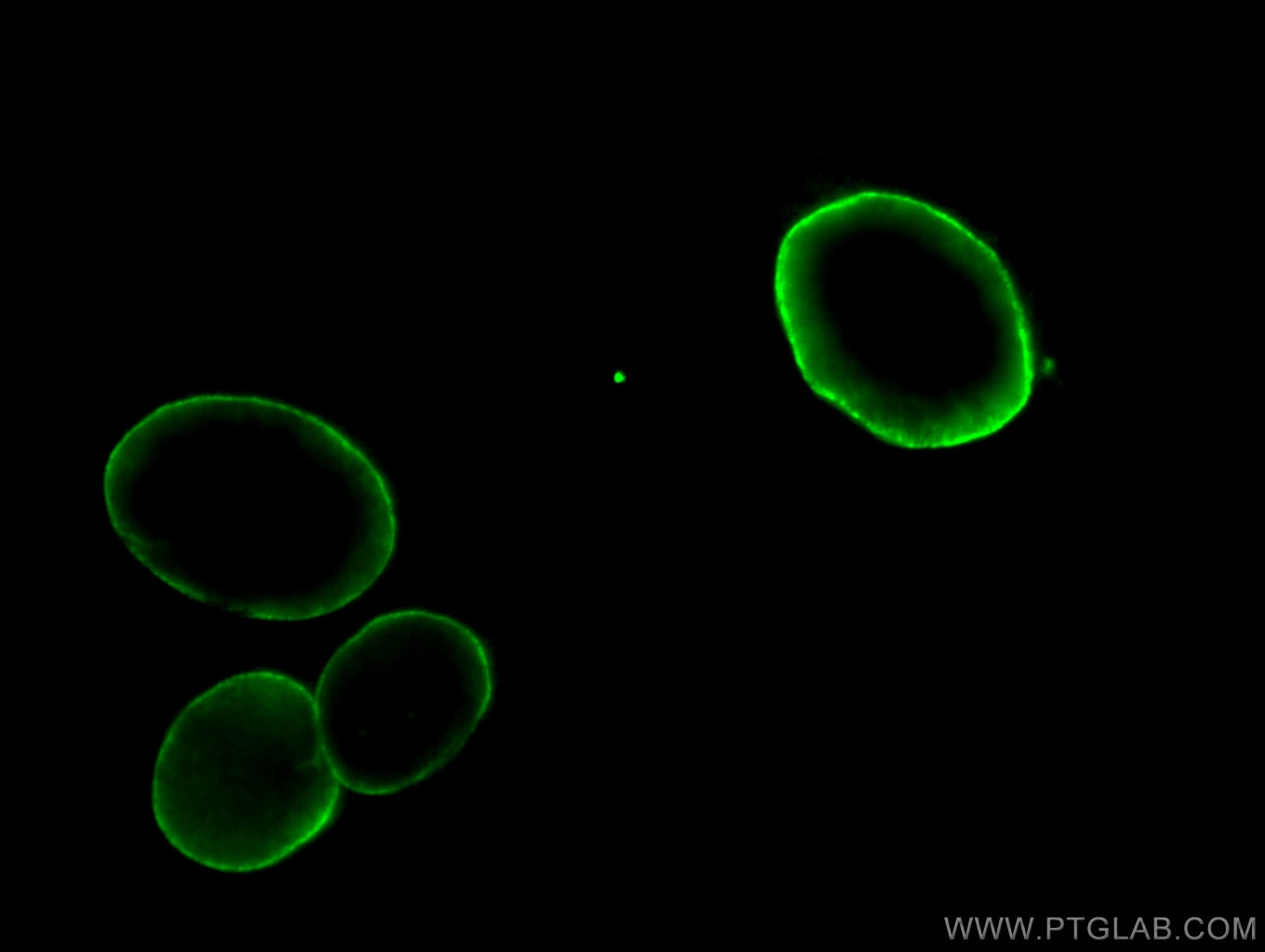
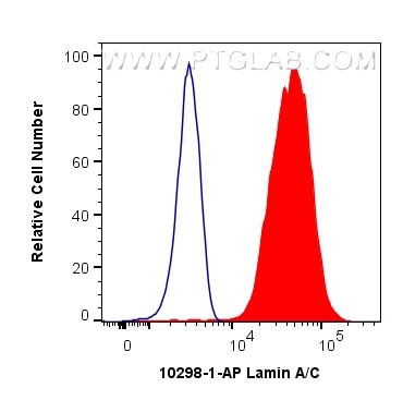
| Positive FC (Intra) detected in | HEK-293T cells |
Recommended dilution
| Application | Dilution |
|---|---|
| Western Blot (WB) | WB : 1:5000-1:50000 |
| Immunoprecipitation (IP) | IP : 0.5-4.0 ug for 1.0-3.0 mg of total protein lysate |
| Immunohistochemistry (IHC) | IHC : 1:2000-1:8000 |
| Immunofluorescence (IF)/ICC | IF/ICC : 1:400-1:1600 |
| Flow Cytometry (FC) (INTRA) | FC (INTRA) : 0.40 ug per 10^6 cells in a 100 µl suspension |
| It is recommended that this reagent should be titrated in each testing system to obtain optimal results. | |
| Sample-dependent, Check data in validation data gallery. | |
Product Information
10298-1-AP targets Lamin A/C in WB, IHC, IF/ICC, FC (Intra), IP, CoIP, ELISA, Western Blot applications and shows reactivity with human, mouse, rat samples.
| Tested Reactivity | human, mouse, rat |
| Cited Reactivity | human, mouse, rat, monkey, zebrafish, bovine, duck |
| Host / Isotype | Rabbit / IgG |
| Class | Polyclonal |
| Type | Antibody |
| Immunogen |
CatNo: Ag0408 Product name: Recombinant human Lamin A/C protein Source: e coli.-derived, PGEX-4T Tag: GST Domain: 26-198 aa of BC003162 Sequence: ITRLQEKEDLQELNDRLAVYIDRVRSLETENAGLRLRITESEEVVSREVSGIKAAYEAELGDARKTLDSVAKERARLQLELSKVREEFKELKARNTKKEGDLIAAQARLKDLEALLNSKEAALSTALSEKRTLEGELHDLRGQVAKLEAALGEAKKQLQDEMLRRVDAENRLQ Predict reactive species |
| Full Name | lamin A/C |
| Calculated Molecular Weight | 65 kDa |
| Observed Molecular Weight | 65 kDa, 70 kDa |
| GenBank Accession Number | BC003162 |
| Gene Symbol | Lamin A/C |
| Gene ID (NCBI) | 4000 |
| ENSEMBL Gene ID | ENSG00000160789 |
| RRID | AB_2296961 |
| Conjugate | Unconjugated |
| Form | Liquid |
| Purification Method | Antigen affinity purification |
| UNIPROT ID | P02545 |
| Storage Buffer | PBS with 0.02% sodium azide and 50% glycerol, pH 7.3. |
| Storage Conditions | Store at -20°C. Stable for one year after shipment. Aliquoting is unnecessary for -20oC storage. 20ul sizes contain 0.1% BSA. |
Background Information
Lamin A/C is also named as LMNA or LMN1. The lamin family of proteins make up the matrix and are highly conserved in evolution. During mitosis, the lamina matrix is reversibly disassembled as the lamin proteins are phosphorylated. Lamin proteins are thought to be involved in nuclear stability, chromatin structure, and gene expression. The lack of lamin A/C can be as a novel marker for undifferentiated embryonic stem cells and lamin A/C expression is an early indicator of differentiation (PMID: 16179429). Mutations in this gene lead to several diseases: Emery-Dreifuss muscular dystrophy, familial partial lipodystrophy, limb-girdle muscular dystrophy, dilated cardiomyopathy, Charcot-Marie-Tooth disease, and Hutchinson-Gilford progeria syndrome. This protein has 4 isoforms produced by alternative splicing with the molecular weight of 74 kDa, 65 kDa, 70 kDa, and 64 kDa. This antibody can recognize 4 isoforms of Lamin A/C.
Protocols
| Product Specific Protocols | |
|---|---|
| FC protocol for Lamin A/C antibody 10298-1-AP | Download protocol |
| IF protocol for Lamin A/C antibody 10298-1-AP | Download protocol |
| IHC protocol for Lamin A/C antibody 10298-1-AP | Download protocol |
| IP protocol for Lamin A/C antibody 10298-1-AP | Download protocol |
| WB protocol for Lamin A/C antibody 10298-1-AP | Download protocol |
| Standard Protocols | |
|---|---|
| Click here to view our Standard Protocols |
Publications
What published studies show
Lamin A/C Polyclonal antibody (Cat# 10298-1-AP) has accumulated 357 citations across high-impact journals including Nature Cell Biology, Nature Communications, Cancer Cell, Cell Reports, Science Advances, Nature Metabolism, Neuron, and EMBO Journal. Research fields span nuclear biology, oncology, aging, immunology, metabolism, neurology, cardiology, and infectious disease. The antibody is primarily used for Western blotting, with additional applications in IF, IHC, and IP.
| Species | Application | Title |
|---|---|---|
Signal Transduct Target Ther GRP75 triggers white adipose tissue browning to promote cancer-associated cachexia | ||
Cancer Cell Inhibiting intracellular CD28 in cancer cells enhances antitumor immunity and overcomes anti-PD-1 resistance via targeting PD-L1 | ||
Cancer Cell SET1A-Mediated Mono-Methylation at K342 Regulates YAP Activation by Blocking Its Nuclear Export and Promotes Tumorigenesis. | ||
Mol Cancer Cell surface CD55 traffics to the nucleus leading to cisplatin resistance and stemness by inducing PRC2 and H3K27 trimethylation on chromatin in ovarian cancer | ||
Nat Metab The flavonoid procyanidin C1 has senotherapeutic activity and increases lifespan in mice. | ||
Nat Aging DNA damage in macrophages drives immune autoreactivity via nuclear antigen presentation. |
Reviews
What customers say
All 8 reviews rate this Lamin A/C antibody 5 stars. Users consistently highlight its high specificity and clean blotting performance, particularly for confirming nuclear fractionation in Western Blot experiments. The antibody reliably detects two bands corresponding to the Lamin A/C isoforms, with no cross-reactivity in cytoplasmic fractions. It also performs well in immunofluorescence and flow cytometry. Reviewers note it is easy to use at dilutions ranging from 1:100 to 1:5000, across diverse cell types including HAP1, NIH-3T3, HEK293T, NSC-34, macrophages, and human brain tissue.
The reviews below have been submitted by verified Proteintech customers who received an incentive for providing their feedback.
Shivali (Verified Customer) (02-11-2026) | I used this lamin antibody for subcellular fractionation during microsome preparation, and it clearly marked the nuclear fraction with strong specificity.
|
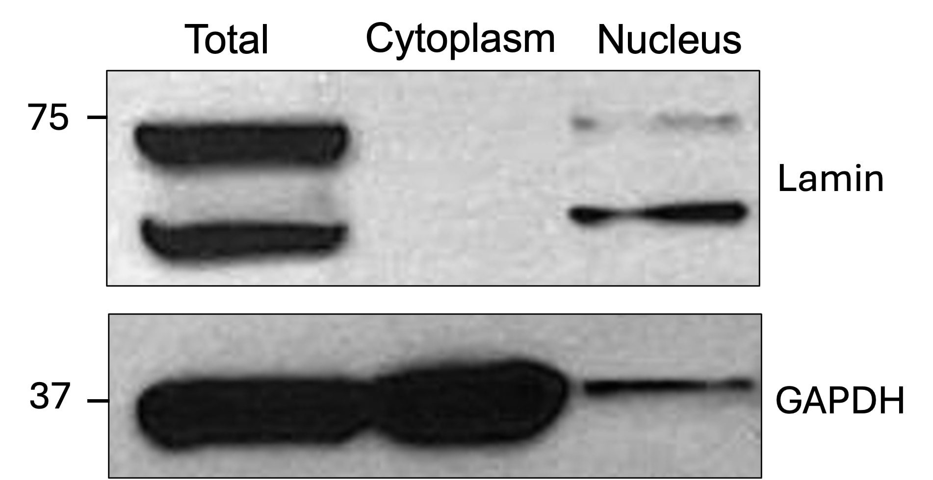
Parijat (Verified Customer) (01-30-2026) | This is a highly specific antibody for Lamin A/C. In subcellular fractionation experiments (cytoplasmic, nuclear, and total fractions), Lamin A/C is detected exclusively in the nuclear and total fractions, with no signal in the cytoplasmic fraction, as expected. The antibody consistently detects two bands corresponding to the two Lamin A/C isoforms.
 |
Zahida (Verified Customer) (04-15-2025) | Easy to reveal
|
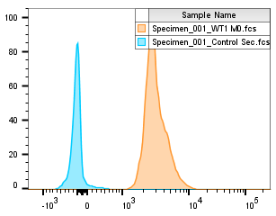
Alejandro (Verified Customer) (08-22-2022) | Shows clear staining in IF and well staining using FACS
 |
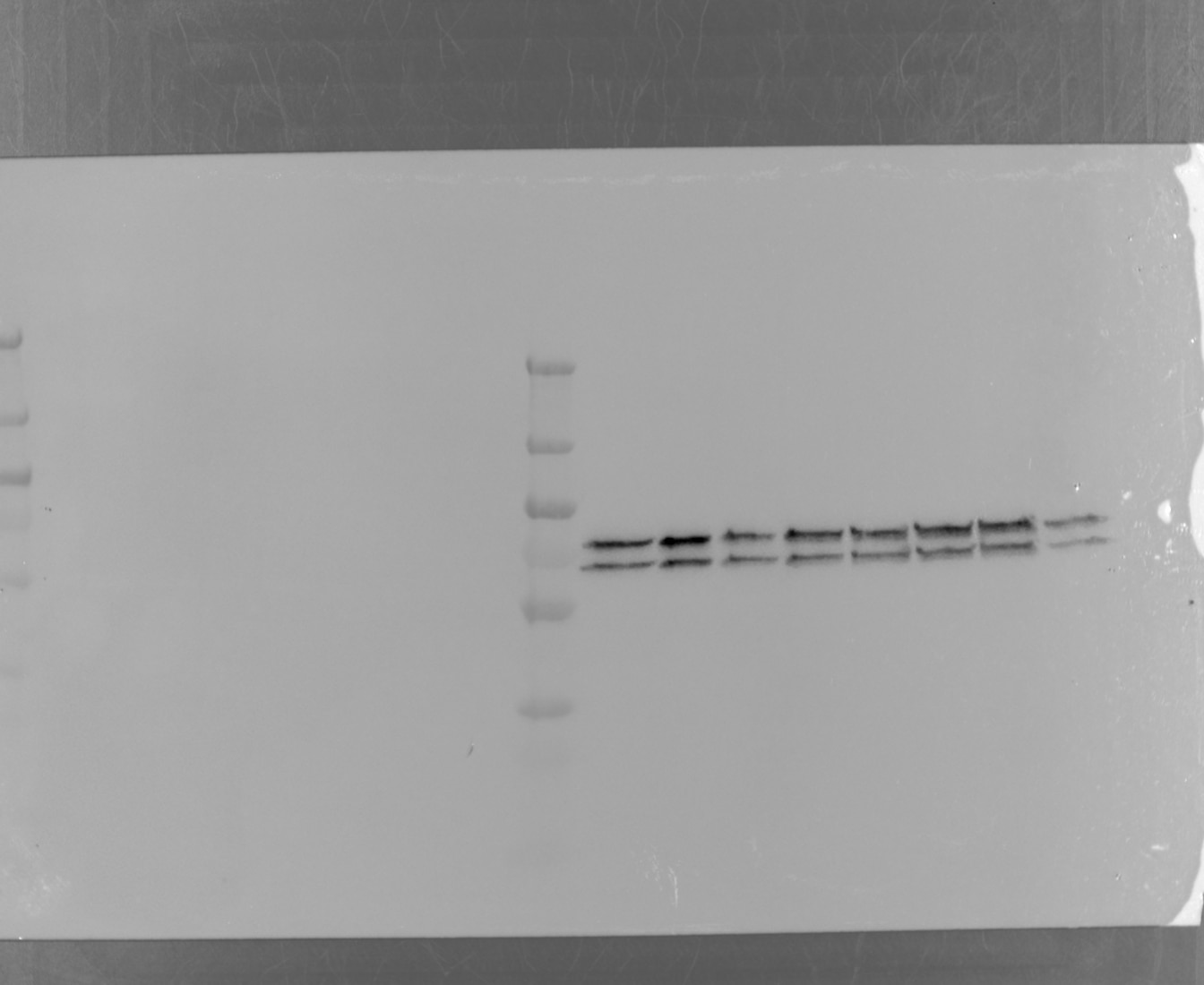
Charlotte (Verified Customer) (07-29-2022) | Cell fraction performed on NIH-3T3 cells to show the nucleus part. Blot super clean. Antibody specific. Easy to reveal.
 |
S (Verified Customer) (12-31-2021) | good antibody.
 |
Azita (Verified Customer) (06-16-2021) | It was used as a marker in WB to confirm proper nuclear fractionation of cell lysate.
|
Declan (Verified Customer) (11-29-2018) |
|


