A Guide to Staining Organelles
Identifying subcellular structures is key for mapping the localization of target proteins. This blog features key markers to identify organelles by immunofluorescence.
Organelles are small subcellular structures located in the cytoplasm of eukaryotic and prokaryotic cells that perform specialized functions. A protein characteristic of an organelle can be used as a marker, and its presence can reflect the location and even shape of the subcellular structure. Additionally, the localization of a target protein and organelle marker can determine the subcellular localization of the target protein and explore the biological processes in which it is involved.
Below are some commonly used markers for identifying organelles by immunofluorescence.
Jump to section:
- Nuclear markers
- Nucleoli markers
- Nuclear membrane markers
- Endoplasmic reticulum markers
- Golgi markers
- Centrosome markers
- Mitochondrial markers
- Ribosome markers
- Lysosomal markers
- Peroxisome markers
- Endosomal markers
- Exosome markers
- Cytoskeleton markers
- Cilia markers
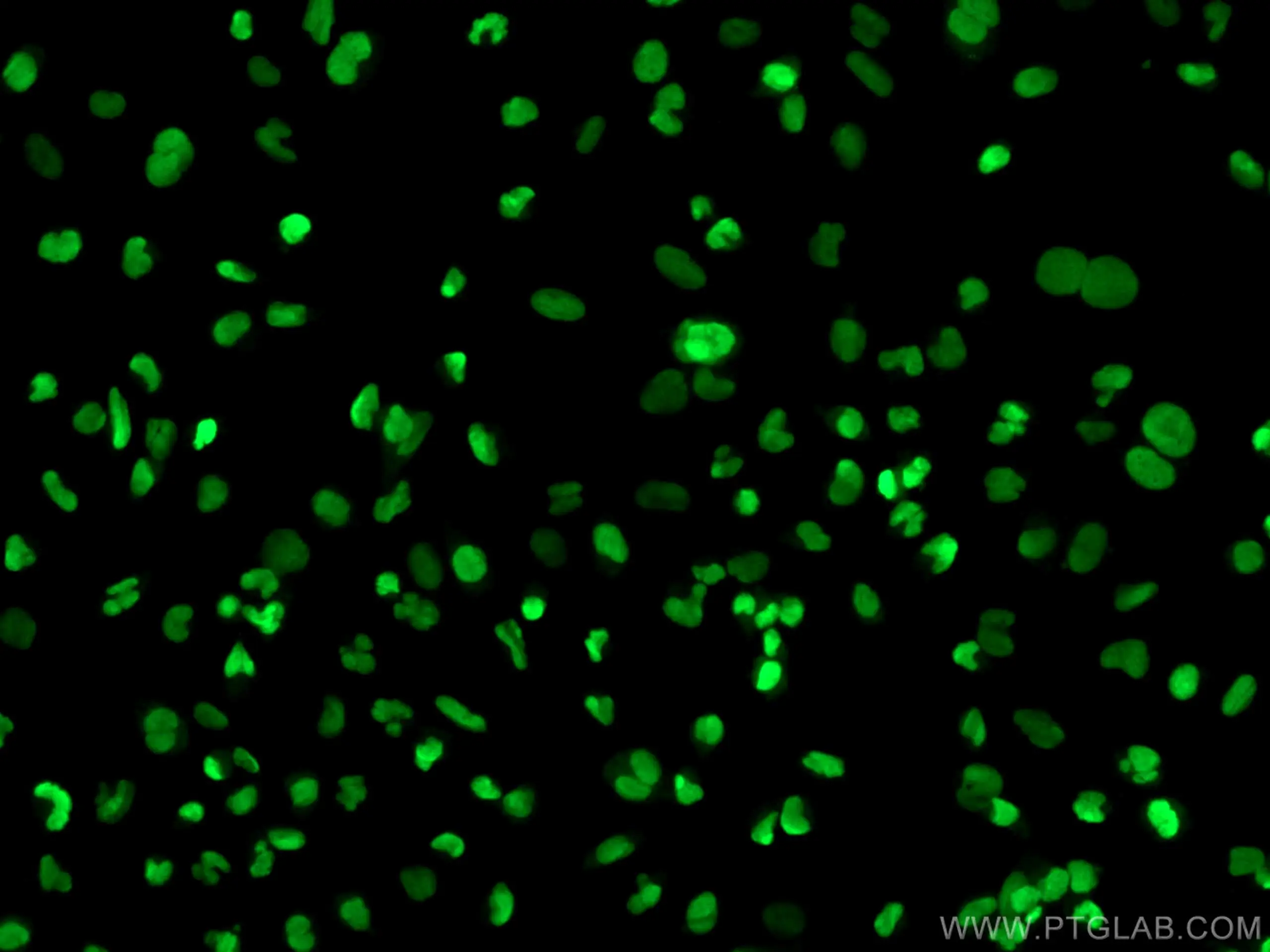
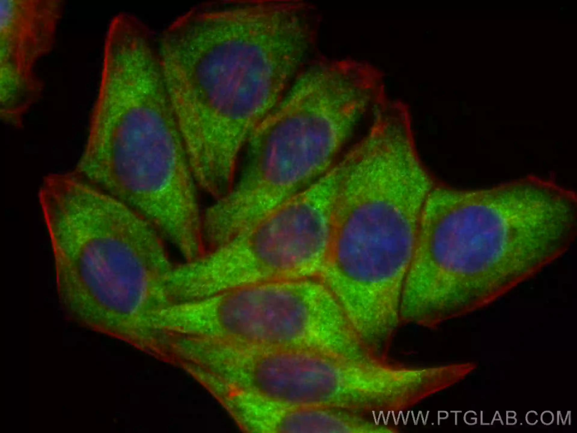
1. Nuclear markers
The nucleus is the main storage site for genetic information and the control center of the entire cell. The nucleus contains a large number of histones and various genetic and epigenetic-related targets, which can be used as nuclear markers.
Among them are histones such as Histone H3 and Histone H2A.X are two of the most commonly and widely used nuclei markers.
|
▲ Histone H3 antibody (17168-1-AP) labeling HeLa nuclei |
▲ Histone H2A.X antibody (10856-1-AP) labeling U-251 nuclei |

2. Nucleoli markers
Nucleoli are dense bodies of nucleic acids and proteins in the nucleus. The vast majority of eukaryotic cells have nucleoli. The main functions of nucleoli are rRNA synthesis, processing and maturation, and assembly of ribosomal subunits.
Nucleolar fibrin (FBL), MKI67 (FHA domain) binding nucleolar phosphoprotein (NIFK), nucleolin (NCL), and NOP2 nucleolar protein (NOP2) are typical and commonly used nucleoli markers.
|
▲ FBL antibody (16021-1-AP) labeling HepG2 cell nucleoli |
▲ NIFK antibody (12615-1-AP) labeling MCF-7 cell nucleoli |


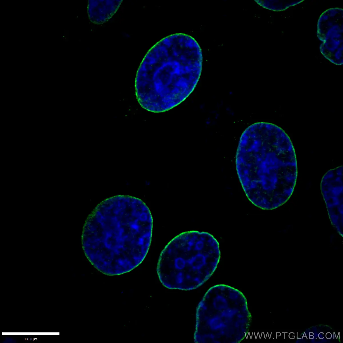
3. Nuclear membrane markers
The presence or absence of a nuclear envelope is one of the main characteristics that distinguishes eukaryotic cells from prokaryotic cells. The nuclear membrane is highly selective for the entry and exit of substances within the nucleus and protects the cell’s genetic material from the cytoplasm.
Lamin B and Lamin A/C are the main components of the inner nuclear membrane and are often used as nuclear membrane markers.
|
▲ Lamin B1 antibody (12987-1-AP) labeling the nuclear membrane of HepG2 cells |
▲ Lamin A/C antibody (10298-1-AP) labeling the nuclear membrane of HepG2 cells |

4. Endoplasmic reticulum markers
The endoplasmic reticulum (ER) is crucial for protein synthesis, folding, assembly, modification, and transport. It is also involved in lipid and carbohydrate metabolism and is the main storage for intracellular Ca2+.
Calnexin, Calreticulin, Endoplasmic Reticulin (ERp72), Glucoregulatory Protein 94 (GRP94), and protein disulfide isomerase (PDI) are mainly localized in the ER and are commonly used as ER markers.
|
▲ Calnexin antibody (66903-1-Ig) labeling HeLa endoplasmic reticulum |
▲ Calreticulin antibody (27298-1-AP) labeling HepG2 endoplasmic reticulum |

5. Golgi markers
The Golgi apparatus’s main function is to process, sort, and transport proteins synthesized by the endoplasmic reticulum, and then secrete or send them to specific parts of the cell.
Golgi protein subfamily A member 2 (GOLGA2/GM130), Golgi weight group stacked protein 2 (GORASP2), Golgi protein 73 (GP73/GOLPH2), Syntaxin 6, and trans-Golgi reticular structural protein 6 (TGOLN2/TGN2) are typical Golgi markers.
|
▲ The GOLGA2/GM130 antibody (11308-1-AP) labeling the Golgi apparatus in HeLa cells |
▲ TGOLN2/TGN46 antibody (13573-1-AP) labeling the Golgi apparatus in HeLa cells |


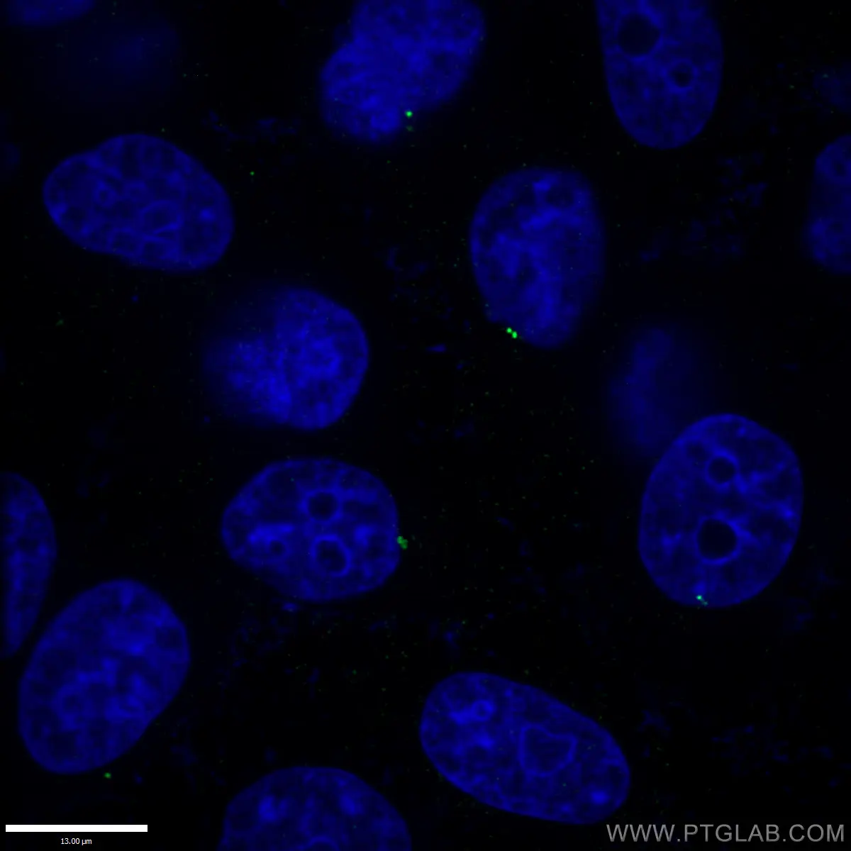
6. Centrosome markers
The centrosome is the main microtubule organizing center of animal cells. It plays an important role in the production of cell polarity, intercellular transport, and cell division.
Centromerin J (CENPJ), centrosome protein 110 (CP110), Pericentrin, and tubulin-γ (Gamma tubulin) are among the most commonly used centrosome markers.
|
▲ CENPJ antibody (11517-1-AP) labeling HepG2 cell centrosomes |
▲ Gamma Tubulin antibody (66320-1-Ig) labeling A549 cell centrosomes |

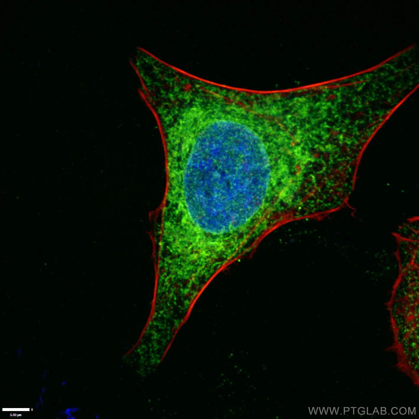
7. Mitochondrial markers
Mitochondria are bilayered membrane organelles found in most cells. It is the main place for aerobic respiration and the main source of energy for cells.
Apoptosis inducible factor (AIF), ATP synthase H+ transport mitochondrial F1 complex α subunit 1 (ATP5A1), Cytochrome C oxidase IV (COXIV), carbamoyl phosphate synthase 1 (CPS1), heat shock protein 60 (HSP60), mitochondrial inner membrane protein (Mitofilin), antiproliferative protein (Prohibitin, also known as inhibin) and voltage-dependent anion channel protein 1 (VDAC1/Porin), are all commonly used as mitochondrial markers.
|
▲ COXIV antibody (11242-1-AP) labeling HepG2 cell mitochondria |
▲ AIF antibody (67791-1-Ig) labeling HeLa cell mitochondria |


8. Ribosome markers
Protein synthesis occurs within ribosomes. Their main function is to translate polypeptide chains and synthesize proteins according to mRNA instructions.
Ribosomal protein S3 (RPS3), ribosomal protein L4 (RPL4), and ribosomal protein L7 (RPL7) are typical ribosomal proteins and are the most commonly used ribosome markers.
|
▲ RPS3 direct antibody (CL594-66046) labeling HepG2 cell ribosomes |
▲ RPL4 recombinant antibody (80959-1-RR) labeling HeLa cell ribosomes |

9. Lysosomal markers
Lysosomes contain a variety of acid hydrolases which can degrade damaged proteins and organelles internally, resist the invasion of bacteria and viruses externally, and engulf and digest foreign substances.
Lysosomal associated membrane proteins 1 and 2 (LAMP1, LAMP2) are the two most commonly used lysosomal markers.
|
▲ LAMP1 direct antibody (CL555-65050) labeling NIH/3T3 cell lysosomes |
▲ LAMP2 antibody (66301-1-Ig) labeling HeLa cell lysosomes |


10. Peroxisome markers
Peroxisomes are highly dynamic, metabolically active organelles that are mainly involved in the metabolism of lipids and the regulation of oxidative stress.
Catalase, peroxisome biosynthetic factors 1, 5 and 14 (PEX1, PEX5, PEX14) are the most common peroxisome markers.
|
▲ Catalase antibody (66765-1-Ig) labeling HepG2 cell peroxisomes |
▲ PEX14 antibody (10594-1-AP) labeling HeLa cell peroxisomes |

11. Endosomal markers
Endosomes are membrane-wrapped vesicles in eukaryotic cells formed by endocytosis. They participate in the sorting, transport, and degradation of macromolecules. According to the different stages of action of endocytosis, endosomes can be divided into early endosomes, late endosomes, and circulating endosomes.
RAB5 and RAB11 are both members of the Rab small molecule GTPase family, which are involved in the intracellular vesicle recycling pathway and mediate the transport of vesicles from endosomes to plasma membranes. Early endosomal antigen 1 (EEA1) is a protein that interacts with RAB5, so they are both commonly used endosomal markers.
|
▲ RAB5A direct antibody (CL488-66339) labeling HeLa cell endosomes |
▲ EEA1 antibody (68065-1-Ig) labeling HepG2 cell endosomes |

12. Exosome markers
Exosomes are single-membrane, secreted vesicles that transfer a variety of bioactive molecules, including nucleic acids, proteins, and lipids, from donor cells to recipient cells, mediating cell-to-cell communication and molecular transport.
Leukocyte differentiation antigens (CD9, CD63, CD81) and tumor susceptibility gene 101 (TSG101) are the more common exosome markers.
Note: A single marker cannot be used to identify exosomes. The International Society for the Study of Extracellular Vesicles (ISEV) recommends the detection of at least three types of proteins (two positive and one negative). Negative markers can include nucleoprotein Histone, Lamin A, endoplasmic reticulum protein calnexin, Golgi protein GM130, mitochondrial protein cytochrome C, and cytoskeleton protein cytokeratin 18.
|
▲ CD63 antibody (25682-1-AP) labeling human malignant melanoma tissue |
▲ TSG101 antibody (67381-1-Ig) staining in HeLa cells |


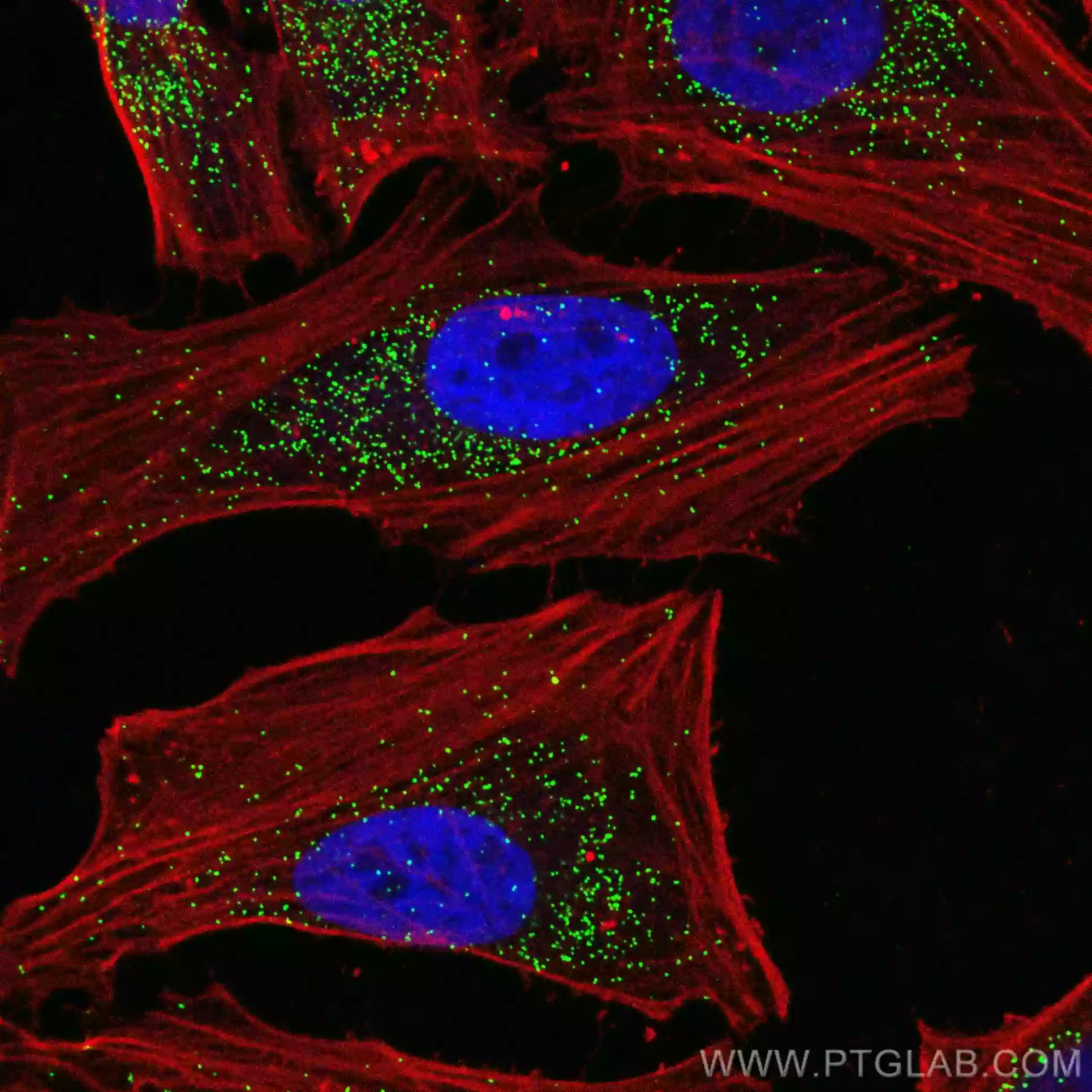
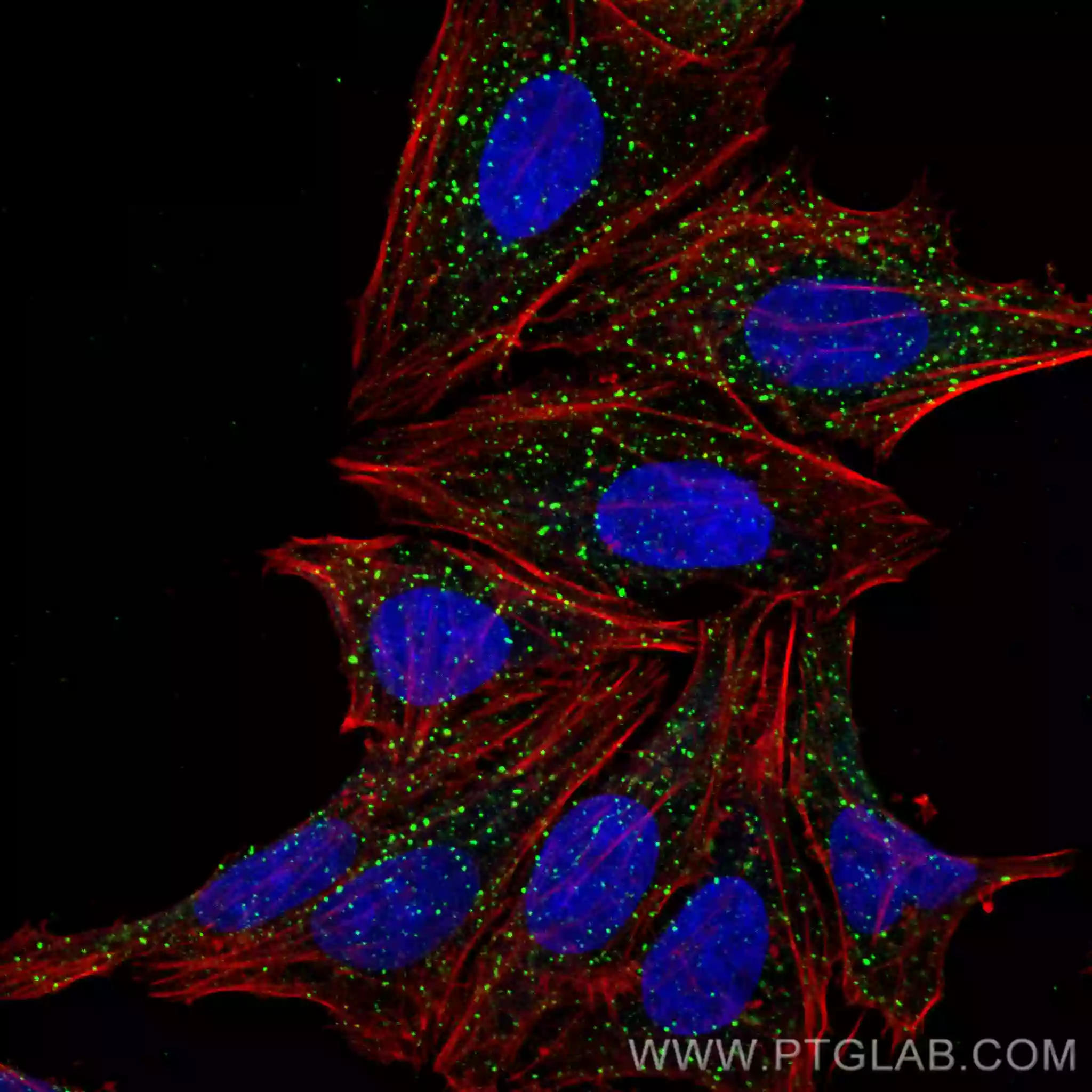
13. Cytoskeleton markers
The cytoskeleton is a network structure composed of microfilaments, microtubules and intermediate fibers. It plays many different roles including supporting cellular morphology, organelle organization, and supporting molecular transport, cell division, and cell signaling.
Alpha/Beta Tubulin and Vimentin are components of microtubule structure and intermediate filament structure, respectively, and are also very frequently used cytoskeleton markers.
|
▲ Alpha Tubulin antibody (80762-1-RR) labels the C2C12 cytoskeleton |
▲ Beta Tubulin antibody (80713-1-RR) labels the C2C12 cytoskeleton |


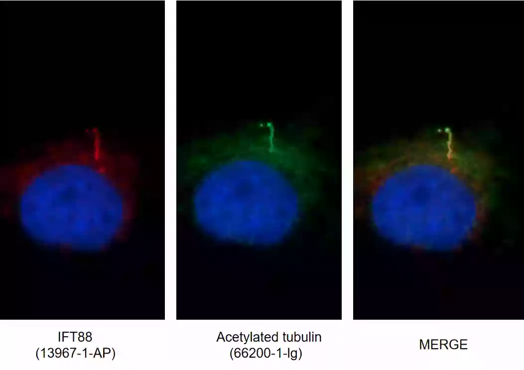
14. Cilia markers
Cilia are evolutionarily conserved hair-like organelles that protrude on the cell surface and consist of microtubules. Cilia play important roles in mechanosensation, mechanotransduction, tissue homeostasis, and animal embryonic development.
Adenylate cyclase 3 (ADCY3), ADP ribosylation factor-like protein 13B (ARL13B), intraciliary transporters 20 and 88 (IFT20, IFT88) and acetylated tubulin (Lys40) can be used as cilia markers.
|
▲ IFT88 antibody (13967-1-AP) and Acetylated Tubulin antibody (66200-1-Ig) label MDCK cell cilia |


Related Content
Dying to see what's inside: Staining Organelles
Organelle Marker Product Focus
IF Workshop: Make your Data Glow
CoraLite® Plus Conjugated Antibodies
Support
Newsletter Signup
Stay up-to-date with our latest news and events. New to Proteintech? Get 10% off your first order when you sign up.