How to Better Study Ubiquitination
Understand the role of Ubiquitin in protein degradation and learn how ChromoTek’s Ubiquitin Trap can help you overcome certain ubiquitin-related research challenges.
Introduction
Ubiquitin is a very small protein (~76aa) that functions as a highly conserved regulator of protein homeostasis. It is ubiquitously expressed in all eukaryotic cells and is part of a larger family of ubiquitin-like proteins and modifiers including SUMO, NEDD8, and ISG15. While ubiquitin plays many roles within the context of autophagy, cell signaling, and endocytosis, its primary function is to serve as an intracellular label for targeted protein degradation.
Ubiquitination Process
Protein ubiquitination is a post-translational modification that consists of three steps, each catalyzed by a different enzymatic component of the ubiquitin proteasome. During the activation step, the E1 enzyme attaches itself to a ubiquitin molecule via ATP hydrolysis. The ubiquitin molecule is then transferred by E1 to the E2 ubiquitin-conjugating enzyme. The E2-ubiquitin complex then associates with the E3 ligase, which catalyzes the attachment of ubiquitin to a lysine residue on the protein substrate. Although less common, ubiquitination has been observed on non-lysine amino acids, such as methionine 1 (M1). A poly-ubiquitin chain is produced when a ubiquitin on a substrate is modified by additional ubiquitin monomers conjugating onto any one of the seven lysine residues of ubiquitin. Ubiquitination can take many forms due to monoubiquitylation vs. polyubiquitination, the different ubiquitin-lysine linkages in chains, and the possibility of chains branching and mixing.
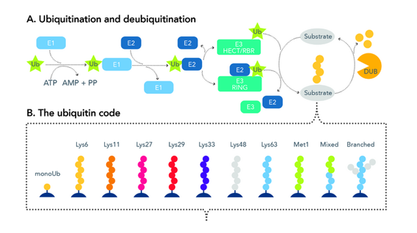
Fig 1. Diagram detailing the process of ubiquitination and deubiquitination.
The Ubiquitin Code
The downstream events triggered by ubiquitination are based on the lysine (K) residues where the ubiquitin is linked as well as the length of the ubiquitin chain itself. Broadly speaking, monomeric ubiquitination usually controls non-proteolytic events such as endocytosis, histone modification, and DNA damage responses. However, K48-linked polyubiquitination is the best known as a signal for downstream, ubiquitin-mediated, proteasomal protein degradation. A summary of common ubiquitination signals is shown in the table below.
|
Linkage Site |
Ubiquitin Chain Length |
Downstream Signaling Event |
|
Substrate-specific lysines |
Monomer |
Endocytosis, histone modification, DNA damage responses |
|
K48 |
Polymeric |
Targeted protein degradation |
|
K63 |
Polymeric |
Immune responses, inflammation, lymphocyte activation |
|
K6 |
Polymeric |
Antiviral responses, autophagy, mitophagy, DNA repair |
|
K11 |
Polymeric |
Cell cycle progression, proteasome-mediated degradation |
|
K27 |
Polymeric |
DNA replication, cell proliferation |
|
K29 |
Polymeric |
Neurodegenerative disorders, Wnt signaling downregulation, autophagy |
|
M1 |
Polymeric |
Cell death and immune signaling |
In addition to some of these classical ubiquitination signals, ubiquitin chains can be further modified through acetylation, phosphorylation, and branching. Ubiquitin chains can also be intermixed with SUMOlyation signals.
Challenges in Studying Ubiquitination
Despite its near “ubiquitous” presence and control of a wide range of cellular functions, researchers face various challenges in attempting to characterize ubiquitination patterns and responses. First, ubiquitin proteins are often weakly immunogenic due to their small size, and as a result, many ubiquitin antibodies are non-specific and bind large amounts of artifacts. The ubiquitination process itself is also highly transient and reversible; the percentage of ubiquitinylated proteins in a cell lysate is often very small and an enrichment method is needed to properly detect them. Additionally, there are over 600 different E3 ligases, and it is possible for multiple ligases to ubiquitinate one protein at the same time, hence requiring a reagent that can detect multiple specificities.
ChromoTek Ubiquitin-Traps for Ubiquitin and Ubiquitinylated Protein Isolation
To overcome some of the challenges with studying ubiquitin, ChromoTek has developed new, high-affinity nano-traps for ubiquitin and ubiquitinylated protein isolation. Consisting of an anti-Ubiquitin nanobody/VHH coupled to either agarose or magnetic agarose beads, the Ubiquitin-Trap can be used to immunoprecipitate monomeric ubiquitin, ubiquitin chains, and ubiquitinylated proteins from a wide range of cell extracts, including mammalian, insect, plants, and yeast. Like our other nano-trap products, the Ubiquitin-Trap is a ready-to-use reagent for fast and easy pulldowns that is stable under harsh washing conditions and results in clean, low-background IPs.
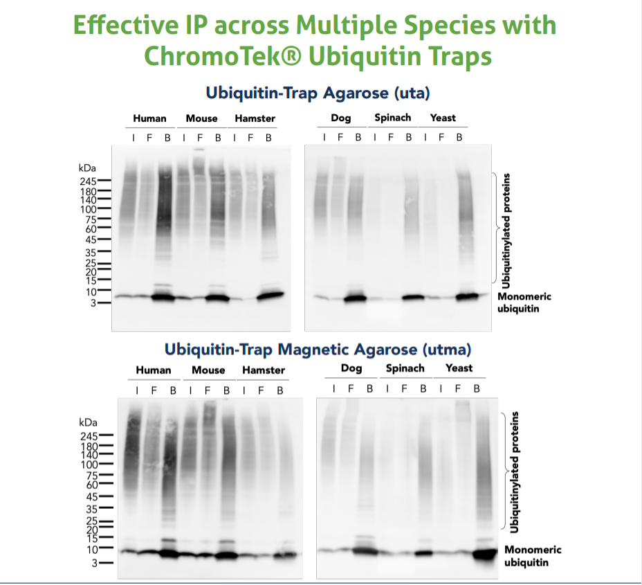
Image Description: The Ubiquitin-Trap Agarose (uta) and Ubiquitin-Trap Magnetic Agarose (utma) were used to immunoprecipitate endogenous ubiquitin and ubiquitinylated proteins from human (HEK293T), mouse (C2C12), hamster (CHO), dog (MDCK), spinach (spinacia oleracea), and baker’s yeast (Saccharomyces cervisiae) cells treated with MG-132. For each IP, samples of the input lysate (I), non-bound flow-through (F), and bound (B) fractions were analyzed using western blot. Ubiquitin recombinant antibody (80992-1-RR) and HRP-conjugated Affinipure Goat Anti-Rabbit (H+L) (SA00001-2) were used in the western blot analysis.
Available Ubiquitin-Trap Products
|
SKU |
Product |
Size/Components |
|
Ubiquitin-Trap Agarose |
10 rxns, 20 rxns, 100 rxns |
|
|
Ubiquitin-Trap Magnetic Agarose |
10 rxns, 20 rxns, 100 rxns, |
|
|
Ubiquitin-Trap Agarose Kit |
20 rxns, also includes lysis, wash, dilution, and elution buffers for IP |
|
|
Ubiquitin-Trap Magnetic Agarose Kit |
20 rxns, also includes lysis, wash, dilution, and elution buffers for IP |
Recommended Antibodies for Western Blot Detection
|
SKU |
Product |
Tested Reactivity |
|
Ubiquitin Polyclonal Antibody |
Human, Mouse, Rat |
|
|
Ubiquitin Recombinant Antibody |
Human, Mouse, Rat, Hamster, Dog, Spinach, Yeast |
|
|
Multi-rAB HRP-Goat Anti-Rabbit Recombinant Secondary Antibody (H+L) |
Rabbit |
|
|
HRP-conjugated Affinipure Goat Anti-Rabbit IgG (H+L) |
Rabbit |
|
|
Biotin-conjugated Affinipure Goat Anti-Rabbit IgG (H+L) |
Rabbit |
|
|
Alkaline Phosphatase-conjugated Affinipure Goat Anti-Rabbit IgG (H+L) |
Rabbit |
FAQ
- Why does ubiquitin show up as a smear on an agarose gel and western blot?
- As the Ubiquitin-Trap can bind monomeric ubiquitin, ubiquitin polymers, and ubiquitinylated proteins, the bound fraction from IP will contain proteins of varying lengths, resulting in a smeared appearance on a gel.
- Can Ubiquitin-Trap differentiate between different ubiquitin linkages?
- Ubiquitin-trap is not linkage specific. Multiple different chains can be bound. Therefore, differentiation between bound linkages is only possible by using a linkage-specific antibody during the western blot.
- What is the binding capacity of Ubiquitin-Trap?
- Due to ubiquitin forming chains of varying lengths, the exact binding of the trap to these chains is not fully defined. Chains can be bound at single or multiple sites, making a determination of total binding capacity difficult.
- Is the Ubiquitin VHH available as a standalone product?
- Yes! Ubiquitin VHH is available in an unconjugated format for custom conjugations and antibody immobilization experiments.
- Can Ubiquitin-Trap be used for IP-MS workflows?
- The trap has been optimized for on-bead digestion. For IP-MS, we recommend this protocol.
- How can I increase/protect the amount of protein ubiquitination in my sample?
- Ubiquitination signals in a sample can be preserved by treating cells with proteasome inhibitors, such as MG-132, prior to harvesting. While the exact conditions must be optimized for each cell type, a good starting point is to incubate cells with 5-25 uM MG-132 for 1–2 hours. Please note that overexposure to MG-132 can lead to cytotoxic effects.
References
Damgaard, R. B. (2021). The ubiquitin system: from cell signalling to disease biology and new therapeutic opportunities. Cell Death & Differentiation, 28(2), 423–426. https://doi.org/10.1038/s41418-020-00703-w
Jahan, A. S., Elbæk, C. R., & Damgaard, R. B. (2021). Met1-linked ubiquitin signalling in health and disease: inflammation, immunity, cancer, and beyond. Cell Death & Differentiation, 28(2), 473–492. https://doi.org/10.1038/s41418-020-00676-w
Madiraju, C., Novack, J. P., Reed, J. C., & Matsuzawa, S. (2022). K63 ubiquitination in immune signaling. Trends in Immunology, 43(2), 148–162. https://doi.org/10.1016/j.it.2021.12.005
Shimizu, Y., Okuda-Shimizu, Y., & Hendershot, L. M. (2010). Ubiquitylation of an ERAD substrate occurs on multiple types of amino acids. Molecular Cell, 40(6), 917–926. https://doi.org/10.1016/j.molcel.2010.11.033
Tracz, M., & Białek, W. (2021). Beyond K48 and K63: non-canonical protein ubiquitination. Cellular & Molecular Biology Letters, 26(1). https://doi.org/10.1186/s11658-020-00245-6
Van Huizen, M., & Kikkert, M. (2020). The role of atypical ubiquitin chains in the regulation of the antiviral innate immune response. Frontiers in Cell and Developmental Biology, 7. https://doi.org/10.3389/fcell.2019.00392
Wang, X., Herr, R. A., Chua, W. J., Lybarger, L., Wiertz, E. J. H. J., & Hansen, T. H. (2007). Ubiquitination of serine, threonine, or lysine residues on the cytoplasmic tail can induce ERAD of MHC-I by viral E3 ligase mK3. Journal of Cell Biology, 177(4), 613–624. https://doi.org/10.1083/jcb.200611063

Related Content
Protein Tags: Advantages and Disadvantages
Tips and Tricks for Immunoprecipitation of low abundant proteins
Ubiquitin-Trap Magnetic Agarose
Support
Newsletter Signup
Stay up-to-date with our latest news and events. New to Proteintech? Get 10% off your first order when you sign up.