


Tested Applications
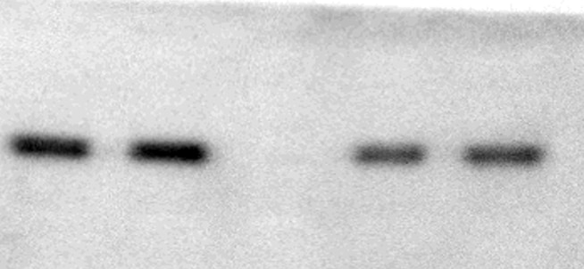
| Positive WB detected in | A431 cells, DU 145 cells, PC-3 cells, Cobalt Chloride treated HeLa cells, LNCaP cells, K-562 cells, A549 cells, SH-SY5Y cells |
| Positive IP detected in | MCF-7 cells |
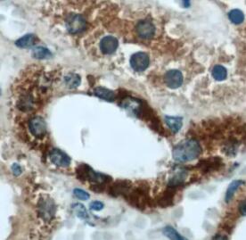
| Positive IHC detected in | human lung cancer tissue, human heart tissue, human liver tissue Note: suggested antigen retrieval with TE buffer pH 9.0; (*) Alternatively, antigen retrieval may be performed with citrate buffer pH 6.0 |
| Positive IF/ICC detected in | LNCaP cells |
Recommended dilution
| Application | Dilution |
|---|---|
| Western Blot (WB) | WB : 1:2000-1:12000 |
| Immunoprecipitation (IP) | IP : 0.5-4.0 ug for 1.0-3.0 mg of total protein lysate |
| Immunohistochemistry (IHC) | IHC : 1:50-1:500 |
| Immunofluorescence (IF)/ICC | IF/ICC : 1:200-1:800 |
| It is recommended that this reagent should be titrated in each testing system to obtain optimal results. | |
| Sample-dependent, Check data in validation data gallery. | |
Published Applications
| KD/KO | See 42 publications below |
| WB | See 279 publications below |
| IHC | See 33 publications below |
| IF | See 19 publications below |
| IP | See 1 publications below |
| CoIP | See 3 publications below |
| ChIP | See 2 publications below |
Product Information
10638-1-AP targets REDD1/DDIT4 specific in WB, IHC, IF/ICC, IP, CoIP, chIP, ELISA applications and shows reactivity with human, mouse samples.
| Tested Reactivity | human, mouse |
| Cited Reactivity | human, mouse, rat, pig, rabbit, squirrel, gerbil |
| Host / Isotype | Rabbit / IgG |
| Class | Polyclonal |
| Type | Antibody |
| Immunogen |
CatNo: Ag0965 Product name: Recombinant human REDD1 protein Source: e coli.-derived, PGEX-4T Tag: GST Domain: 1-232 aa of BC007714 Sequence: MPSLWDRFSSSSTSSSPSSLPRTPTPDRPPRSAWGSATREEGFDRSTSLESSDCESLDSSNSGFGPEEDTAYLDGVSLPDFELLSDPEDEHLCANLMQLLQESLAQARLGSRRPARLLMPSQLVSQVGKELLRLAYSEPCGLRGALLDVCVEQGKSCHSVGQLALDPSLVPTFQLTLVLRLDSRLWPKIQGLFSSANSPFLPGFSQSLTLSTGFRVIKKKLYSSEQLLIEEC Predict reactive species |
| Full Name | DNA-damage-inducible transcript 4 |
| Calculated Molecular Weight | 25 kDa |
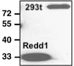
| Observed Molecular Weight | 32-35 kDa |
| GenBank Accession Number | BC007714 |
| Gene Symbol | REDD1/DDIT4 |
| Gene ID (NCBI) | 54541 |
| RRID | AB_2245711 |
| Conjugate | Unconjugated |
| Form | Liquid |
| Purification Method | Antigen affinity purification |
| UNIPROT ID | Q9NX09 |
| Storage Buffer | PBS with 0.02% sodium azide and 50% glycerol, pH 7.3. |
| Storage Conditions | Store at -20°C. Stable for one year after shipment. Aliquoting is unnecessary for -20oC storage. 20ul sizes contain 0.1% BSA. |
Background Information
REDD1, also named as RTP801 and DDIT4, belongs to the DDIT4 family. REDD1 promotes neuronal cell death. It is a novel transcriptional target of p53 implicated ROS in the p53-dependent DNA damage response. REDD1 controlled cell growth under energy stress, as an essential regulator of TOR activity through the TSC1/2 complex. REDD-1 expression has also been linked to apoptosis, Aβ toxicity and the pathogenesis of ischemic diseases. As an HIF-1-responsive gene, REDD-1 exhibits strong hypoxia-dependent upregulation in ischemic cells of neuronal origin[PMID: 19996311]. In response to stress due to DNA damage and glucocorticoid treatment, REDD-1 is upregulated at the transcriptional level[PMID: 21733849]. REDD-1 negatively regulates the mammalian target of Rapamycin, a serine/threonine kinase often referred to as mTOR[PMID: 22951983]. It is crucial in the coupling of extra- and intracellular cues to mTOR regulation. The absence of REDD-1 is associated with the development of retinopathy, a major cause of blindness[PMID: 22304497]. REDD1 is a new host defense factor, and chemical activation of REDD1 expression represents a potent antiviral intervention strategy[PMID: 21909097]. The calculated molecular weight of REDD1 is 25 kDa. Because of multiple lysines in the proteins, REDD1 offen migrates around 35 kDa on Western blot[PMID: 19221489]. This antibody is a rabbit polyclonal antibody raised against full length human REDD1 antigen. This antibody is specific to the REDD1 from siRNA experiment (PMID:24713927)
Protocols
| Product Specific Protocols | |
|---|---|
| IF protocol for REDD1/DDIT4 specific antibody 10638-1-AP | Download protocol |
| IHC protocol for REDD1/DDIT4 specific antibody 10638-1-AP | Download protocol |
| IP protocol for REDD1/DDIT4 specific antibody 10638-1-AP | Download protocol |
| WB protocol for REDD1/DDIT4 specific antibody 10638-1-AP | Download protocol |
| Standard Protocols | |
|---|---|
| Click here to view our Standard Protocols |
Publications
| Species | Application | Title |
|---|---|---|
Cell Metab Disrupted methionine cycle triggers muscle atrophy in cancer cachexia through epigenetic regulation of REDD1 | ||
Cancer Cell A UBE2O-AMPKα2 Axis that Promotes Tumor Initiation and Progression Offers Opportunities for Therapy. | ||
Nat Immunol The transcription factor Rfx7 limits metabolism of NK cells and promotes their maintenance and immunity. |
Reviews
The reviews below have been submitted by verified Proteintech customers who received an incentive for providing their feedback.
FH Kristen (Verified Customer) (03-30-2026) | Great, very little background via Western. Used as marker for hypoxia in cell line panel of human melanoma cell lines
|
FH Haley (Verified Customer) (12-01-2025) | Strong signal with minimal background. Would recommend.
|
FH jelan (Verified Customer) (11-25-2025) | The REDD1 antibody from Proteintech generally has clean bands, making it reliable for detecting stress response related changes in REDD1 expression. It performs consistently across Western blot and immunofluorescence with minimal background.
|
FH Abdulwahab (Verified Customer) (11-07-2025) | This antibody works very well for many leukemic cell lines
 |
FH Diana (Verified Customer) (10-17-2025) |
|
FH Haley (Verified Customer) (10-13-2025) | High sensitivity with minimal background
|
FH Sonam (Verified Customer) (10-02-2025) | The antibody worked well.
|
FH Yuki (Verified Customer) (11-10-2022) | This product was used for IHC in human tissue.
 |
FH Stephane (Verified Customer) (11-26-2020) | The antibody performs great on human cell extract. It detects specific bands with the REDD1 band resolving around 30-35 kD.
 |
FH Jamal (Verified Customer) (09-17-2019) | This product was used for Western Blotting in mouse cortex tissue.
|
FH Bradley (Verified Customer) (08-19-2019) | I have used this antibody on numerous occasions with success. The antibody performs great on human cell extract, as evidenced by a single immunoreactive band. The antibody performs moderately well on mouse skeletal muscle extract as it detects many non-specific bands with the REDD1 band resolving around 32-35 kD (specificity determined using REDD1-/- samples). The antibody is capable of detecting large changes in REDD1 protein in mouse skeletal muscle (i.e. induction following dexamethasone treatment), but smaller changes are difficult to assess in this tissue. This antibody will detect smaller changes in human cell lines such as HEK 293 or Hela cells.
|


