

Tested Applications
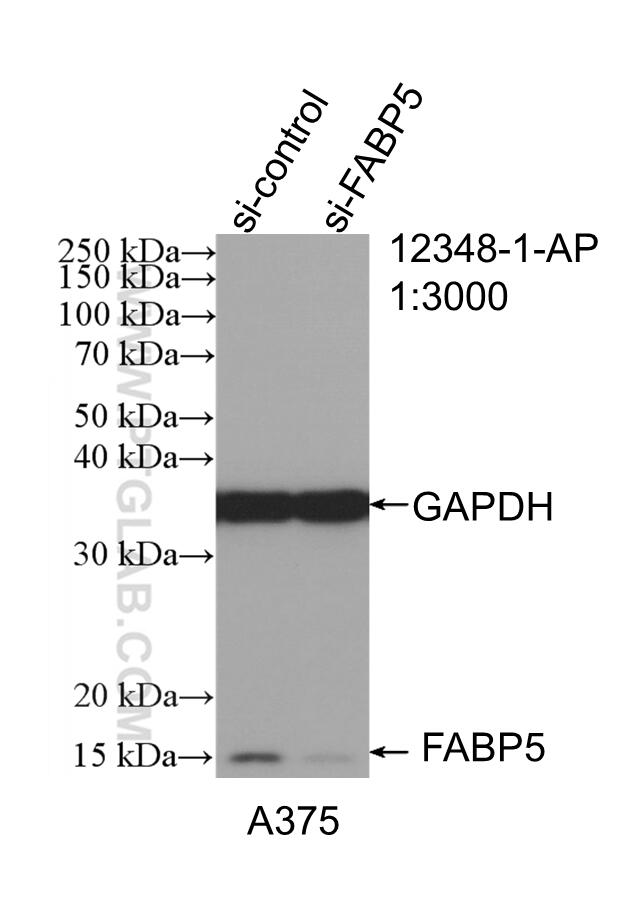
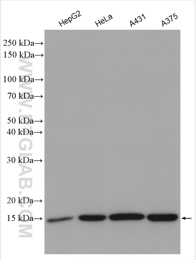
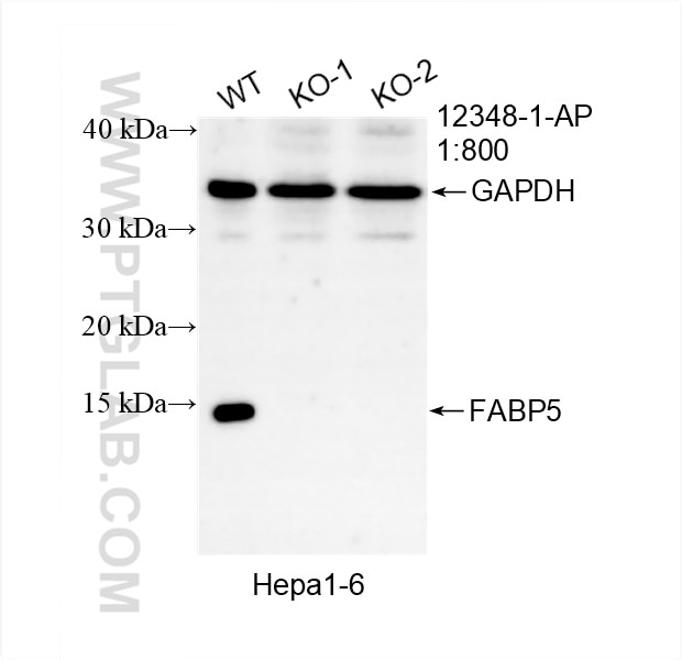
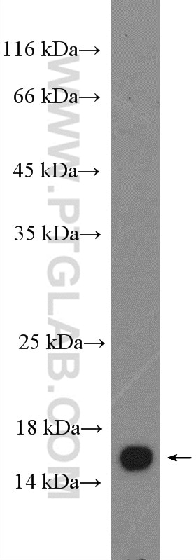
| Positive WB detected in | HepG2 cells, mouse skin tissue, A375 cells, Hepa1-6 cells, HeLa cells, A431 cells |
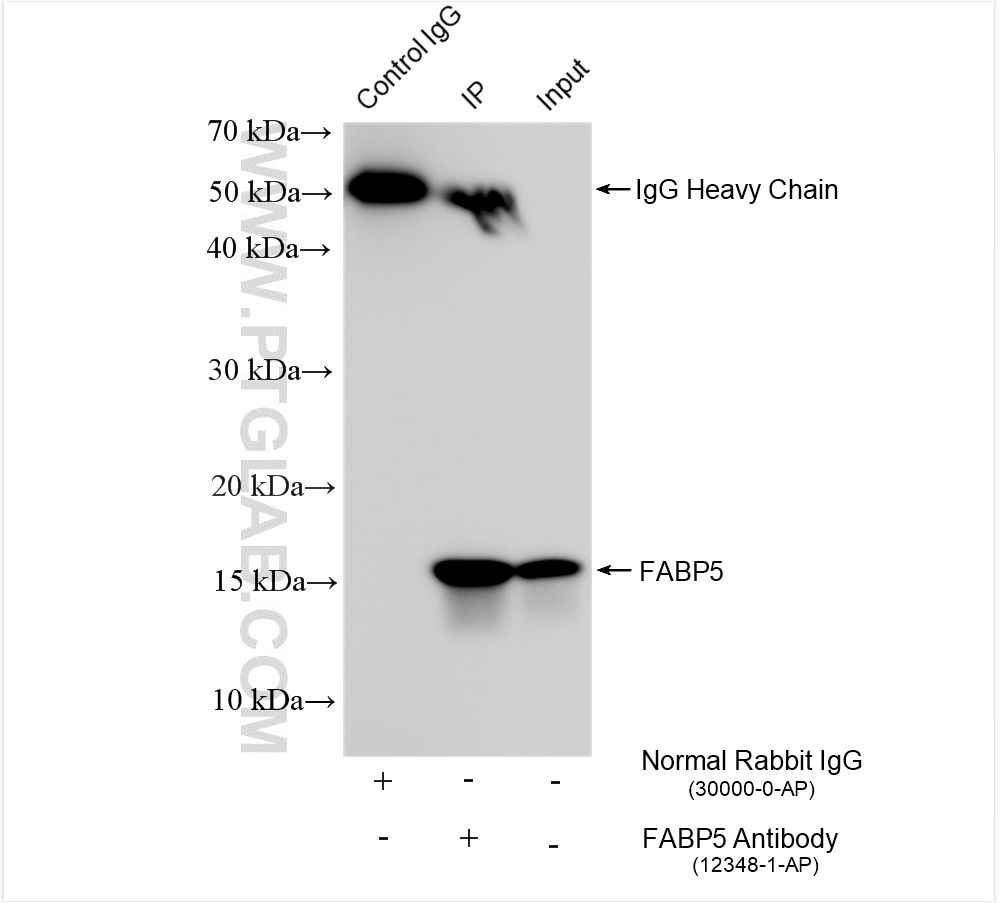
| Positive IP detected in | A375 cells |
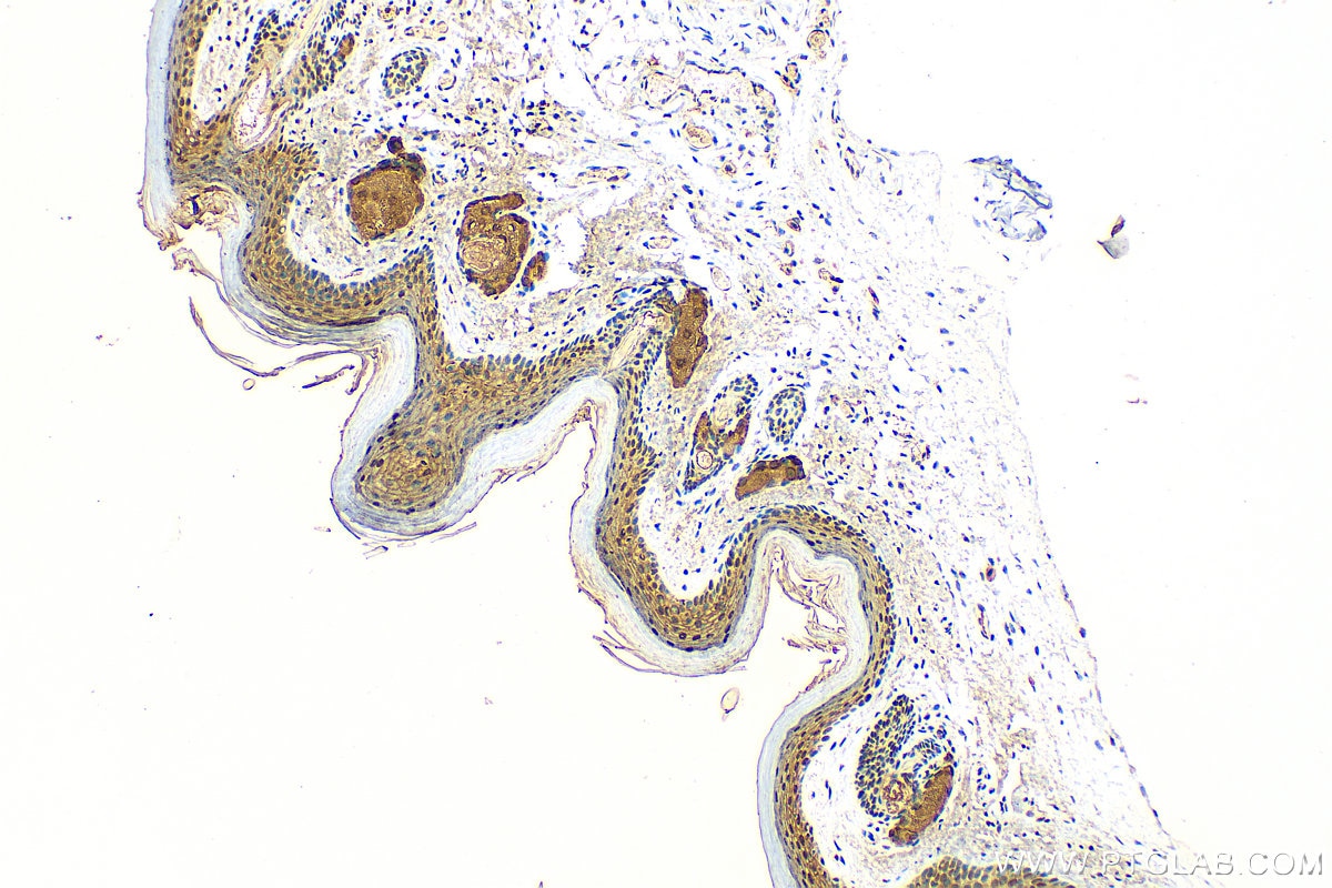
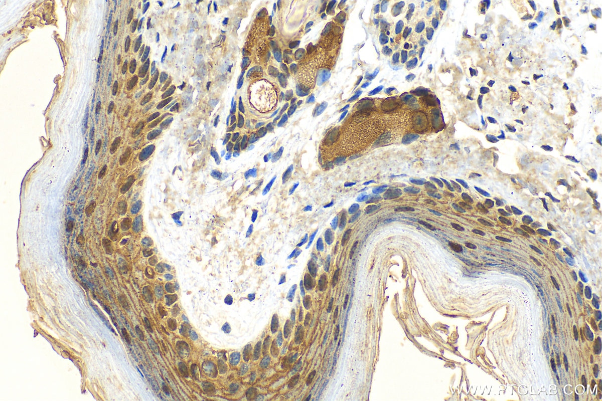
| Positive IHC detected in | mouse skin tissue Note: suggested antigen retrieval with TE buffer pH 9.0; (*) Alternatively, antigen retrieval may be performed with citrate buffer pH 6.0 |
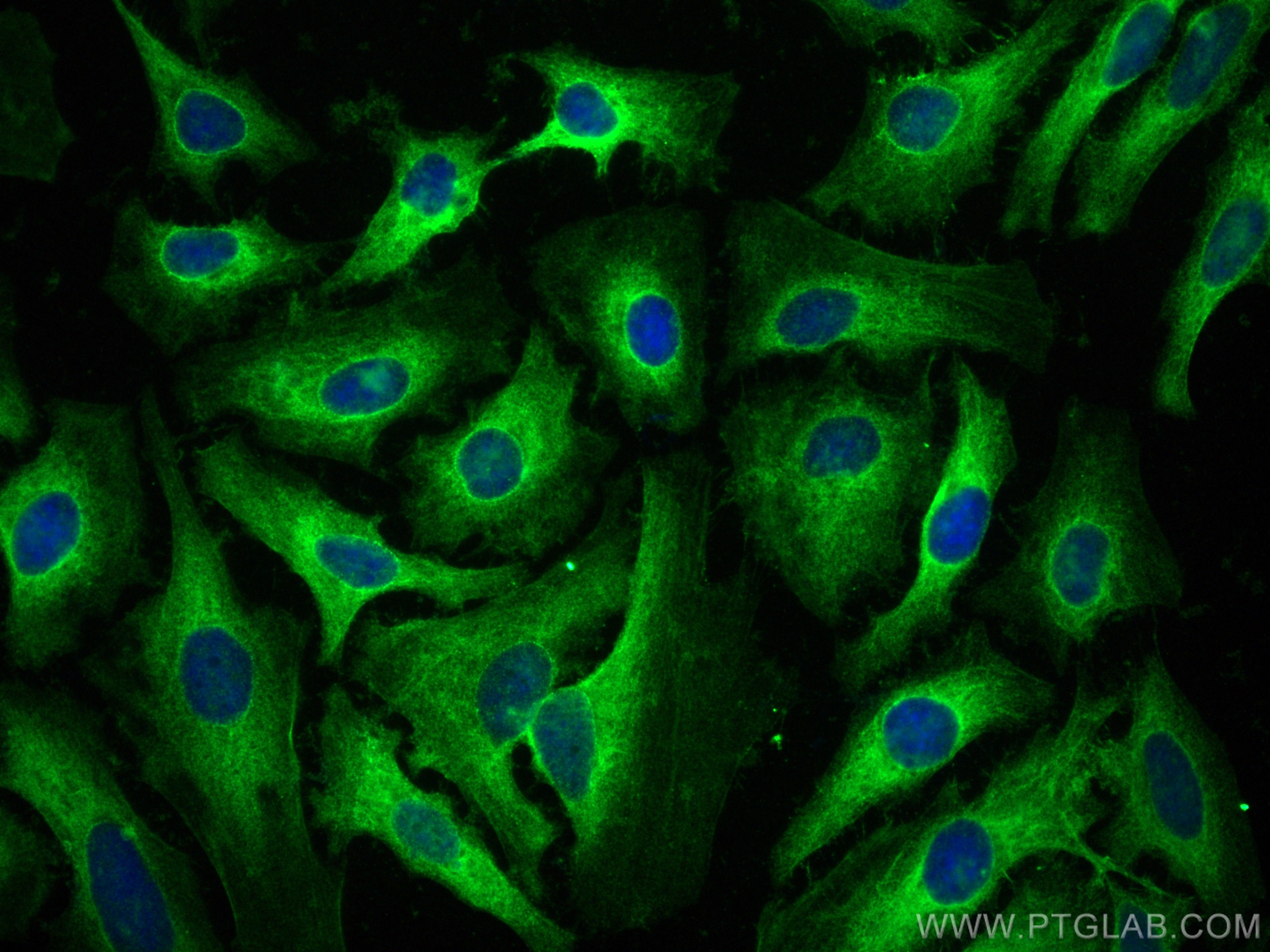
| Positive IF/ICC detected in | HeLa cells |
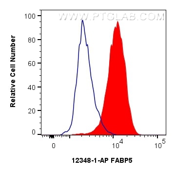
| Positive FC (Intra) detected in | HeLa cells |
Recommended dilution
| Application | Dilution |
|---|---|
| Western Blot (WB) | WB : 1:2000-1:10000 |
| Immunoprecipitation (IP) | IP : 0.5-4.0 ug for 1.0-3.0 mg of total protein lysate |
| Immunohistochemistry (IHC) | IHC : 1:50-1:500 |
| Immunofluorescence (IF)/ICC | IF/ICC : 1:50-1:500 |
| Flow Cytometry (FC) (INTRA) | FC (INTRA) : 0.40 ug per 10^6 cells in a 100 µl suspension |
| It is recommended that this reagent should be titrated in each testing system to obtain optimal results. | |
| Sample-dependent, Check data in validation data gallery. | |
Product Information
12348-1-AP targets FABP5 in WB, IHC, IF/ICC, FC (Intra), IP, CoIP, ELISA applications and shows reactivity with human, mouse, rat samples.
| Tested Reactivity | human, mouse, rat |
| Cited Reactivity | human, mouse, rat, pig |
| Host / Isotype | Rabbit / IgG |
| Class | Polyclonal |
| Type | Antibody |
| Immunogen |
CatNo: Ag3005 Product name: Recombinant human FABP5 protein Source: e coli.-derived, PGEX-4T Tag: GST Domain: 1-135 aa of BC019385 Sequence: MATVQQLEGRWRLVDSKGFDEYMKELGVGIALRKMGAMAKPDCIITCDGKNLTIKTESTLKTTQFSCTLGEKFEETTADGRKTQTVCNFTDGALVQHQEWDGKESTITRKLKDGKLVVECVMNNVTCTRIYEKVE Predict reactive species |
| Full Name | fatty acid binding protein 5 (psoriasis-associated) |
| Calculated Molecular Weight | 135 aa, 15 kDa |
| Observed Molecular Weight | 15 kDa |
| GenBank Accession Number | BC019385 |
| Gene Symbol | FABP5 |
| Gene ID (NCBI) | 2171 |
| RRID | AB_2100341 |
| Conjugate | Unconjugated |
| Form | Liquid |
| Purification Method | Antigen affinity purification |
| UNIPROT ID | Q01469 |
| Storage Buffer | PBS with 0.02% sodium azide and 50% glycerol, pH 7.3. |
| Storage Conditions | Store at -20°C. Stable for one year after shipment. Aliquoting is unnecessary for -20oC storage. 20ul sizes contain 0.1% BSA. |
Background Information
FABP5, also named as PA-FABP and E-FABP, belongs to the calycin superfamily and Fatty-acid binding protein (FABP) family. It is high specificity for fatty acids. FABP5 is highest affinity for C18 chain length. It may be involved in keratinocyte differentiation. FABP5 is a fatty acid-binding protein and is expressed in epidermis and endothelial cells of the microvasculature of different organs. FABP5 has also been identified as a tumor-associated antigen, which is highly expressed in various cancers. FABP5 was detected in the sera of HNSCC patients with early stage cancer. Antibodies specific for FABP5 were significantly increased in a substantial amount in patients, suggesting that FABP5 may be a potential diagnostic biomarker for HNSCC. FABP5 may serve as a biomarker for HNSCC.(PMID:19602232)
Protocols
| Product Specific Protocols | |
|---|---|
| FC protocol for FABP5 antibody 12348-1-AP | Download protocol |
| IF protocol for FABP5 antibody 12348-1-AP | Download protocol |
| IHC protocol for FABP5 antibody 12348-1-AP | Download protocol |
| IP protocol for FABP5 antibody 12348-1-AP | Download protocol |
| WB protocol for FABP5 antibody 12348-1-AP | Download protocol |
| Standard Protocols | |
|---|---|
| Click here to view our Standard Protocols |
Publications
What published studies show
The anti-FABP5 antibody (Cat# 12348-1-AP) has 75 citations across journals including Nature Communications, Immunity, Cell Reports, Advanced Science, Redox Biology, Journal of Allergy and Clinical Immunology, EMBO Journal, and Autophagy. Research fields span lipid metabolism, cancer (liver, breast, colorectal, thyroid, renal), ferroptosis, inflammation, psoriasis, cardiovascular disease, neurology, obesity, liver disease, and dermatology.
| Species | Application | Title |
|---|---|---|
Immunity Direct microglia replacement reveals pathologic and therapeutic contributions of brain macrophages to a monogenic neurological disease | ||
Nat Metab Intestinal TM6SF2 protects against metabolic dysfunction-associated steatohepatitis through the gut-liver axis | ||
Nat Commun Gut microbiota-induced perturbation in bile acids alter keratinocyte lipid metabolism via FXR-NQO1 signaling in psoriasis. | ||
Nat Commun Oxidative stress-induced FABP5 S-glutathionylation protects against acute lung injury by suppressing inflammation in macrophages. | ||
J Adv Res Fatty acid-binding protein 5 aggravates pulmonary artery fibrosis in pulmonary hypertension secondary to left heart disease via activating wnt/β-catenin pathway |
Reviews
What customers say
Users report strong performance across multiple applications. One reviewer achieved excellent Western blot and immunofluorescence results on BMDMs at 1:400. Another used it successfully for IHC and IF on 100 µm frozen mouse brain sections, noting strong, consistent labeling with good penetration, though mild background in some cortical areas was manageable with adjusted blocking. A third user confirmed robust immunoreactivity in mouse nerve tissue by IF at 1:500. Overall, the antibody is considered reliable and reproducible, with only minor background as a noted limitation.
The reviews below have been submitted by verified Proteintech customers who received an incentive for providing their feedback.
Jeroen (Verified Customer) (03-19-2026) | The antibody works perfectly for nice westernblots (on a special gel for small proteins) and ICC images
|
Vanessa (Verified Customer) (11-12-2025) | I used the FAB antibody for immunolabeling 100 µm frozen mouse brain sections collected at postnatal day 12 (P12) and postnatal day 30 (P30). The antibody performed well overall, showing strong and specific labeling of the expected structures in both age groups. Signal penetration through the thick frozen sections was good, and labeling was consistent across samples. There was a mild level of background staining, particularly in some cortical regions, but this did not interfere with interpretation of the specific signal. Adjusting blocking conditions and antibody dilution helped reduce the background. In general, the FAB antibody is reliable for use in thick frozen brain sections. It produces clear, reproducible staining with manageable background, making it suitable for developmental and comparative studies. Pros: Good penetration and signal in thick (100 µm) frozen sections Strong, consistent labelling at P12 and P30 Reproducible results Cons: Slight nonspecific background in some areas
|
Kenzo (Verified Customer) (01-09-2023) | Robust immunoreactivity detected.
|

