Tested Applications
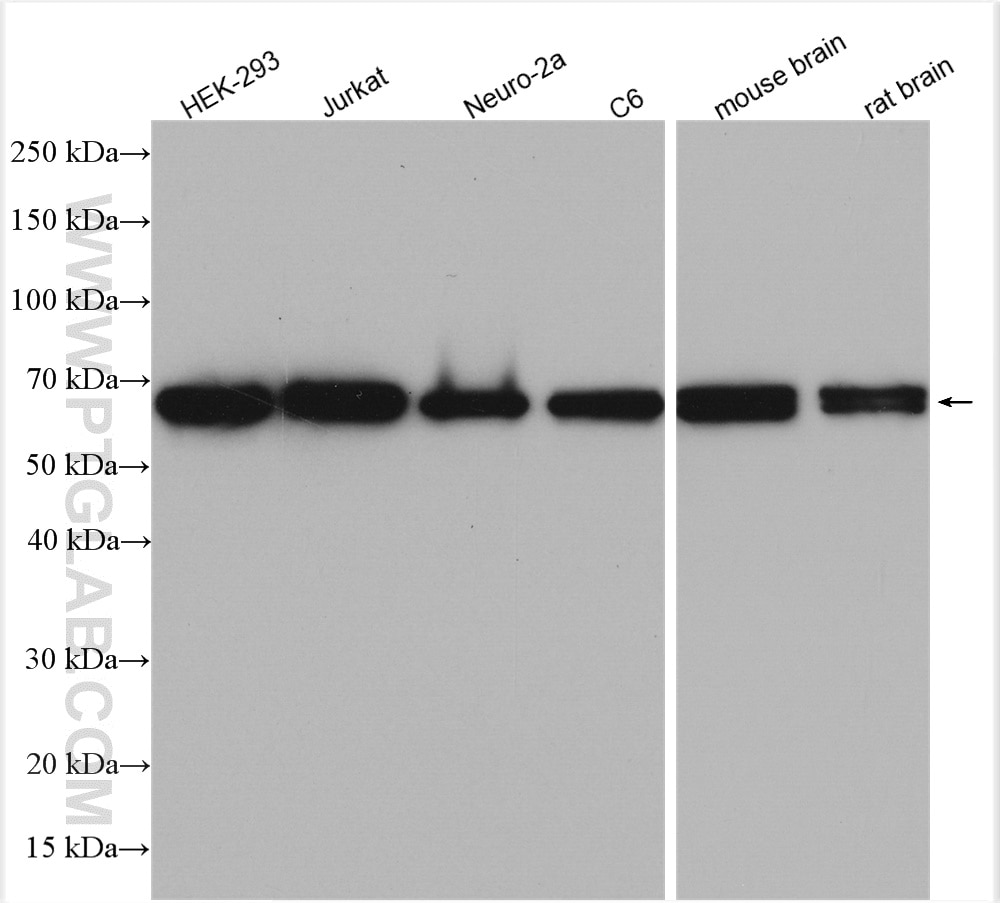
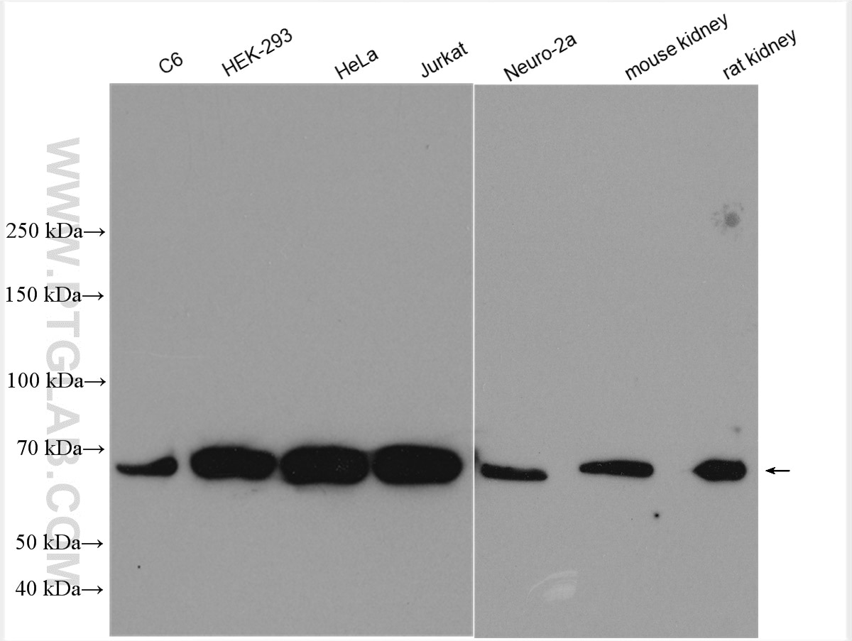
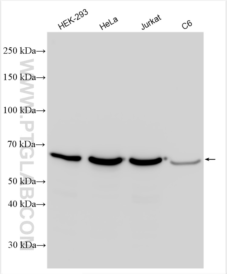
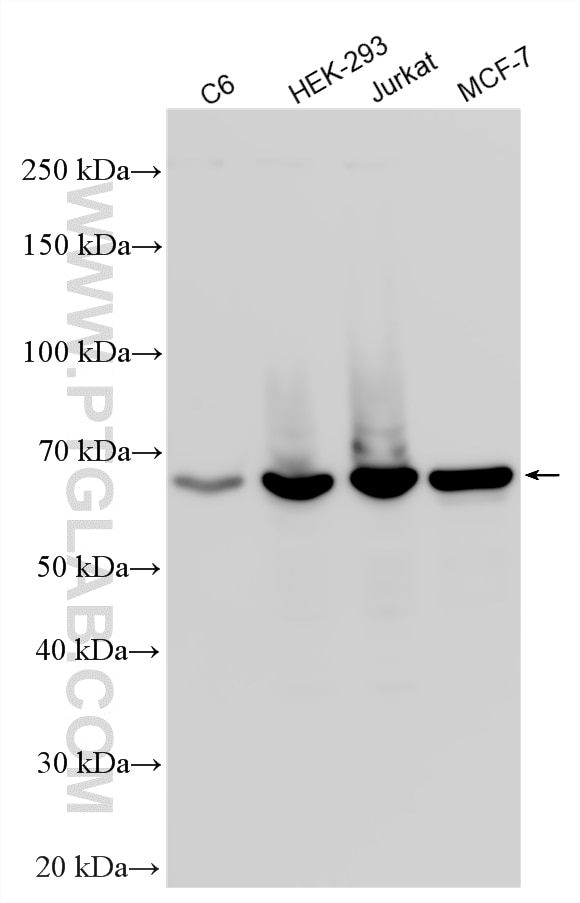
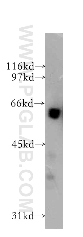
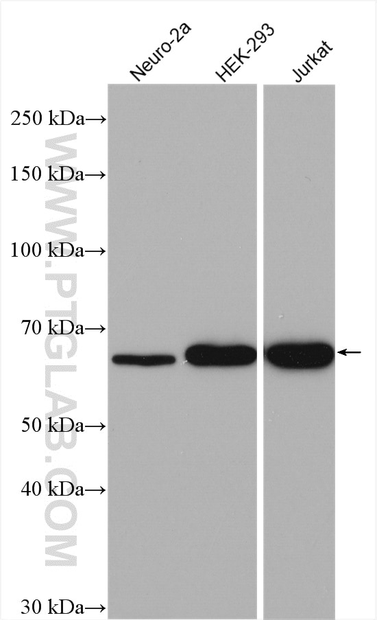
| Positive WB detected in | C6 cells, HEK-293 cells, human brain tissue, Neuro-2a cells, Jurkat cells, MCF-7 cells, HeLa cells, mouse kidney tissue, rat kidney tissue, mouse brain tissue, rat brain tissue |
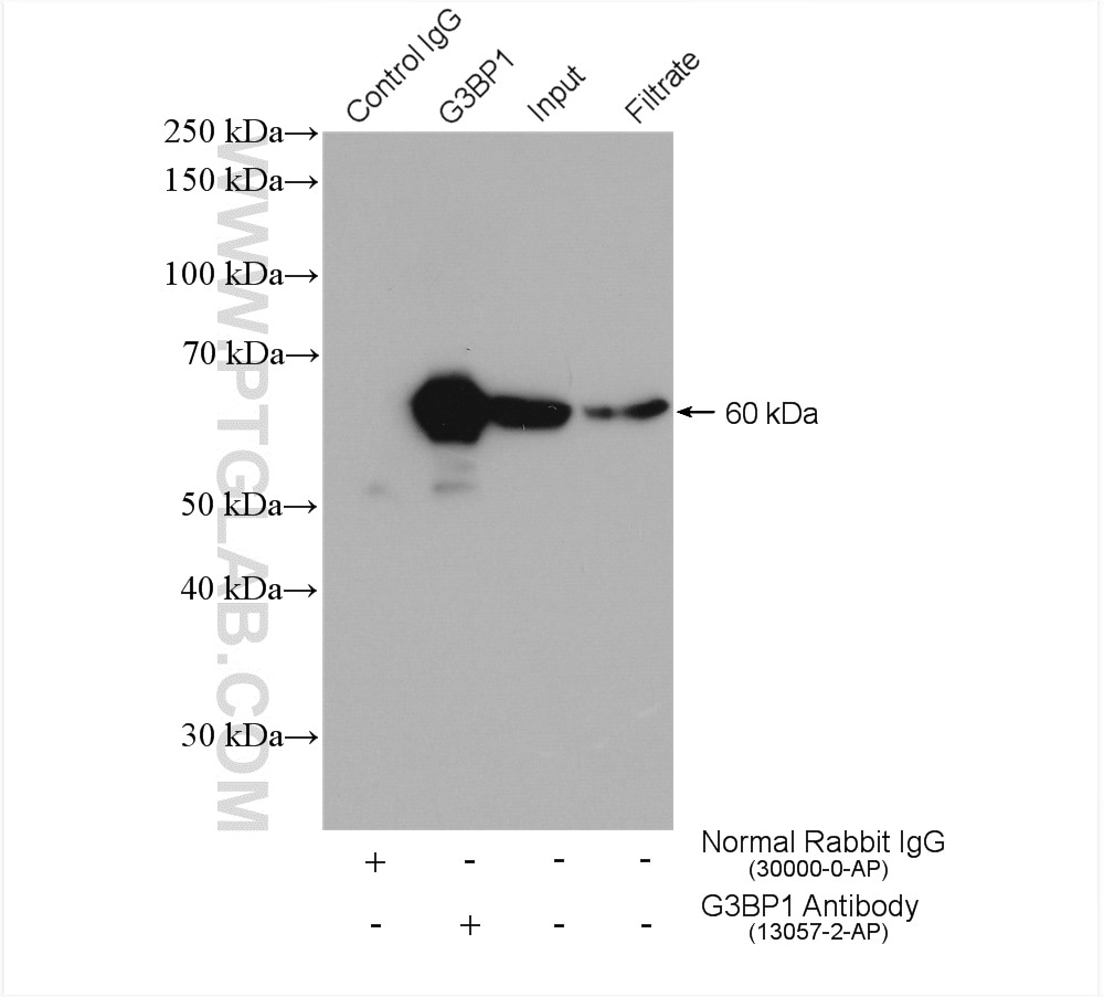
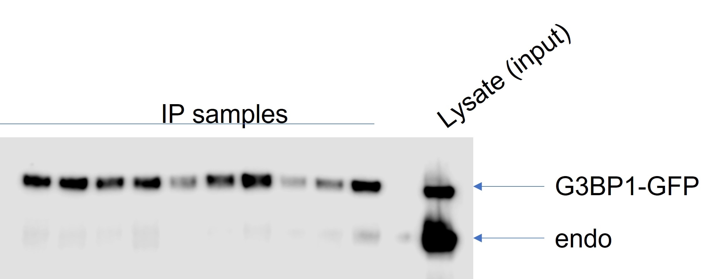
| Positive IP detected in | HEK-293 cells |
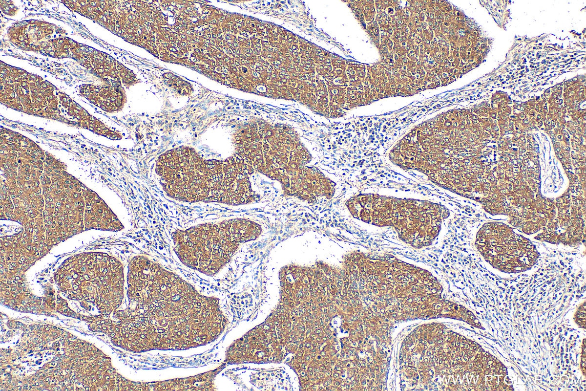
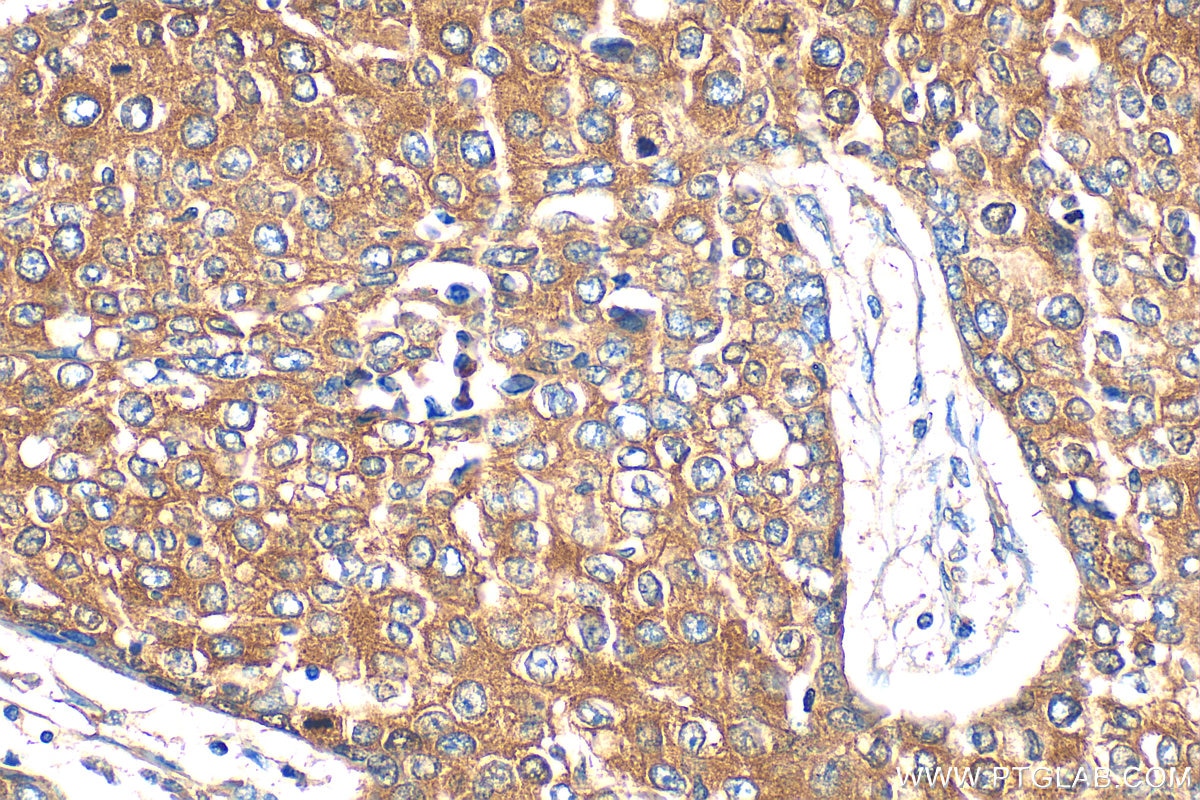
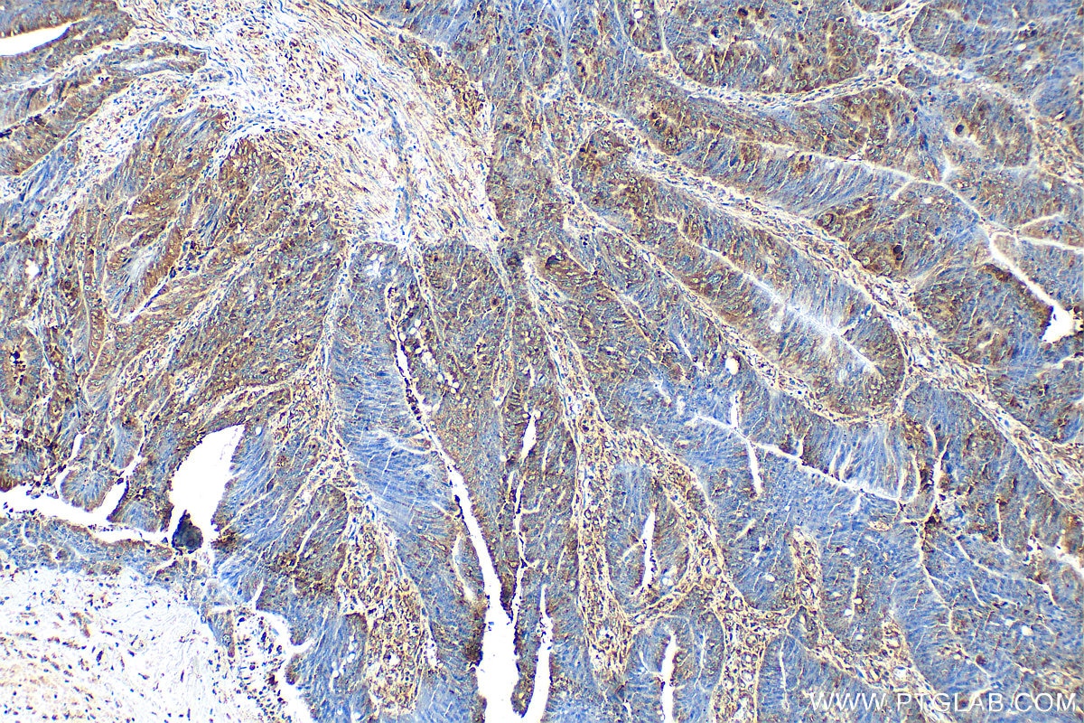
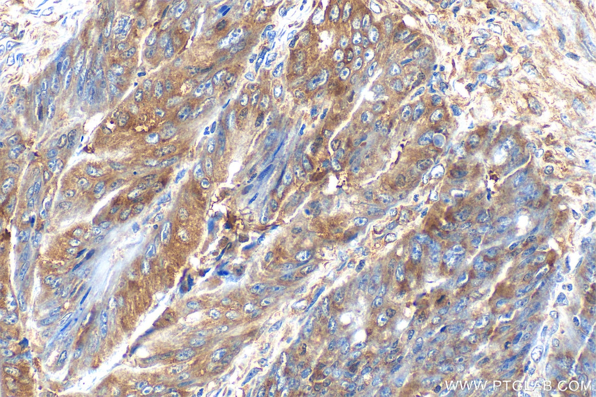
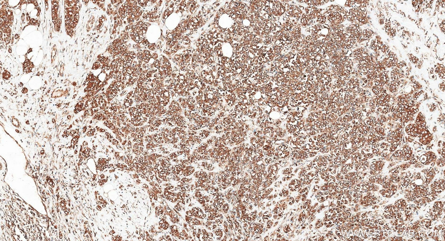
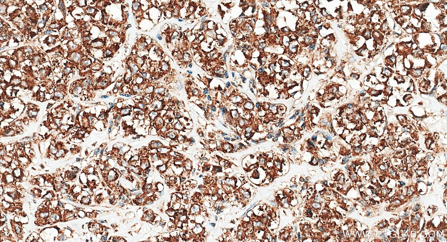
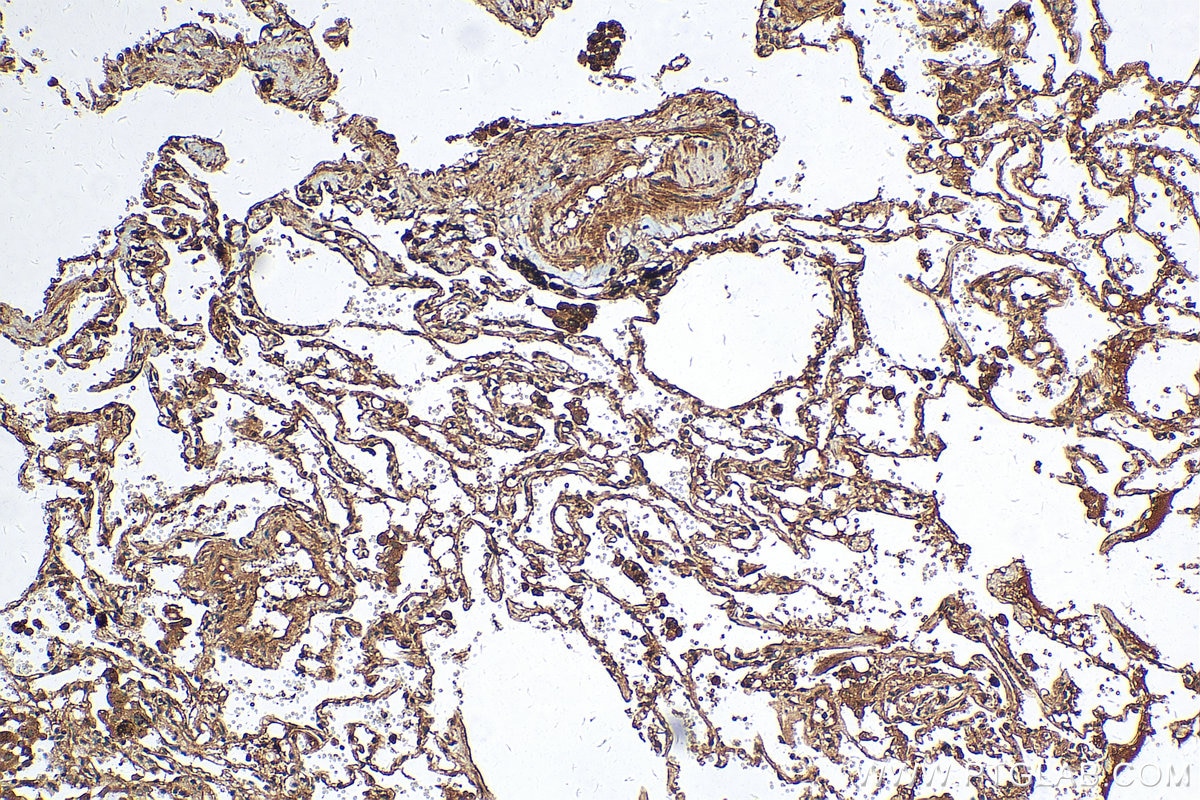
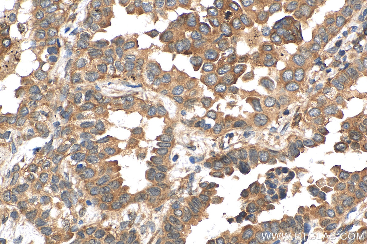
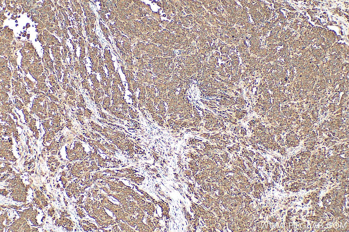
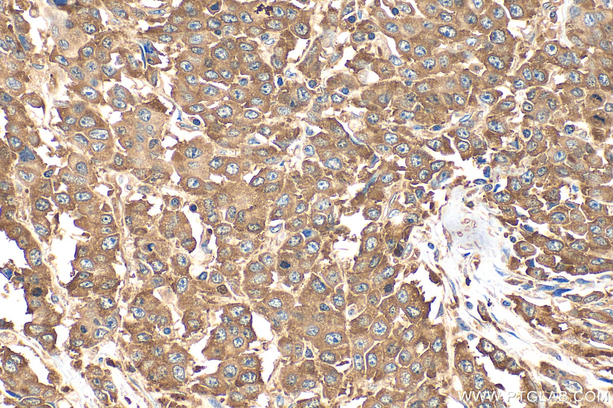
| Positive IHC detected in | human lung cancer tissue, human breast cancer tissue, human colon cancer tissue Note: suggested antigen retrieval with TE buffer pH 9.0; (*) Alternatively, antigen retrieval may be performed with citrate buffer pH 6.0 |
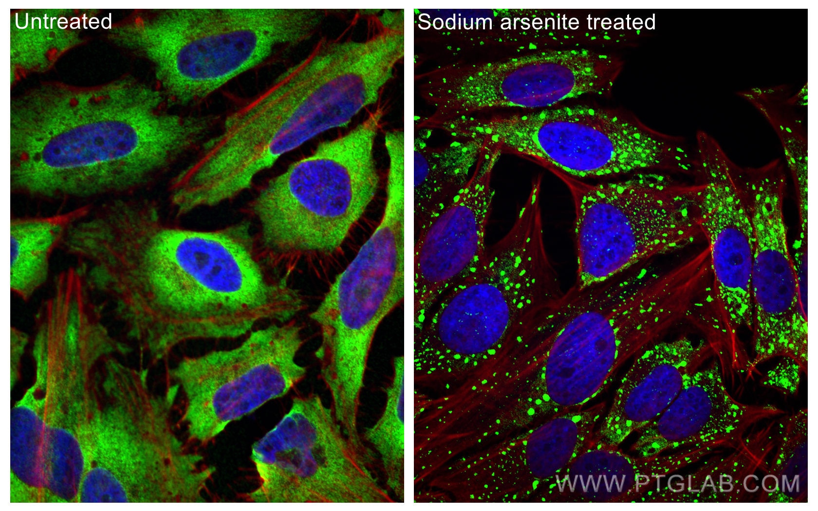
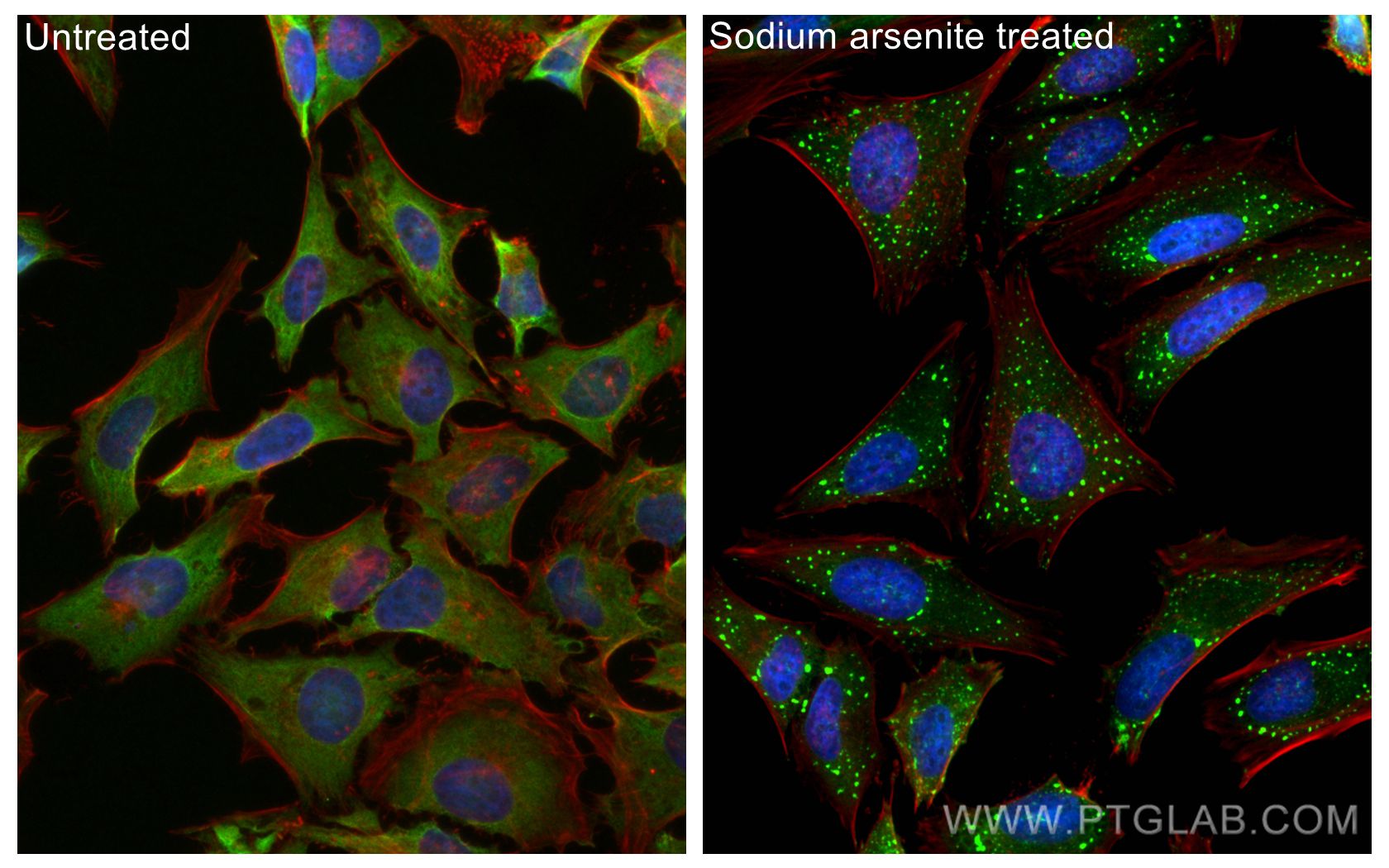
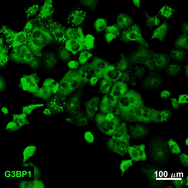
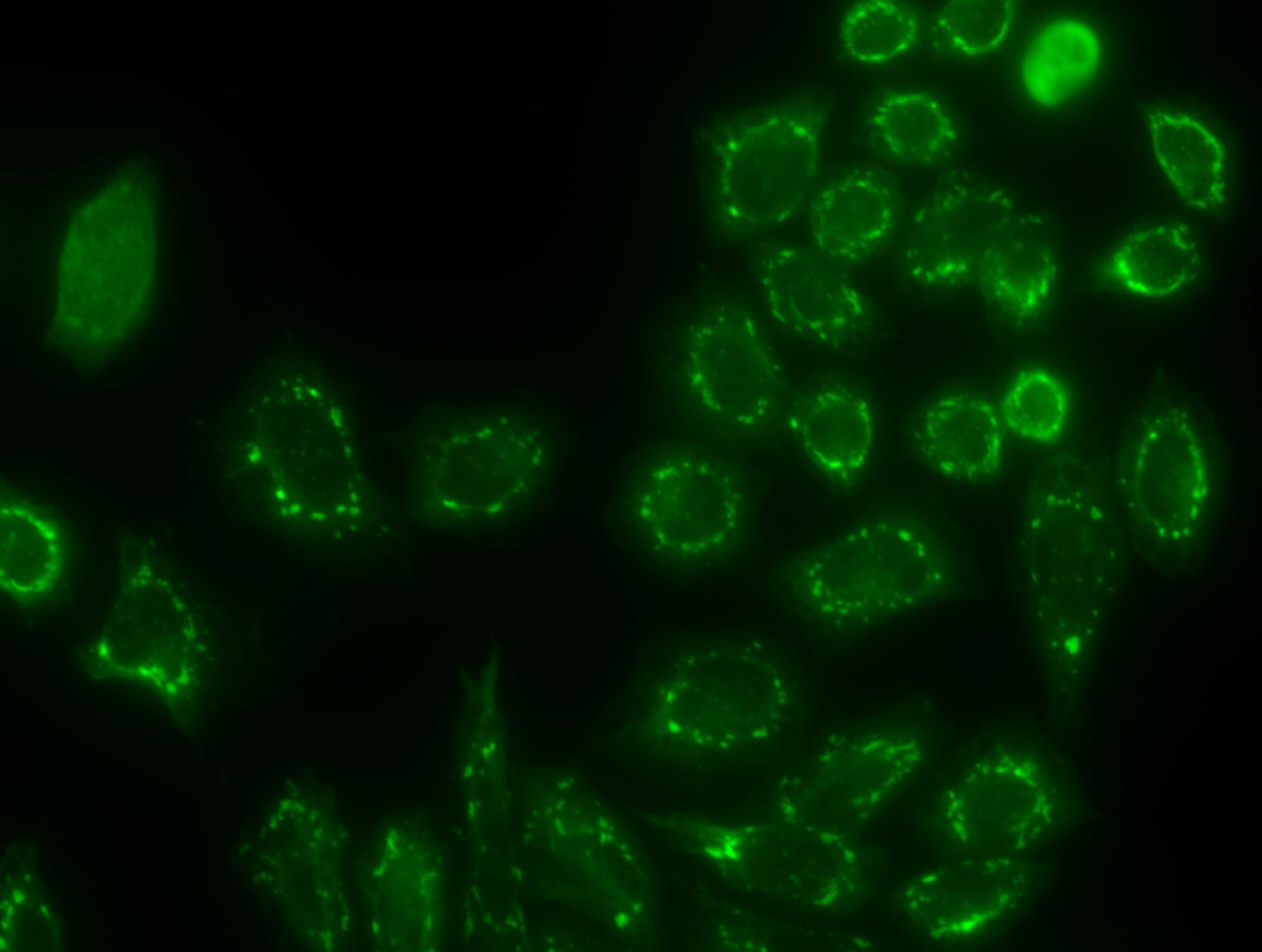
| Positive IF/ICC detected in | sodium arsenite treated HeLa cells |
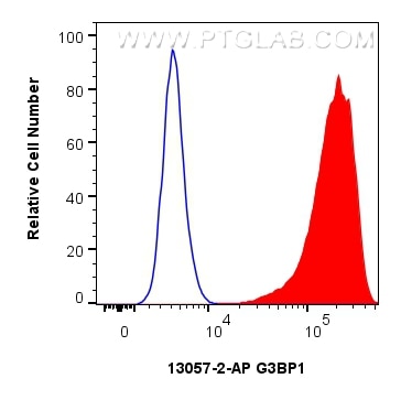
| Positive FC (Intra) detected in | HeLa cells |
Recommended dilution
| Application | Dilution |
|---|---|
| Western Blot (WB) | WB : 1:2000-1:16000 |
| Immunoprecipitation (IP) | IP : 0.5-4.0 ug for 1.0-3.0 mg of total protein lysate |
| Immunohistochemistry (IHC) | IHC : 1:50-1:500 |
| Immunofluorescence (IF)/ICC | IF/ICC : 1:1000-1:4000 |
| Flow Cytometry (FC) (INTRA) | FC (INTRA) : 0.25 ug per 10^6 cells in a 100 µl suspension |
| It is recommended that this reagent should be titrated in each testing system to obtain optimal results. | |
| Sample-dependent, Check data in validation data gallery. | |
Product Information
13057-2-AP targets G3BP1 in WB, IHC, IF/ICC, FC (Intra), IP, CoIP, RIP, ELISA applications and shows reactivity with human, mouse, rat samples.
| Tested Reactivity | human, mouse, rat |
| Cited Reactivity | human, mouse, rat, pig, monkey, chicken, zebrafish, drosophila melanogaster (fruit fly) |
| Host / Isotype | Rabbit / IgG |
| Class | Polyclonal |
| Type | Antibody |
| Immunogen |
CatNo: Ag3728 Product name: Recombinant human G3BP1 protein Source: e coli.-derived, PGEX-4T Tag: GST Domain: 167-466 aa of BC006997 Sequence: PDDSGTFYDQAVVSNDMEEHLEEPVAEPEPDPEPEPEQEPVSEIQEEKPEPVLEETAPEDAQKSSSPAPADIAQTVQEDLRTFSWASVTSKNLPPSGAVPVTGIPPHVVKVPASQPRPESKPESQIPPQRPQRDQRVREQRINIPPQRGPRPIREAGEQGDIEPRRMVRHPDSHQLFIGNLPHEVDKSELKDFFQSYGNVVELRINSGGKLPNFGFVVFDDSEPVQKVLSNRPIMFRGEVRLNVEEKKTRAAREGDRRDNRLRGPGGPRGGLGGGMRGPPRGGMVQKPGFGVGRGLAPRQ Predict reactive species |
| Full Name | GTPase activating protein (SH3 domain) binding protein 1 |
| Calculated Molecular Weight | 466 aa, 52 kDa |
| Observed Molecular Weight | 68 kDa |
| GenBank Accession Number | BC006997 |
| Gene Symbol | G3BP1 |
| Gene ID (NCBI) | 10146 |
| RRID | AB_2232034 |
| Conjugate | Unconjugated |
| Form | Liquid |
| Purification Method | Antigen affinity purification |
| UNIPROT ID | Q13283 |
| Storage Buffer | PBS with 0.02% sodium azide and 50% glycerol, pH 7.3. |
| Storage Conditions | Store at -20°C. Stable for one year after shipment. Aliquoting is unnecessary for -20oC storage. 20ul sizes contain 0.1% BSA. |
Background Information
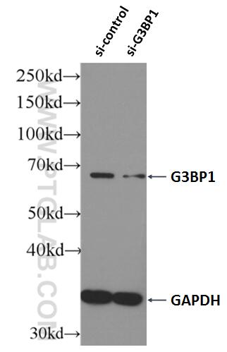
GAP SH3 Binding Protein 1 (G3BP1), also named as G3BP, is an effector of stress granule (SG) assembly. SG biology plays an important role in the pathophysiology of TDP-43 in ALS and FTLD-U. G3BP1 can be used as a marker of SG. It has been shown to function downstream of Ras and play a role in RNA metabolism, signal transduction, and proliferation. G3BP1 is a ubiquitously expressed protein that localizes to the cytoplasm in proliferating cells and to the nucleus in non-proliferating cells. G3BP1 has recently been implicated in cancer biology.
Protocols
| Product Specific Protocols | |
|---|---|
| FC protocol for G3BP1 antibody 13057-2-AP | Download protocol |
| IF protocol for G3BP1 antibody 13057-2-AP | Download protocol |
| IHC protocol for G3BP1 antibody 13057-2-AP | Download protocol |
| IP protocol for G3BP1 antibody 13057-2-AP | Download protocol |
| WB protocol for G3BP1 antibody 13057-2-AP | Download protocol |
| Standard Protocols | |
|---|---|
| Click here to view our Standard Protocols |
Publications
| Species | Application | Title |
|---|---|---|
Cell Diverse CMT2 neuropathies are linked to aberrant G3BP interactions in stress granules | ||
Science Ubiquitination of G3BP1 mediates stress granule disassembly in a context-specific manner. | ||
Cell ELAVL4, splicing, and glutamatergic dysfunction precede neuron loss in MAPT mutation cerebral organoids. | ||
Cell RNA Granules Hitchhike on Lysosomes for Long-Distance Transport, Using Annexin A11 as a Molecular Tether. | ||
Cell Phase Separation of FUS Is Suppressed by Its Nuclear Import Receptor and Arginine Methylation. |
Reviews
The reviews below have been submitted by verified Proteintech customers who received an incentive for providing their feedback.
FH Vasudevarao (Verified Customer) (04-09-2026) | Antibody works well for western blot in 1:1000 dilution, incubated overnight in 3%BSA/PBS on human lung cancer cell lines.
|
FH Prakash (Verified Customer) (10-17-2025) | working very well in western blot
|
FH lu (Verified Customer) (09-18-2025) | Good antibody for flow cytometry
|
FH Nikolett (Verified Customer) (07-29-2025) | Antibody works well for western blot in 1:1000 dilution, incubated overnight in 3%BSA/PBS on human lung cancer cell lines.
|
FH Xiaochen (Verified Customer) (11-11-2024) |
 |
FH Xiaochen (Verified Customer) (11-11-2024) |
 |
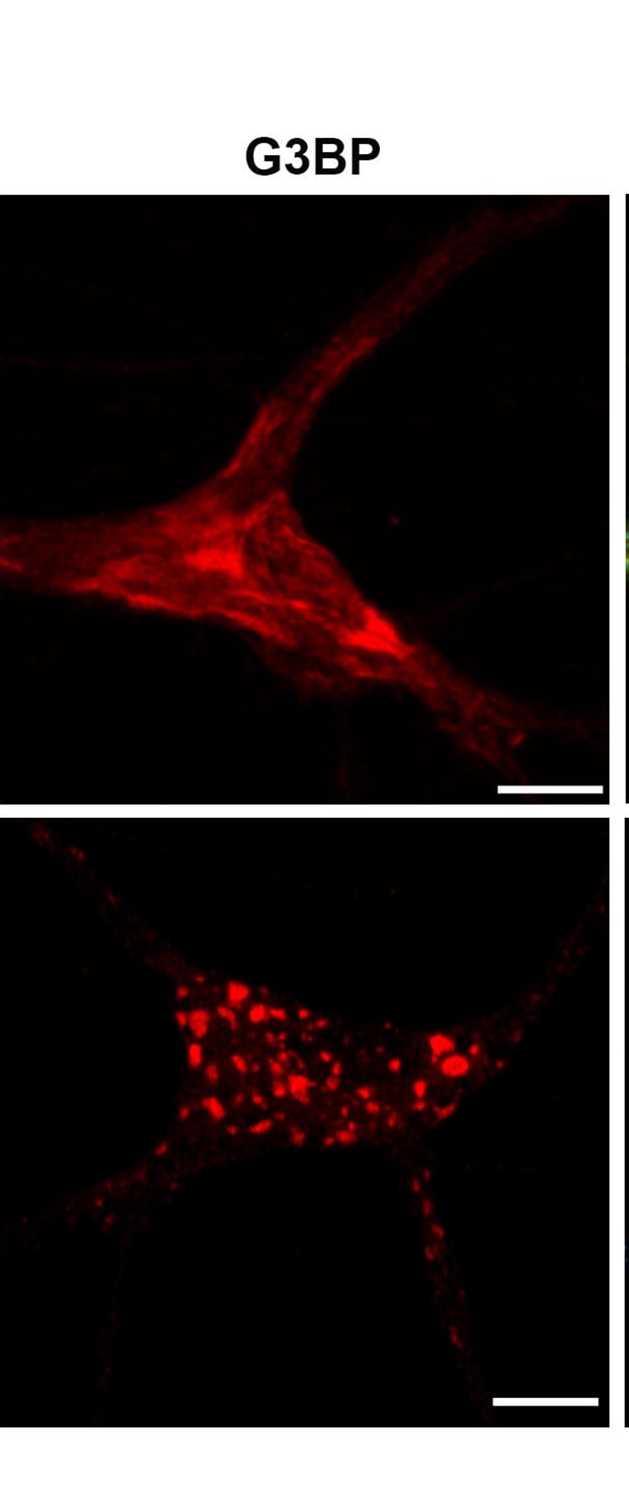
FH Roy (Verified Customer) (06-12-2024) | Works great on WB (1/1000 - Overnight 4°C) - Very beautiful IF staining in non stressed (diffuse staining) and Sodium Arsenite-mediated Stress induction (Punctate staining corresponding to stress granules) in HeLa cells (1/250 - 1h at RT).
|
FH Vinny (Verified Customer) (02-20-2024) | Good product.
|
FH Andrea (Verified Customer) (10-05-2023) | Good and strong signal.
|
FH Tatyana (Verified Customer) (01-21-2023) | Used for IP and WB of GFP-tagged overexpressed human G3BP1 (HEK293 cells). Lysates were subjected to IP using GFP-Trap beads. WB was done using semi-dry transfer. Antibody was used in 4% milk in TBST at 1:1,000 dilution. It could be reused up to 3 times. As the image shows, the antibody can successfully detect both endogenous and OE protein.
 |
FH Tobias (Verified Customer) (10-25-2022) |
|
FH Peter (Verified Customer) (10-24-2022) | Best G3BP1 antibody I've used for Western, IF and IP
|
FH Patryk (Verified Customer) (03-18-2021) | I used the antibody for immunofluorescence imaging to label stress granules induced by treating the cells with with 50µM sodium arsenite. Used the antibody at a dilution 1:100 overnight at 4°C. Worked perfectly well, strong and specific signal. I am very satisfied of this antibody and strongly recommend if for immunofluorescence.
 |
FH Kun (Verified Customer) (03-23-2020) | Very specific and sensitive
|
FH Biao (Verified Customer) (03-11-2020) | This antibody is very specific and good quality.
|
FH Joshua (Verified Customer) (12-28-2019) | PANC1 cells fixed in 4% paraformaldehdye. Bright localization to stress granules.
 |
FH Yuan (Verified Customer) (11-02-2019) | Very bright staining for stress granule on NaAsO2 treated Hela cells. 1:500 should be sufficient for IF staining.
 |
FH Zeinab (Verified Customer) (08-19-2019) | It worked great
|
FH Erica (Verified Customer) (05-15-2019) | Our lab has been using this antibody for IP, WB and IF for many years and it always worked well. I highly recommend this antibody, especially for IP for stress granules.
|
FH Karthik (Verified Customer) (04-24-2019) | Upon induction of sodium arsenite stress in neurons G3BP1 positive stress granules formed in 90 minutes.Cells permeabilized with 0.2% triton for 10 minutes
 |
FH Tian (Verified Customer) (01-23-2019) | I used it for ICC and it worked Great.
|