
Tested Applications
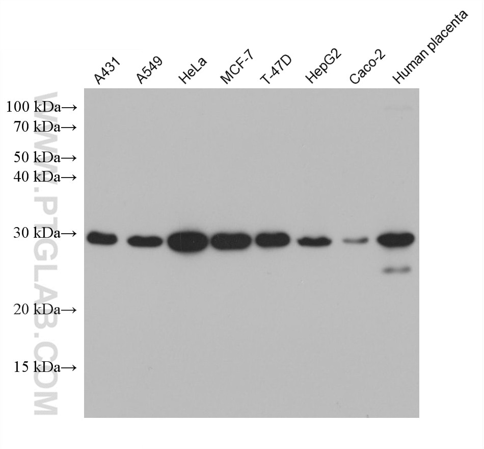
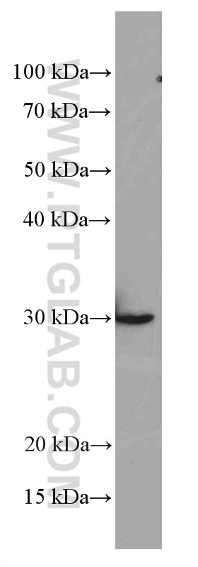
| Positive WB detected in | A431 cells, HSC-T6 cells, HeLa cells, A549 cells, MCF-7 cells, T-47D cells, HepG2 cells, Caco-2 cells, human placenta tissue |
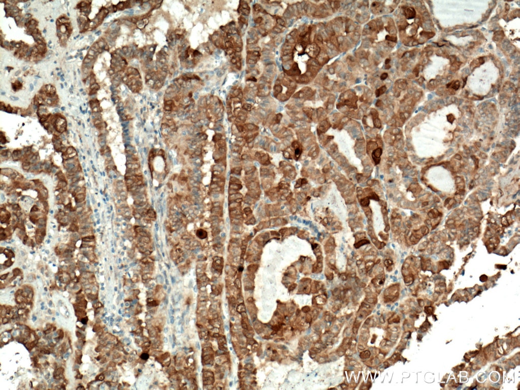
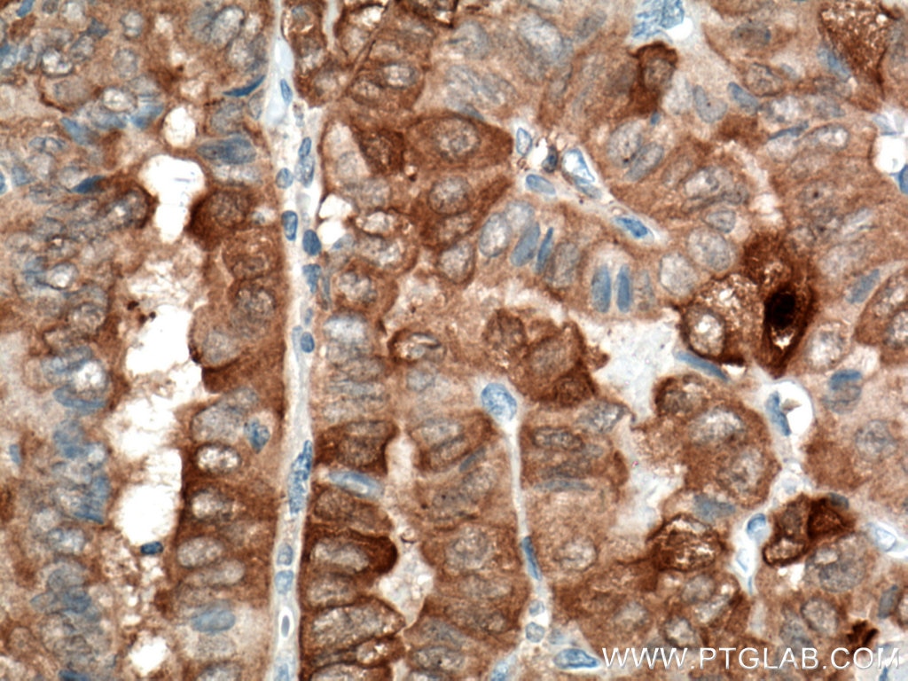
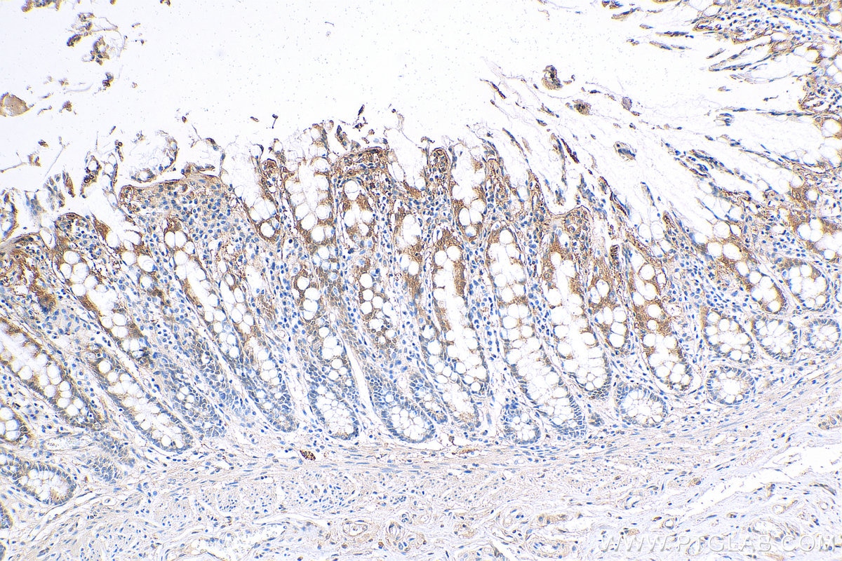
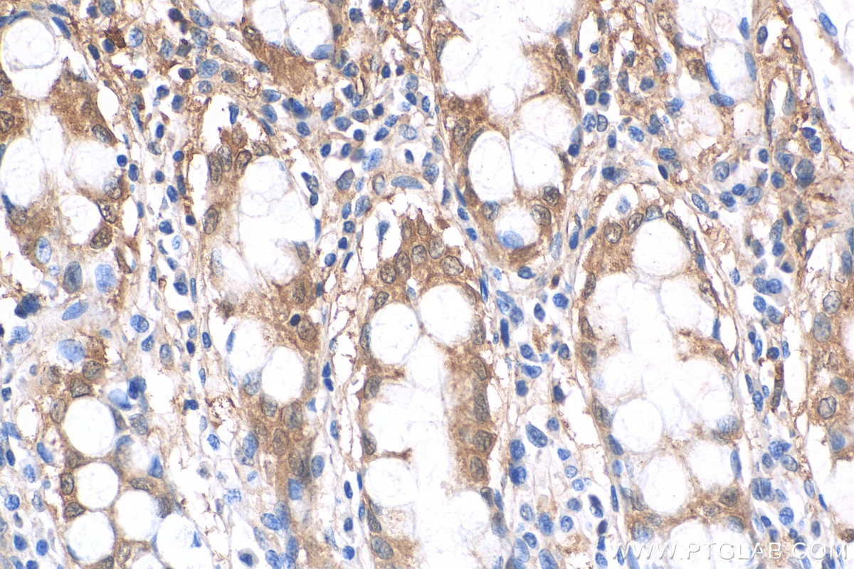
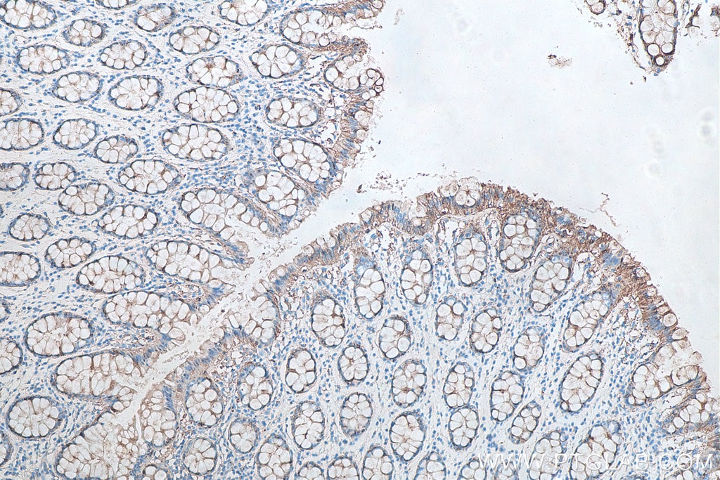
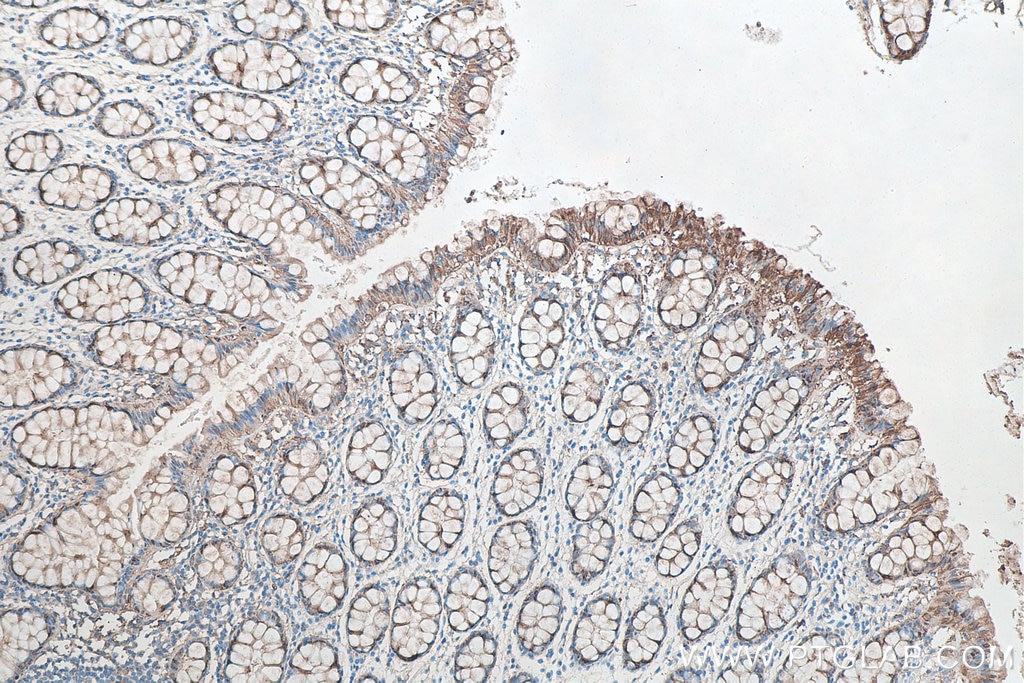
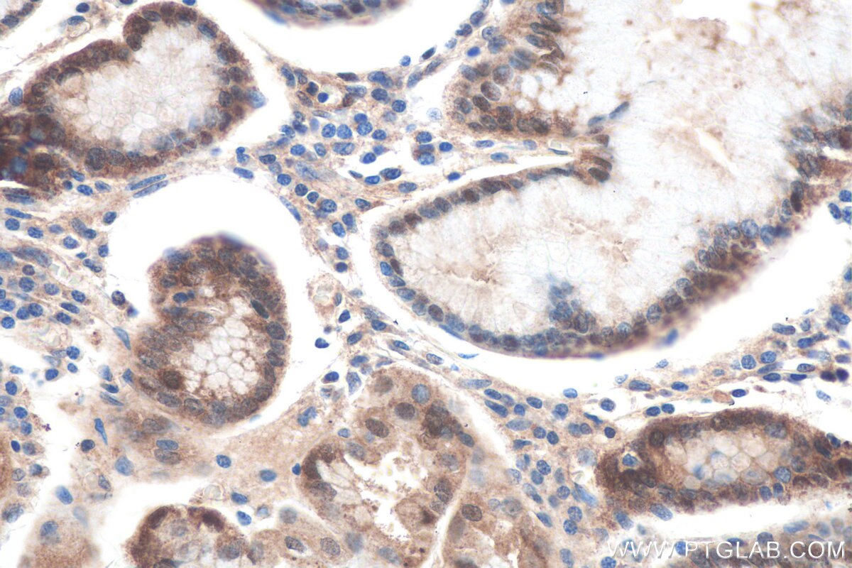
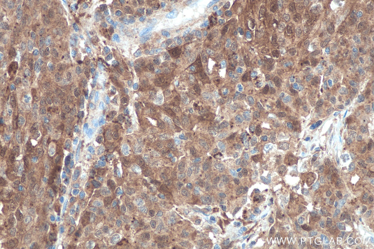
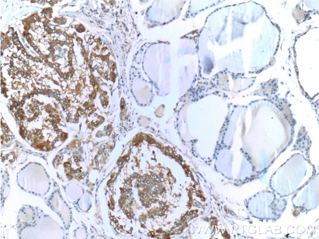
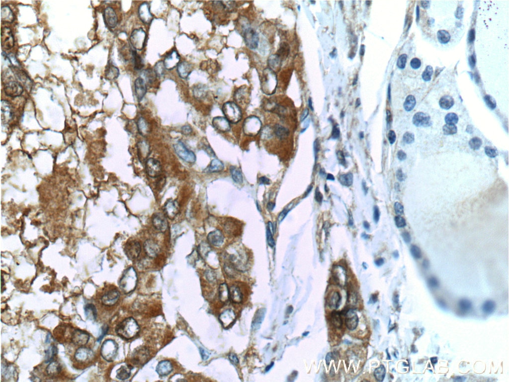
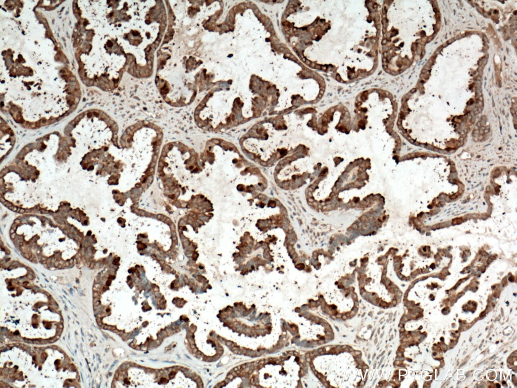
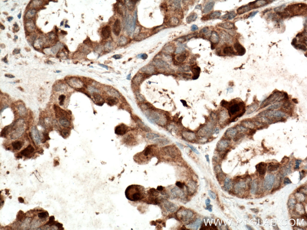
| Positive IHC detected in | human thyroid cancer tissue, human colon tissue, human ovary tumor tissue Note: suggested antigen retrieval with TE buffer pH 9.0; (*) Alternatively, antigen retrieval may be performed with citrate buffer pH 6.0 |
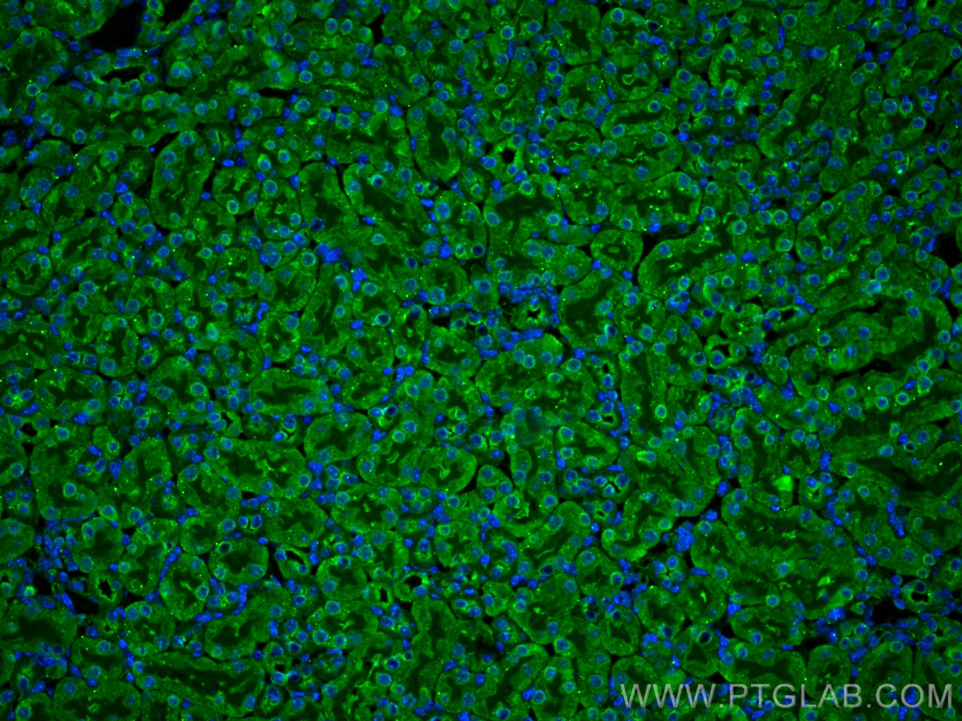
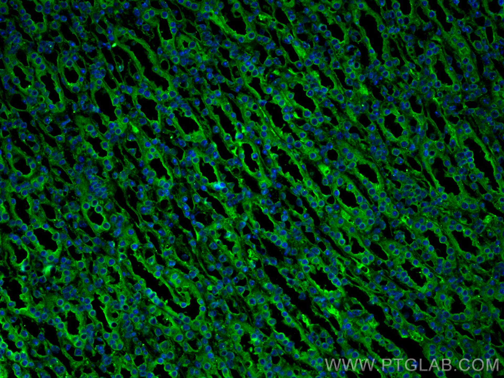
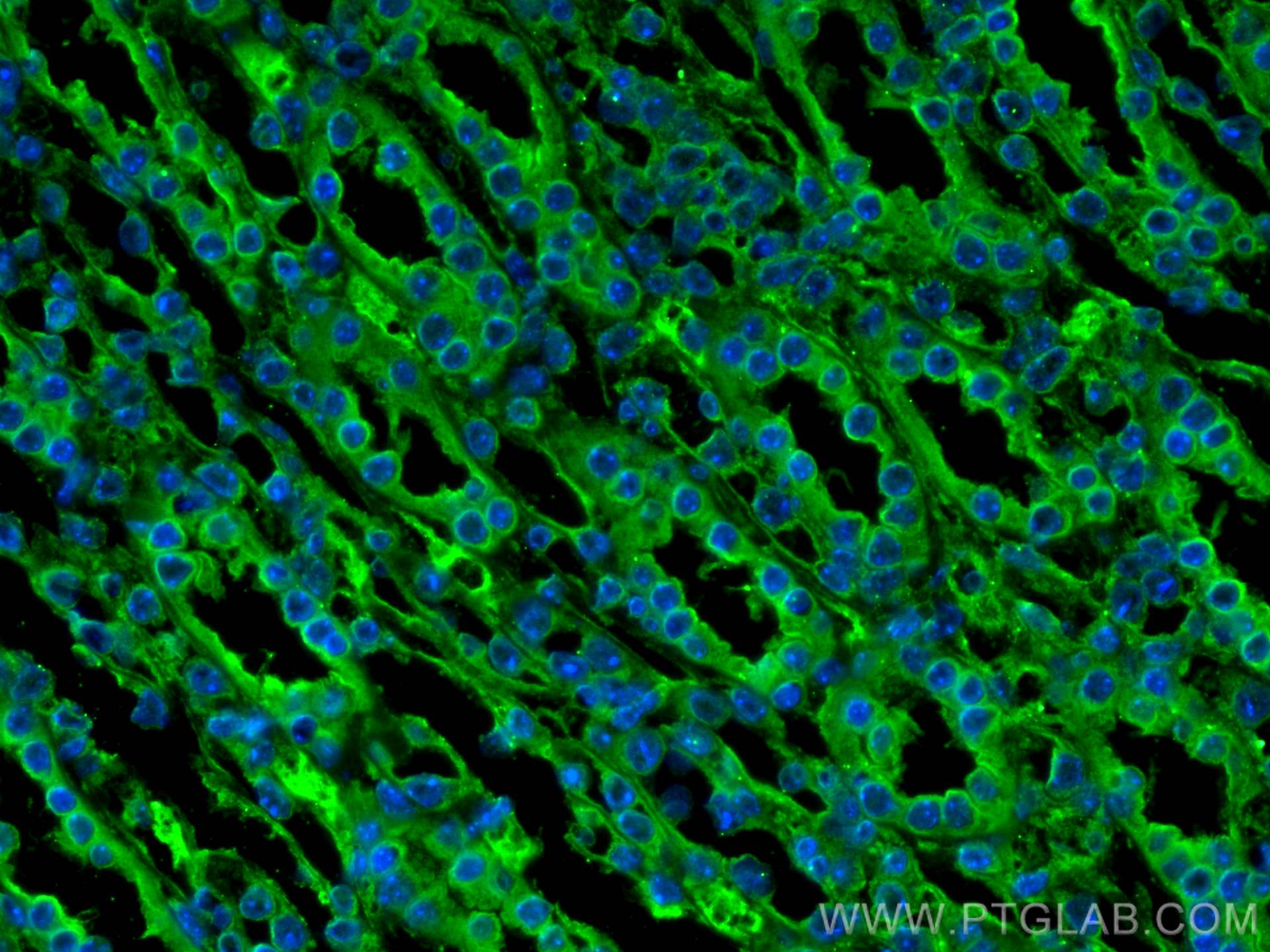
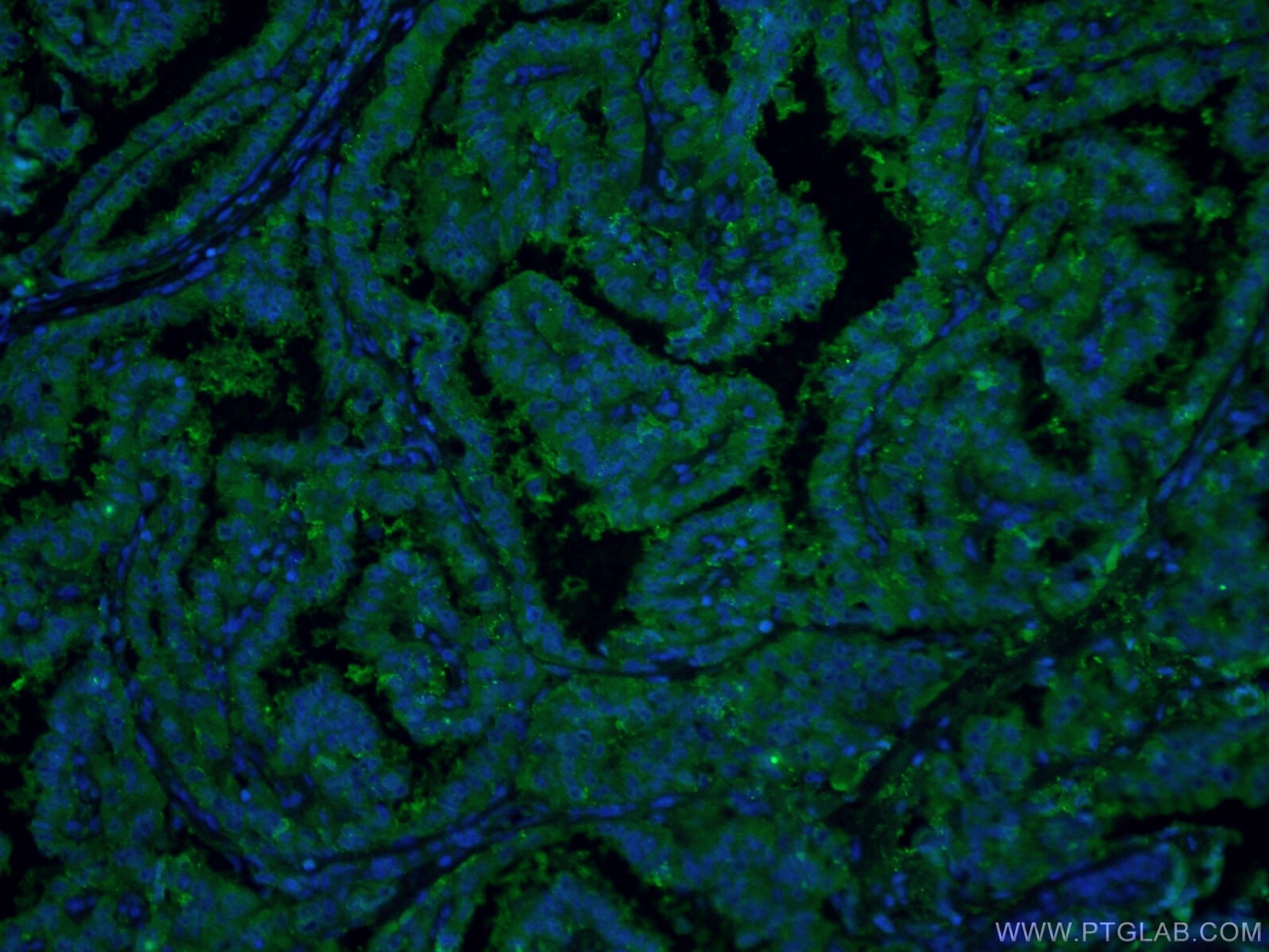
| Positive IF-P detected in | mouse kidney tissue, human thyroid cancer tissue |
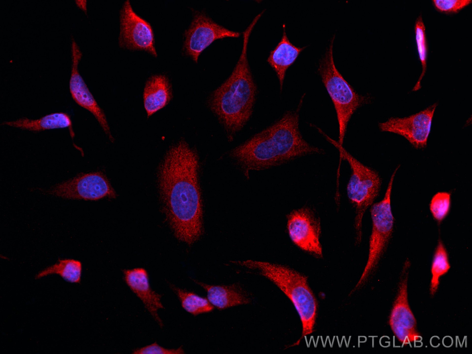
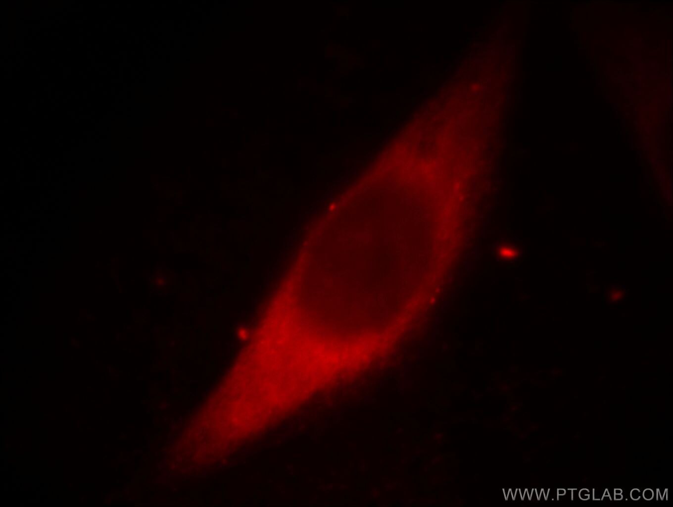
| Positive IF/ICC detected in | HeLa cells, mouse kidney tissue, MCF-7 cells, human thyroid cancer tissue |
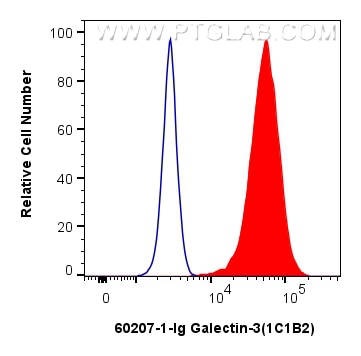
| Positive FC (Intra) detected in | HeLa cells |
Recommended dilution
| Application | Dilution |
|---|---|
| Western Blot (WB) | WB : 1:5000-1:50000 |
| Immunohistochemistry (IHC) | IHC : 1:1000-1:4000 |
| Immunofluorescence (IF)-P | IF-P : 1:200-1:800 |
| Immunofluorescence (IF)/ICC | IF/ICC : 1:400-1:1600 |
| Flow Cytometry (FC) (INTRA) | FC (INTRA) : 0.80 ug per 10^6 cells in a 100 µl suspension |
| It is recommended that this reagent should be titrated in each testing system to obtain optimal results. | |
| Sample-dependent, Check data in validation data gallery. | |
Product Information
60207-1-Ig targets Galectin-3 in WB, IHC, IF/ICC, IF-P, FC (Intra), IP, CoIP, ELISA applications and shows reactivity with human, mouse samples.
| Tested Reactivity | human, mouse |
| Cited Reactivity | human, mouse, rat |
| Host / Isotype | Mouse / IgG2b |
| Class | Monoclonal |
| Type | Antibody |
| Immunogen |
CatNo: Ag6753 Product name: Recombinant human Galectin 3 protein Source: e coli.-derived, PET28a Tag: 6*His Domain: 1-250 aa of BC001120 Sequence: MADNFSLHDALSGSGNPNPQGWPGAWGNQPAGAGGYPGASYPGAYPGQAPPGAYPGQAPPGAYPGAPGAYPGAPAPGVYPGPPSGPGAYPSSGQPSATGAYPATGPYGAPAGPLIVPYNLPLPGGVVPRMLITILGTVKPNANRIALDFQRGNDVAFHFNPRFNENNRRVIVCNTKLDNNWGREERQSVFPFESGKPFKIQVLVEPDHFKVAVNDAHLLQYNHRVKKLNEISKLGISGDIDLTSASYTMI Predict reactive species |
| Full Name | lectin, galactoside-binding, soluble, 3 |
| Calculated Molecular Weight | 26 kDa |
| Observed Molecular Weight | 31 kDa |
| GenBank Accession Number | BC001120 |
| Gene Symbol | Galectin-3 |
| Gene ID (NCBI) | 3958 |
| RRID | AB_10951109 |
| Conjugate | Unconjugated |
| Form | Liquid |
| Purification Method | Protein A purification |
| UNIPROT ID | P17931 |
| Storage Buffer | PBS with 0.02% sodium azide and 50% glycerol, pH 7.3. |
| Storage Conditions | Store at -20°C. Stable for one year after shipment. Aliquoting is unnecessary for -20oC storage. 20ul sizes contain 0.1% BSA. |
Background Information
Galectins are a family of animal lectins defined by shared characteristic amino-acid sequences and affinity for β-galactose-containing oligosac-charides (PMID: 8063692). Galectin-3, a member of the β-galactoside-binding proteins, contains one carbohydrate recognition domain (CRD) and a proline- and glycine-rich N-terminal domain through which is able to form oligomers (PMID: 14758078). Galectin-3 is widely expressed in many normal tissues and a variety of tumors. It is found intracellularly in nucleus and cytoplasm or secreted outside of cell, being present on the cell surface or in the extracellular space (PMID: 16478649). Galectin-3 is involved in various biological processes including cell growth, adhesion, differentiation, apoptosis, angiogenesis, immune response, neoplastic transformation and metastasis (PMID: 16478649; 14758078).
Protocols
| Product Specific Protocols | |
|---|---|
| FC protocol for Galectin-3 antibody 60207-1-Ig | Download protocol |
| IF protocol for Galectin-3 antibody 60207-1-Ig | Download protocol |
| IHC protocol for Galectin-3 antibody 60207-1-Ig | Download protocol |
| WB protocol for Galectin-3 antibody 60207-1-Ig | Download protocol |
| Standard Protocols | |
|---|---|
| Click here to view our Standard Protocols |
Publications
What published studies show
| Species | Application | Title |
|---|---|---|
Nat Commun Surplus fatty acid synthesis increases oxidative stress in adipocytes and lnduces lipodystrophy | ||
Cell Death Differ NLRX1 mediated impaired microglial phagocytosis of NETs in cerebral ischemia and reperfusion injury | ||
Cancer Res CD248 Regulates Wnt Signaling in Pericytes to Promote Angiogenesis and Tumor Growth in Lung Cancer | ||
Autophagy SMURF1 controls the PPP3/calcineurin complex and TFEB at a regulatory node for lysosomal biogenesis | ||
Circ. Res. Inhibition of KLF5-Myo9b-RhoA Pathway-Mediated Podosome Formation in Macrophages Ameliorates Abdominal Aortic Aneurysm. | ||
Acta Biomater Tumor cell membrane-based peptide delivery system targeting the tumor microenvironment for cancer immunotherapy and diagnosis. |
Reviews
What customers say
The reviews below have been submitted by verified Proteintech customers who received an incentive for providing their feedback.
Boyan (Verified Customer) (06-19-2026) | good for both imaging and WB
|
Marco (Verified Customer) (07-09-2024) | works perfectly fine
|
Kenzo (Verified Customer) (08-29-2023) | I used this antibody in mouse kidney tissues and it worked well.
|
Muhammad (Verified Customer) (09-13-2022) | Work best in HUVECs
|
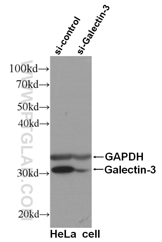
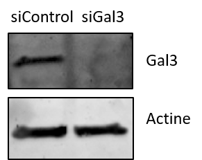
Noémie (Verified Customer) (10-20-2021) | I used this antibody for Western Blot against Gal3 in mamalian cells (U2OS), and did a validation by siRNA.
 |

