

Tested Applications
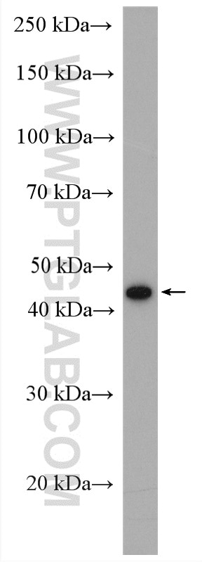
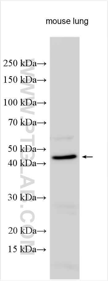
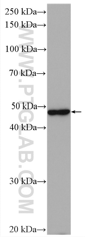
| Positive WB detected in | mouse lung tissue |
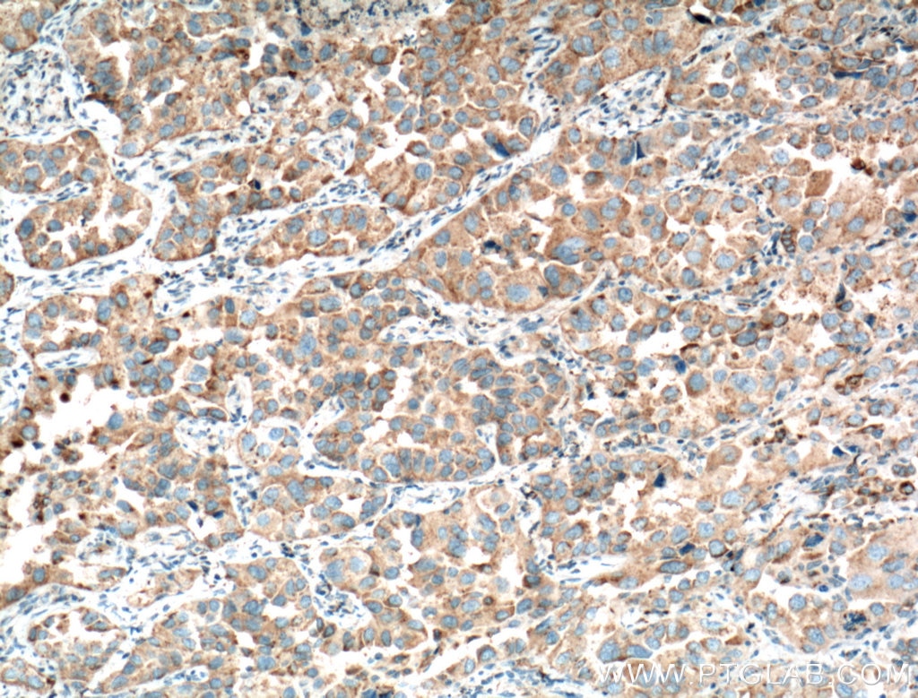
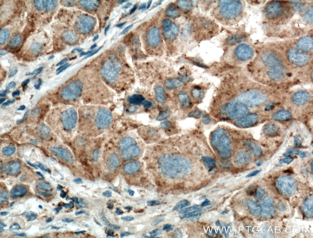
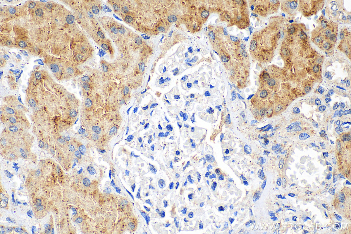
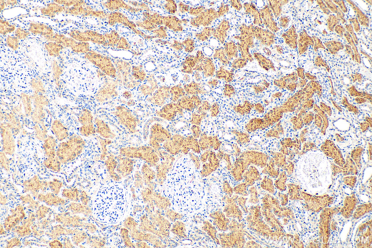
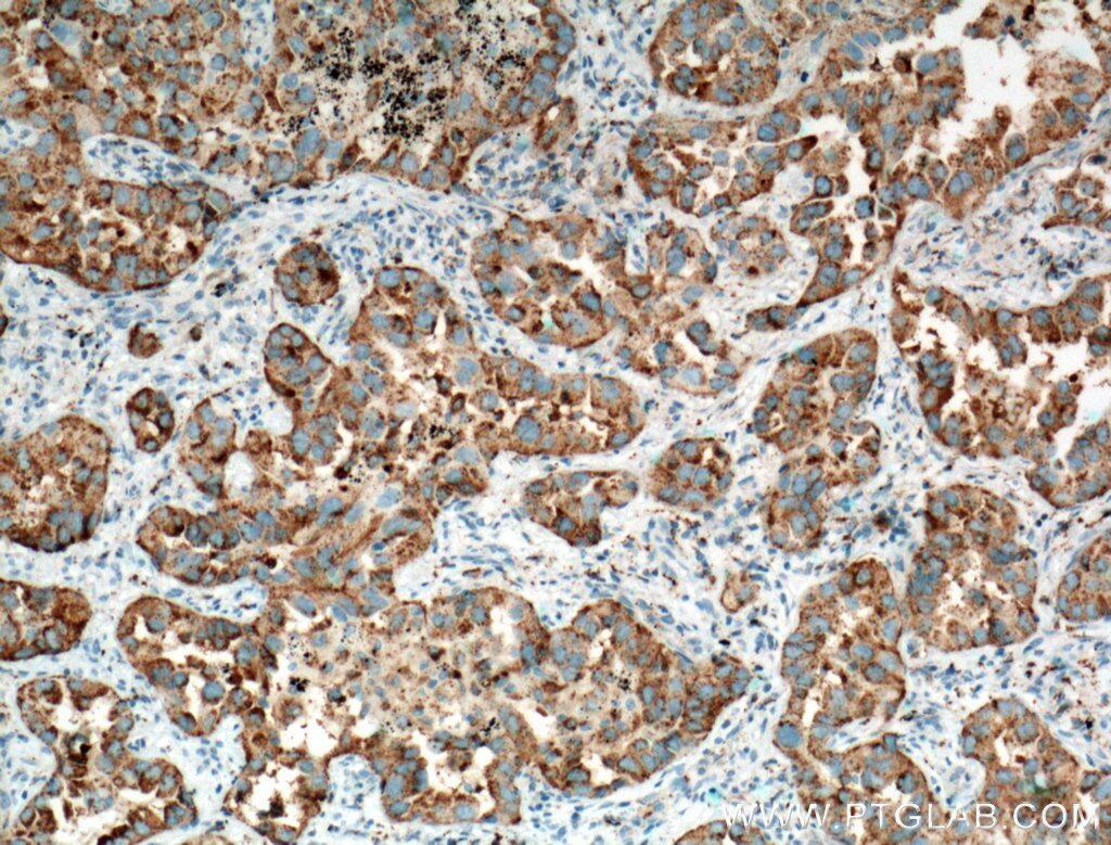
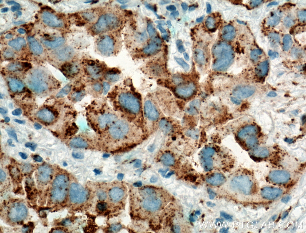
| Positive IHC detected in | human lung cancer tissue, human kidney tissue Note: suggested antigen retrieval with TE buffer pH 9.0; (*) Alternatively, antigen retrieval may be performed with citrate buffer pH 6.0 |
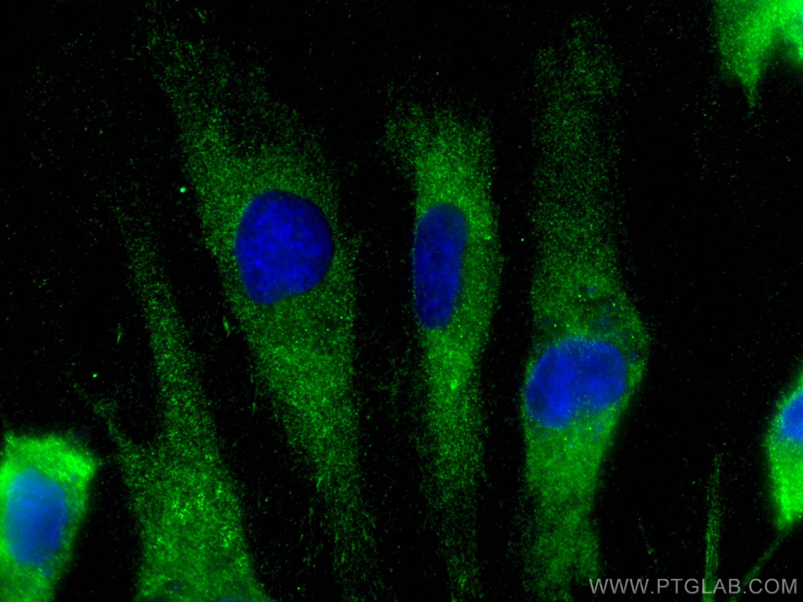
| Positive IF/ICC detected in | HUVEC cells |
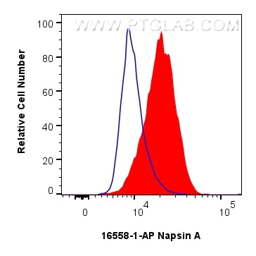
| Positive FC (Intra) detected in | A549 cells |
Recommended dilution
| Application | Dilution |
|---|---|
| Western Blot (WB) | WB : 1:500-1:1000 |
| Immunohistochemistry (IHC) | IHC : 1:50-1:500 |
| Immunofluorescence (IF)/ICC | IF/ICC : 1:50-1:500 |
| Flow Cytometry (FC) (INTRA) | FC (INTRA) : 4.00 ug per 10^6 cells in a 100 ul suspension |
| It is recommended that this reagent should be titrated in each testing system to obtain optimal results. | |
| Sample-dependent, Check data in validation data gallery. | |
Product Information
16558-1-AP targets Napsin A in WB, IHC, IF/ICC, FC (Intra), ELISA applications and shows reactivity with human, mouse samples.
| Tested Reactivity | human, mouse |
| Cited Reactivity | human |
| Host / Isotype | Rabbit / IgG |
| Class | Polyclonal |
| Type | Antibody |
| Immunogen |
CatNo: Ag9786 Product name: Recombinant human NAPSA protein Source: e coli.-derived, PGEX-4T Tag: GST Domain: 1-420 aa of BC017842 Sequence: MSPPPLLQPLLLLLPLLNVEPSGATLIRIPLHRVQPGRRTLNLLRGWREPAELPKLGAPSPGDKPIFVPLSNYRDVQYFGEIGLGTPPQNFTVAFDTGSSNLWVPSRRCHFFSVPCWLHHRFDPKASSSFQANGTKFAIQYGTGRVDGILSEDKLTIGGIKGASVIFGEALWEPSLVFAFAHFDGILGLGFPILSVEGVRPPMDVLVEQGLLDKPVFSFYLNRDPEEPDGGELVLGGSDPAHYIPPLTFVPVTVPAYWQIHMERVKVGPGLTLCAKGCAAILDTGTSLITGPTEEIRALHAAIGGIPLLAGEYIILCSEIPKLPAVSFLLGGVWFNLTAHDYVIQTTRNGVRLCLSGFQALDVPPPAGPFWILGDVFLGTYVAVFDRGDMKSSARVGLARARTRGADLGWGETAQAQFPG Predict reactive species |
| Full Name | napsin A aspartic peptidase |
| Calculated Molecular Weight | 420 aa, 45 kDa |
| Observed Molecular Weight | 40-45 kDa |
| GenBank Accession Number | BC017842 |
| Gene Symbol | Napsin A |
| Gene ID (NCBI) | 9476 |
| RRID | AB_2878278 |
| Conjugate | Unconjugated |
| Form | Liquid |
| Purification Method | Antigen affinity purification |
| UNIPROT ID | O96009 |
| Storage Buffer | PBS with 0.02% sodium azide and 50% glycerol, pH 7.3. |
| Storage Conditions | Store at -20°C. Stable for one year after shipment. Aliquoting is unnecessary for -20oC storage. 20ul sizes contain 0.1% BSA. |
Background Information
Napsin is found in two isoforms, napsin A and B, with highly homologous nucleotide sequences (91.2%). Napsin A appears to be a functional proteinase, predominantly expressed in lung and kidney. Napsin B is transcribed exclusively in cells related to the immune system and lacks an in-frame stop codon and is believed to be a pseudogene.(PMID:12698189). Napsin A is superior to TTF-1 in distinguishing primary lung ACA from other carcinomas (except kidney), particularly primary lung small cell carcinoma, and primary thyroid carcinoma.(PMID:22288963).
Protocols
| Product Specific Protocols | |
|---|---|
| FC protocol for Napsin A antibody 16558-1-AP | Download protocol |
| IF protocol for Napsin A antibody 16558-1-AP | Download protocol |
| IHC protocol for Napsin A antibody 16558-1-AP | Download protocol |
| WB protocol for Napsin A antibody 16558-1-AP | Download protocol |
| Standard Protocols | |
|---|---|
| Click here to view our Standard Protocols |
Publications
What published studies show
The Napsin A Polyclonal antibody (Cat# 16558-1-AP) has 2 total citations. These appear in Cell Biology and Toxicology and Cell Death and Differentiation. The research fields include lung adenocarcinoma prognosis and immunotherapy prediction using multiomics integration, and pancreatic ductal adenocarcinoma (PDAC) senolysis involving EGFR signaling and CDK4/6 inhibition.
| Species | Application | Title |
|---|---|---|
Cell Biol Toxicol Harnessing machine learning-driven multiomics integration: deciphering programmed cell death networks for prognostication and immunotherapy prediction in lung adenocarcinoma. | ||
Cell Death Differ Deprivation of EGFR signal causes senolysis in PDAC with CDK4/6 inhibition. |

