


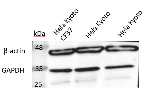
Tested Applications
| Positive WB detected in | HeLa cells, A549 cells, Jurkat cells, mouse pancreas, rat brain, rat pancreas, HEK-293 cells, HepG2 cells, K-562 cells, HSC-T6 cells, NIH/3T3 cells, 4T1 cells, CHO cells, HSC-T6 cells. NIH/3T3 cells, Pig brain, Rabbit brain, Mouse brain, Chicken brain |
| Positive IP detected in | HeLa cells |
| Positive IHC detected in | human kidney tissue, human brain tissue, human colon cancer tissue, human heart tissue Note: suggested antigen retrieval with TE buffer pH 9.0; (*) Alternatively, antigen retrieval may be performed with citrate buffer pH 6.0 |
| Positive IF-P detected in | mouse brain tissue |
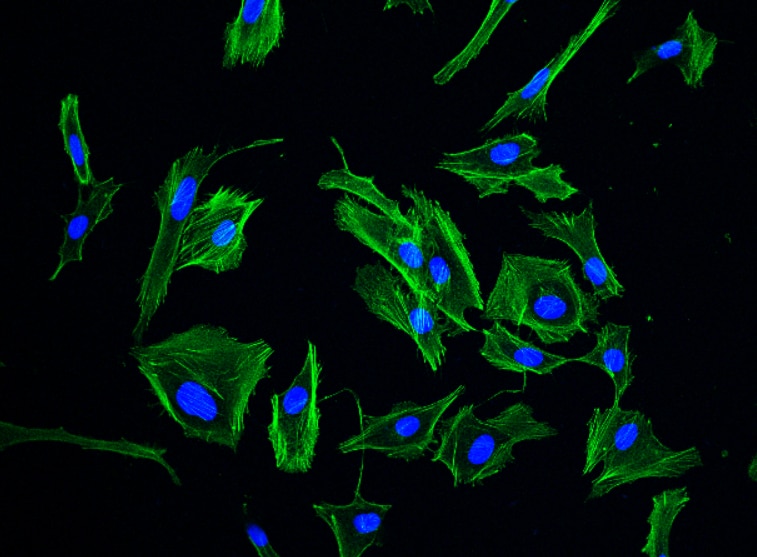
| Positive IF/ICC detected in | MDCK cells, HeLa cells |
| Positive FC (Intra) detected in | HeLa cells |
Recommended dilution
| Application | Dilution |
|---|---|
| Western Blot (WB) | WB : 1:20000-1:100000 |
| Immunoprecipitation (IP) | IP : 0.5-4.0 ug for 1.0-3.0 mg of total protein lysate |
| Immunohistochemistry (IHC) | IHC : 1:20-1:2000 |
| Immunofluorescence (IF)-P | IF-P : 1:500-1:2000 |
| Immunofluorescence (IF)/ICC | IF/ICC : 1:500-1:2000 |
| Flow Cytometry (FC) (INTRA) | FC (INTRA) : 0.40 ug per 10^6 cells in a 100 µl suspension |
| It is recommended that this reagent should be titrated in each testing system to obtain optimal results. | |
| Sample-dependent, Check data in validation data gallery. | |
Product Information
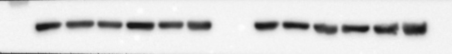
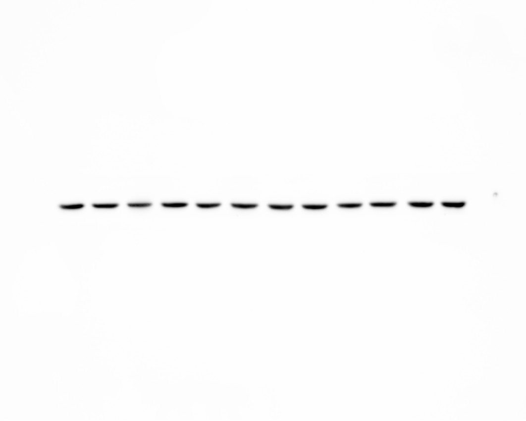
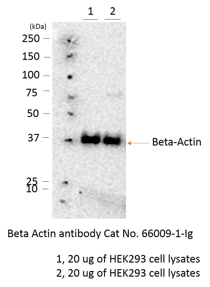
66009-1-Ig targets Beta Actin in WB, IHC, IF/ICC, IF-P, FC (Intra), IP, CoIP, ChIP, RIP, ELISA applications and shows reactivity with human, mouse, rat, pig, rabbit, canine, monkey, chicken, zebrafish, hamster samples.
| Tested Reactivity | human, mouse, rat, pig, rabbit, canine, monkey, chicken, zebrafish, hamster |
| Cited Reactivity | canine, chicken, bovine, cyprinus carpio, drosophila, cat, a.fumigatus strains, aedes albopictus, arabidopsis |
| Host / Isotype | Mouse / IgG2b |
| Class | Monoclonal |
| Type | Antibody |
| Immunogen |
Recombinant protein Predict reactive species |
| Full Name | actin, beta |
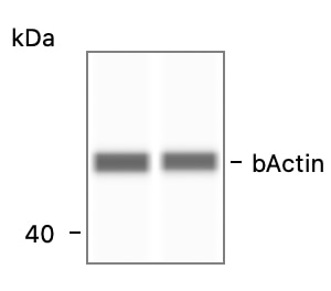
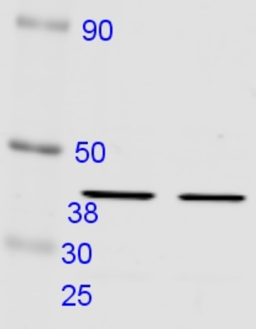
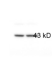
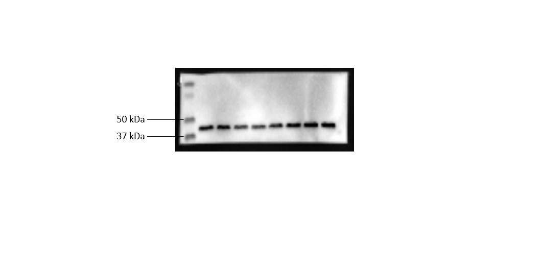
| Calculated Molecular Weight | 42 kDa |
| Observed Molecular Weight | 42 kDa |
| GenBank Accession Number | NM_001101 |
| Gene Symbol | Beta Actin |
| Gene ID (NCBI) | 60 |
| RRID | AB_2687938 |
| Conjugate | Unconjugated |
| Form | Liquid |
| Purification Method | Protein A purification |
| UNIPROT ID | P60709 |
| Storage Buffer | PBS with 0.02% sodium azide and 50% glycerol, pH 7.3. |
| Storage Conditions | Store at -20°C. Stable for one year after shipment. Aliquoting is unnecessary for -20oC storage. 20ul sizes contain 0.1% BSA. |
Background Information
Actins are highly conserved globular proteins that are involved in various types of cell motility and are ubiquitously expressed in all eukaryotic cells. At least six isoforms of actins are known in mammals and other vertebrates. Beta-actin (or β-actin) is a cytoskeletal actin isoform, highly conserved and ubiquitously expressed in eukaryotic organisms, making it a commonly used "housekeeping gene" in many laboratory techniques. It has been widely used as the internal control in RT-PCR and Western Blotting. The isotype of this antibody is IgG2b.
What is the molecular weight of beta-actin?
42 kDa
What is the function of beta-actin?
Actins are a key part of the cytoskeleton and are involved in cell motility, structure, and signaling, with beta-actin as part of the contractile apparatus. Beta-actin is located in the cytoplasm of non-muscle cells, particularly where the plasma membrane meets the cytoskeleton (PMID: 1993736) and is found co-distributed with alpha-actin (PMID: 9490838).
What are the applications for beta-actin?
As it is ubiquitously and constitutively expressed, beta-actin is a useful loading control in techniques such as RT-PCR and Western Blots to measure baseline expression and degradation. Beta-actin can be used as a reference gene to normalize mRNA or protein levels in quantitative studies of expression. Although some evidence suggests variation in tissue types and developing embryos (PMID: 23099774) if expression is stable across conditions, beta-actin is still considered by many to be a suitable gene for normalization.
Beyond its role as a housekeeping gene, it has been shown that beta-actin associates with endothelial nitric-oxide synthase (eNOS) and can therefore regulate nitric oxide activity in endothelial cells (PMID: 12734108).
What are the post-transcriptional modifications (PTMs) to beta actin?
Alterations to actin proteins can modulate their properties and function. While small molecules are known to alter actin, post-translational modifications such as phosphorylation and acetylation can also affect actin (PMID: 23195437). Modifications to actin have been associated with diseases including but not limited to age-related macular degeneration (PMID: 17652714), cardiovascular disease (PMID: 21232667), and Alzheimer's disease (PMID: 11246152).
For murine tissue sample, conjugated antibody (HRP-66009) or rabbit antibody (20536-1-AP) is preferable.
Protocols
| Product Specific Protocols | |
|---|---|
| FC protocol for Beta Actin antibody 66009-1-Ig | Download protocol |
| IF protocol for Beta Actin antibody 66009-1-Ig | Download protocol |
| IHC protocol for Beta Actin antibody 66009-1-Ig | Download protocol |
| IP protocol for Beta Actin antibody 66009-1-Ig | Download protocol |
| WB protocol for Beta Actin antibody 66009-1-Ig | Download protocol |
| Standard Protocols | |
|---|---|
| Click here to view our Standard Protocols |
Publications
| Species | Application | Title |
|---|---|---|
Cancer Cell KMT2C deficiency drives transdifferentiation of double-negative prostate cancer and confer resistance to AR-targeted therapy | ||
Cell Res Mannose antagonizes GSDME-mediated pyroptosis through AMPK activated by metabolite GlcNAc-6P | ||
Reviews
The reviews below have been submitted by verified Proteintech customers who received an incentive for providing their feedback.
FH rashi (Verified Customer) (12-11-2025) | The Pan-Actin Antibody from Proteintech delivers consistent, high-specificity detection of actin across multiple species and applications (WB, IHC, IF/ICC, IP, FC). It produces strong, reliable signals even at higher dilutions, making it a solid choice as a housekeeping/loading control reagent in cell and tissue studies.
|
FH Prakash (Verified Customer) (10-17-2025) | Its working very good
|
FH JIN-FENG (Verified Customer) (10-08-2025) | The beta actin antibody works perfectly fine in my result. the intensity is very strong. I can even used 1:10000 dilution and got nice signal.
 |
FH Liangze (Verified Customer) (10-06-2025) | Nice product
|
FH Nathalie (Verified Customer) (09-26-2025) | Antibody does not stand up to freezing and must be much less diluted than expected
 |
FH Harvey (Verified Customer) (09-10-2025) | 1:1000 Housekeeper for mouse tissue western blot
|
FH Danyan (Verified Customer) (09-04-2025) | very stable, we ordered all antibodies from proteintech
|
FH Jimmy (Verified Customer) (05-20-2025) | Nice results for western blot application!
|
FH Pallavi (Verified Customer) (04-02-2025) | Worked well with 1:25000 dilution.
|
FH Karthik (Verified Customer) (03-03-2025) | This lot of the antibody was offered in replacement of an earlier one and works well albeit at a slightly higher concentration than the lowest recommended concentration.
|
FH kis (Verified Customer) (02-21-2025) | Works nicely for human, rat, and mouse tissues and cells for WB analysis.
|
FH Rashmi (Verified Customer) (09-25-2024) | Used for Western Blot
|
FH Rashmi (Verified Customer) (09-25-2024) | Used for WB, Highly Recommended
|
FH Saurabh (Verified Customer) (08-07-2024) | Used on Simple Western (CE-SDS); Highly Recommended!
 |
FH Vikas (Verified Customer) (07-11-2024) | Used for WB, excellent product.
|
FH Vikas (Verified Customer) (07-11-2024) | Used for western blotting , very nice product. Highly recommended.
|
FH PK (Verified Customer) (07-05-2024) |
 |
FH Molly (Verified Customer) (06-11-2024) | Worked really well when loading 30ug protein and incubating for 1.5h at RT. Clear signal and single band.
|
FH S (Verified Customer) (05-16-2024) | Excellent
 |
FH Samantha (Verified Customer) (05-10-2024) | Could not detect B-actin in the sample although MS analysis confirmed it was there.
|
FH MANOHAR (Verified Customer) (03-06-2024) |
|
FH Ziqiao (Verified Customer) (09-11-2023) | The beta actin works very well for 40ug total protein loaded
 |
FH Xiaoyu (Verified Customer) (06-23-2023) | Excellent antibody, can be re-used multiple times without reduction in signal.
 |
FH Priya (Verified Customer) (06-21-2023) | Used this antibody for Caco2 cells andmice tissue
|
FH Christos (Verified Customer) (04-04-2023) | Separating Gel: 10% poly-acrylamide Transfer at 400mA for 2hr onto nitrocellulose membrane at 4oC Blocking and antibodies dilutions: 5% non-fat milk in PBS-Tween20 for 1 hr at RT Incubation with primary Abs: overnight at 4oC Incubation with secondary Abs: 1hr at 4oC
 |
FH Cynthia (Verified Customer) (03-29-2023) | Everything was fine
|
FH Huw (Verified Customer) (01-18-2023) | Excellent antibody, can be re-used multiple times without reduction in signal
|
FH Wu (Verified Customer) (12-05-2022) | Works good for Elisa assay
 |
FH Balawant (Verified Customer) (07-25-2022) | I will recommend this antibody for Immunoblotting. it is working ar 1:6000 dilution. Even it will work at 1:8000 dilution but I just used at 1:6000.
|
FH Susmita (Verified Customer) (06-13-2022) | This B-Actin antibody works excellently. The dilution also significantly high. I use 1:20,000 and this works great for me.
|
FH Susmita (Verified Customer) (06-13-2022) | This B-Actin antibody works excellently. The dilution also significantly high. I use 1:20,000 and this works great for me.
|
FH Xin (Verified Customer) (04-17-2022) | A very good and convincing antibody to detect beta-actin in mouse tissues and various cell lines lysate.
 |
FH Iram (Verified Customer) (04-13-2022) | Very good signal.
|
FH Weihao (Verified Customer) (04-11-2022) | Very specific antibody!
|
FH Raul (Verified Customer) (03-10-2022) | Very good and strong staining
 |
FH Ashish (Verified Customer) (02-09-2022) | We ordered this actin antibody to detect actin as internal control in the cell lysates during the western blot, and the antibody is very sensitive.
 |
FH James (Verified Customer) (10-07-2021) | Consistently works antibody
|
FH Jinwei (Verified Customer) (10-01-2021) | The Beta Actin antibody works fine.
 |
FH Eric (Verified Customer) (06-25-2021) | We ordered this actin antibody to detect actin in the cell lysates during the western blot, and it works very well, and the antibody is very sensitive. We highly recommend it.
 |
FH Elisa (Verified Customer) (06-14-2021) | This antibody work really well in cells and tissues (mouse). I have also used in human skeletal muscle with good results as well.
 |
FH Ambra (Verified Customer) (01-20-2021) | Works nicely.Used on Human brain lysates (temporal cortex).
 |
FH V (Verified Customer) (01-12-2021) | Very good product; signal is good
|
FH Yu Siong (Verified Customer) (07-30-2020) | Generate very consistent result for my porcine brain endothelial cells. The antibody at dilution of 1:5000 can be reused for multiple times.
 |
FH Magda (Verified Customer) (12-31-2019) | I needed to run a housekeeping gene control for my protein expression test. I was testing HEK293T cell lysates. My Lab did not have a proper AB, but a colleague from a lab next door borrowed me a sample from Proteintech. I was very happy with the results, they were consistent among my triplicate. I would purchase this AB but as a HRP conjugate, as it saves more time and provides cleaner results.
|
FH Elisa (Verified Customer) (10-21-2019) | The antibody works very nicely, very clean and I used only 10 uL of protein.
|
FH Tracy Chih-Ting (Verified Customer) (09-24-2019) | I used this antibody for Hydra actin immunostaining. The outcome is quite good.
|
FH Rui (Verified Customer) (09-02-2019) | Very specific and strong labeling in whole cell lysates.
|
FH Huai-Chin (Verified Customer) (06-19-2019) | Very good anitbody. Works well in Western for various type of cells.
|
FH Francesco (Verified Customer) (06-12-2019) | in the testing staining it kind of worked, even if with higher dilution it could work better.anyway further test should be done to confirm it.if results are reproducible it might be a valid alternative to more expensive antibody
|
FH Kyosuke (Verified Customer) (06-05-2019) | This antibody works very well for Western blot using vascular tissue or cell lysates.
|


