
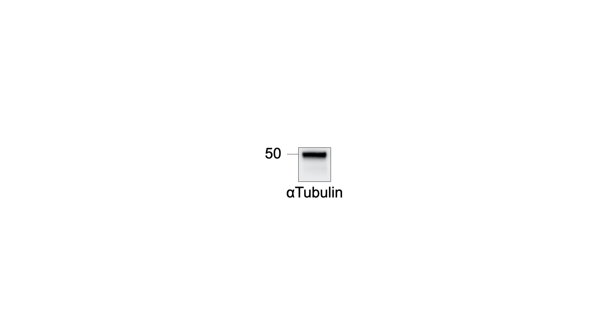
 at dilution of 1:200000 incubated at room temperature for 1.5 hours.")
 at dilution of 1:100000 incubated at room temperature for 1.5 hours.")
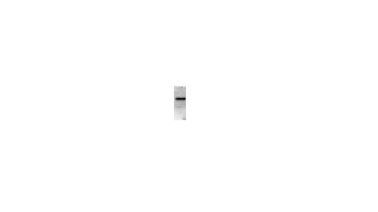
 with HeLa cells lysate 880ug.")
.")
.")
.")
.")
.")
.")
 at dilution of 1:10000 (under 10x lens). Heat mediated antigen retrieval with Tris-EDTA buffer (pH 9.0).")
 fixed paraffin-embedded mouse eye tissue using Beta Tubulin antibody (66240-1-Ig, Clone: 1D4A4 ) at dilution of 1:800 and CoraLite®488-Conjugated Goat Anti-Mouse IgG(H+L) (SA00013-1). Heat mediated antigen retrieval with Tris-EDTA buffer (pH 9.0).")
 fixed frozen OCT-embedded mouse brain tissue using Beta Tubulin antibody (66240-1-Ig, Clone: 1D4A4 ) at dilution of 1:400 and CoraLite®594-Conjugated Goat Anti-Mouse IgG(H+L) (SA00013-3).")
 fixed HeLa cells using 66240-1-Ig (beta Tubulin antibody), at dilution of 1:400 and CoraLite®488-Conjugated AffiniPure Goat Anti-Mouse IgG(H+L). Red: staning with CoraLite555-Phalloidin.")
 fixed HeLa cells using Beta Tubulin antibody (66240-1-Ig, Clone: 1D4A4 ) at dilution of 1:400 and CoraLite®488-Conjugated AffiniPure Goat Anti-Mouse IgG(H+L), CL594-Phalloidin (red).")
 at dilution of 1:50 and Alexa Fluor 488-conjugated AffiniPure Goat Anti-Mouse IgG (H+L).")
 at dilution of 1:50 and Alexa Fluor 488-conjugated AffiniPure Goat Anti-Mouse IgG (H+L).")
"Beta Tubulin Antibodies" Comparison
View side-by-side comparison of Beta Tubulin antibodies from other vendors to find the one that best suits your research needs.
Tested Applications
| Positive WB detected in | HeLa cells, HEK-293 cells, HepG2 cells, Jurkat cells, HSC-T6 cells, MCF-7 cells, NCCIT cells, HT-1080 cells, LNCaP cells, ROS1728 cells, NIH/3T3 cells |
| Positive IP detected in | HeLa cells |
| Positive IHC detected in | human brain tissue, human breast cancer tissue, rat brain tissue Note: suggested antigen retrieval with TE buffer pH 9.0; (*) Alternatively, antigen retrieval may be performed with citrate buffer pH 6.0 |
| Positive IF-P detected in | mouse eye tissue |
| Positive IF-Fro detected in | mouse brain tissue |
| Positive IF/ICC detected in | HeLa cells, HepG2 cells |
Recommended dilution
| Application | Dilution |
|---|---|
| Western Blot (WB) | WB : 1:20000-1:100000 |
| Immunoprecipitation (IP) | IP : 0.5-4.0 ug for 1.0-3.0 mg of total protein lysate |
| Immunohistochemistry (IHC) | IHC : 1:20-1:1000 |
| Immunofluorescence (IF)-P | IF-P : 1:400-1:1600 |
| Immunofluorescence (IF)-FRO | IF-FRO : 1:200-1:800 |
| Immunofluorescence (IF)/ICC | IF/ICC : 1:200-1:800 |
| It is recommended that this reagent should be titrated in each testing system to obtain optimal results. | |
| Sample-dependent, Check data in validation data gallery. | |
Published Applications
| WB | See 485 publications below |
| IHC | See 1 publications below |
| IF | See 32 publications below |
| IP | See 4 publications below |
Product Information
66240-1-Ig targets Beta Tubulin in WB, IHC, IF/ICC, IF-P, IF-Fro, IP, ELISA applications and shows reactivity with human, mouse, rat, pig, zebrafish, nematode samples.
| Tested Reactivity | human, mouse, rat, pig, zebrafish, nematode |
| Cited Reactivity | human, mouse, rat, pig, rabbit, monkey, chicken, zebrafish, hamster, sheep |
| Host / Isotype | Mouse / IgG2a |
| Class | Monoclonal |
| Type | Antibody |
| Immunogen |
CatNo: Ag0117 Product name: Recombinant human Tubulin-beta protein Source: e coli.-derived, PGEX-4T Tag: GST Domain: 44-259 aa of BC000748 Sequence: LERISVYYNEASSHKYVPRAILVDLEPGTMDSVRSGAFGHLFRPDNFIFGQSGAGNNWAKGHYTEGAELVDSVLDVVRKECENCDCLQGFQLTHSLGGGTGSGMGTLLISKVREEYPDRIMNTFSVVPSPKVSDTVVEPYNATLSIHQLVENTDETYCIDNEALYDICFRTLKLATPTYGDLNHLVSATMSGVTTSLRFPGQLNADLRKLAVNMVP Predict reactive species |
| Full Name | tubulin, beta 3 |
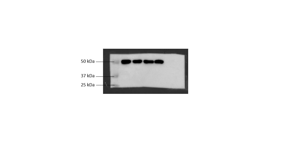
| Calculated Molecular Weight | 450 aa, 50 kDa |
| Observed Molecular Weight | 50-55 kDa |
| GenBank Accession Number | BC000748 |
| Gene Symbol | TUBB3 |
| Gene ID (NCBI) | 10381 |
| RRID | AB_2881629 |
| Conjugate | Unconjugated |
| Form | Liquid |
| Purification Method | Protein A purification |
| UNIPROT ID | Q13509 |
| Storage Buffer | PBS with 0.02% sodium azide and 50% glycerol, pH 7.3. |
| Storage Conditions | Store at -20°C. Stable for one year after shipment. Aliquoting is unnecessary for -20oC storage. 20ul sizes contain 0.1% BSA. |
Background Information
There are five tubulins in human cells: alpha, beta, gamma, delta, and epsilon. Tubulins are conserved across species. They form heterodimers, which multimerize to form a microtubule filament. An alpha and beta tubulin heterodimer is the basic structural unit of microtubules. The alpha and beta tubulins (+/- 55 kDa MW) are homologous but are not identical. Beta tubulins have been widely used as loading control.
What is the molecular weight of beta-tubulin? Are there any isoforms of beta-tubulin?
The molecular weight of tubulin is 50-52 kDa. Humans have eight beta-tubulin isotypes, encoded by different genes, that differ in their C-terminal sequences. They have different tissue expression profiles and can rise to microtubules of different properties (PMID: 20191564).
How to use beta-tubulin as a loading control
Beta-tubulin is one of the most commonly used references as a loading control for cell lysates in western blotting. It is abundantly expressed across various tissues and developmental stages and highly conserved across species. However, since some variability has been observed in the expression levels of commonly used housekeeping genes (PMID: 15627964), it is recommended that more than one loading control antibody is used while developing new assays. More information can be found here: https://www.ptglab.com/news/blog/loading-control-antibodies-for-western-blotting/.
What drugs can influence beta-tubulin and organization of microtubules?
Many drugs that affect microtubule dynamics target beta-tubulin, mainly by interfering with the GTP hydrolysis (PMID: 21381049). Paclitaxel (Taxol) is used to stabilize microtubules by slowing down their depolymerization, while colchicine and vinca alkaloids (vinblastine) destabilize microtubules. They are used in research and also in the clinic as anti-cancer agents.
Is beta-tubulin post-translationally modified?
Yes, tubulins are subject to extensive post-translational modifications (PTMs) that affect the organization of microtubules and their dynamics. The most common modifications include polyglutamylation, polyglycylation, polyamination, glycososylation, glycation, phosphorylation, and acetylation (PMID: 24801181 and 25468068).
Protocols
| Product Specific Protocols | |
|---|---|
| IF protocol for Beta Tubulin antibody 66240-1-Ig | Download protocol |
| IHC protocol for Beta Tubulin antibody 66240-1-Ig | Download protocol |
| IP protocol for Beta Tubulin antibody 66240-1-Ig | Download protocol |
| WB protocol for Beta Tubulin antibody 66240-1-Ig | Download protocol |
| Standard Protocols | |
|---|---|
| Click here to view our Standard Protocols |
Publications
| Species | Application | Title |
|---|---|---|
Nat Biotechnol Magnify is a universal molecular anchoring strategy for expansion microscopy | ||
Science A prometastatic splicing program regulated by SNRPA1 interactions with structured RNA elements. | ||
Adv Mater A Physiologically Responsive Nanocomposite Hydrogel for Treatment of Head and Neck Squamous Cell Carcinoma via Proteolysis-targeting Chimeras Enhanced Immunotherapy | ||
Gut Integrative multiomics enhancer activity profiling identifies therapeutic vulnerabilities in cholangiocarcinoma of different etiologies |
Reviews
The reviews below have been submitted by verified Proteintech customers who received an incentive for providing their feedback.
FH Aditya (Verified Customer) (11-01-2024) | great with flourescence, our standard housekeeping control
|
FH P (Verified Customer) (09-23-2024) | Excellent
 |
FH Moritz (Verified Customer) (09-12-2024) | The antibody worked very well and gave a very strong signal (can be diluted more). Used it in HEK293T cell lysate analysed with WB.
 |
FH Tsimafei (Verified Customer) (08-04-2024) | Excellent antibody! The membrane was incubated for 1h at RT
 |
FH Andrea (Verified Customer) (01-11-2023) | Antibody were incubated for 2h at room temperature. Provide a specific tubulin signal
 |
FH Emma (Verified Customer) (11-29-2021) | Very clean antibody and runs at the right size by western blot.
|
FH Raul (Verified Customer) (08-20-2021) | Very good antibody producing a single strong band at ~55kDa
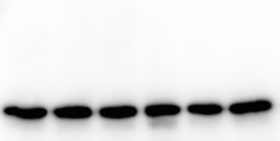 |
FH Yu Siong (Verified Customer) (08-18-2020) | Western blot of 4 bands: 4 different treatments.
 |
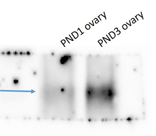
FH Kathryn (Verified Customer) (12-10-2019) | Used 1:20000 in 5% BSA on a small amount of neonatal mouse ovary. Very clean and specific, and great that it works at such a low concentration.
 |
FH Sara (Verified Customer) (10-09-2019) | This antibody works great in WB. We use it as loading control in whole cell lysates (mice B cell line), 1:10.000 O/N at 4°C.
|
FH VLADIMIR (Verified Customer) (09-25-2019) | B-tubulin IgG2a were used to prepare Fab' conjugated with Alexa 647-maleimide
|
FH Jeffery (Verified Customer) (08-01-2019) | Very clean antibody. Works exceptionally well. Runs right at the 50kd marker.
 |
FH Shashirekha (Verified Customer) (05-09-2019) | Works great!
 |
FH Zunair (Verified Customer) (12-03-2018) |
 |


