- Featured Product
- KD/KO Validated
TPD52L2 Polyclonal antibody
TPD52L2 Polyclonal Antibody for WB, IP, IF, IHC, ELISA
Host / Isotype
Rabbit / IgG
Reactivity
human, mouse, rat
Applications
WB, IP, IF, IHC, ELISA
Conjugate
Unconjugated
Cat no : 11795-1-AP
Synonyms
Validation Data Gallery
 at dilution of 1:800 incubated at room temperature for 1.5 hours.")
 at dilution of 1:1000 incubated at room temperature for 1.5 hours.")
 at dilution of 1:1000 incubated at room temperature for 1.5 hours.")
 with <a class='green' href='/productredirect?CatalogNo=HEK-293' target='_blank'>HEK-293</a> cells lysate 2400ug.")
 at dilution of 1:200 (under 10x lens). Heat mediated antigen retrieval with Tris-EDTA buffer (pH 9.0).")
 at dilution of 1:200 (under 40x lens). Heat mediated antigen retrieval with Tris-EDTA buffer (pH 9.0).")
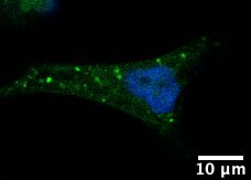
 fixed HepG2 cells using 11795-1-AP (TPD52L2 antibody), at dilution of 1:200 and CoraLite®488-Conjugated AffiniPure Goat Anti-Rabbit IgG(H+L).")
Tested Applications
| Positive WB detected in | HEK-293 cells, mouse brain tissue, rat brain tissue, MCF-7 cells |
| Positive IP detected in | HEK-293 cells |
| Positive IHC detected in | human breast cancer tissue Note: suggested antigen retrieval with TE buffer pH 9.0; (*) Alternatively, antigen retrieval may be performed with citrate buffer pH 6.0 |
| Positive IF detected in | HepG2 cells |
Recommended dilution
| Application | Dilution |
|---|---|
| Western Blot (WB) | WB : 1:500-1:1000 |
| Immunoprecipitation (IP) | IP : 0.5-4.0 ug for 1.0-3.0 mg of total protein lysate |
| Immunohistochemistry (IHC) | IHC : 1:50-1:500 |
| Immunofluorescence (IF) | IF : 1:50-1:500 |
| It is recommended that this reagent should be titrated in each testing system to obtain optimal results. | |
| Sample-dependent, Check data in validation data gallery. | |
Published Applications
| KD/KO | See 3 publications below |
| WB | See 7 publications below |
| IHC | See 2 publications below |
| IF | See 1 publications below |
| IP | See 1 publications below |
Product Information
11795-1-AP targets TPD52L2 in WB, IP, IF, IHC, ELISA applications and shows reactivity with human, mouse, rat samples.
| Tested Reactivity | human, mouse, rat |
| Cited Reactivity | human, mouse |
| Host / Isotype | Rabbit / IgG |
| Class | Polyclonal |
| Type | Antibody |
| Immunogen | TPD52L2 fusion protein Ag2364 |
| Full Name | tumor protein D52-like 2 |
| Calculated Molecular Weight | 206 aa, 22 kDa |
| Observed Molecular Weight | 25-30 kDa |
| GenBank Accession Number | BC006804 |
| Gene Symbol | TPD52L2 |
| Gene ID (NCBI) | 7165 |
| RRID | AB_2207431 |
| Conjugate | Unconjugated |
| Form | Liquid |
| Purification Method | Antigen affinity purification |
| Storage Buffer | PBS with 0.02% sodium azide and 50% glycerol pH 7.3. |
| Storage Conditions | Store at -20°C. Stable for one year after shipment. Aliquoting is unnecessary for -20oC storage. 20ul sizes contain 0.1% BSA. |
Background Information
Tumor protein D52-like proteins (TPD52) are small coiled-coil motif bearing proteins that were first identified in breast carcinoma. Three human TPD52 members had been identified, named hD52 (TPD52), hD53 (TPD52L1), and hD54 (TPD52L2). The most important characteristic of the protein family is a highly conserved coiled-coil motif that is required for homo- and heteromeric interaction with other TPD52-like proteins. TPD52 and related proteins have been implicated in cell proliferation, apoptosis, and vesicle trafficking. TPD52L2 has five isoforms produced by alternative splicing, and its multiple sites have been identified to be phosphorylated. Interaction of TPD52L2 with MAL2, a novel member of the MAL proteolipid family, may be required for the role of TPD52L2 in vesicle transport.
Protocols
| Product Specific Protocols | |
|---|---|
| WB protocol for TPD52L2 antibody 11795-1-AP | Download protocol |
| IHC protocol for TPD52L2 antibody 11795-1-AP | Download protocol |
| IF protocol for TPD52L2 antibody 11795-1-AP | Download protocol |
| IP protocol for TPD52L2 antibody 11795-1-AP | Download protocol |
| Standard Protocols | |
|---|---|
| Click here to view our Standard Protocols |
Publications
| Species | Application | Title |
|---|---|---|
Carcinogenesis TPD52L2 impacts proliferation, invasiveness and apoptosis of glioblastoma cells via modulation of wnt/β-catenin/snail signaling.
| ||
Cell Biosci Tumor protein D52 is upregulated in oral squamous carcinoma cells under hypoxia in a hypoxia-inducible-factor-independent manner and is involved in cell death resistance. | ||
J Biol Chem Tumor protein D54 binds intracellular nanovesicles via an extended amphipathic region. | ||
Int J Oncol Opposite effects of tumor protein D (TPD) 52 and TPD54 on oral squamous cell carcinoma cells. | ||
Biomed Res Int Tumor Proteins D52 and D54 Have Opposite Effects on the Terminal Differentiation of Chondrocytes. | ||
Neoplasma Silencing of TPD52 inhibits proliferation, migration, invasion but induces apoptosis of pancreatic cancer cells by deactivating Akt pathway.
|
Reviews
The reviews below have been submitted by verified Proteintech customers who received an incentive forproviding their feedback.
FH Jerome (Verified Customer) (01-05-2020) | Puncta in the cytoplasm
 |

