- Featured Product
- KD/KO Validated
DHODH Polyclonal antibody
DHODH Polyclonal Antibody for WB, IP, IF, FC, IHC, ELISA
Host / Isotype
Rabbit / IgG
Reactivity
human, mouse, rat and More (1)
Applications
WB, IP, IF, FC, IHC, ELISA
Conjugate
Unconjugated
Cat no : 14877-1-AP
Synonyms
Validation Data Gallery
 at dilution of 1:8000 incubated at room temperature for 1.5 hours.")
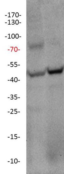
 with mouse spleen tissue lysate 4000ug.")
 at dilution of 1:200 (under 10x lens). Heat mediated antigen retrieval with Tris-EDTA buffer (pH 9.0).")
 at dilution of 1:200 (under 40x lens). Heat mediated antigen retrieval with Tris-EDTA buffer (pH 9.0).")
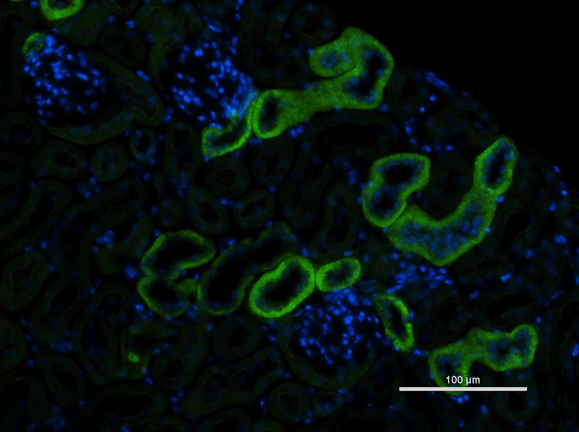
 fixed mouse kidney tissue using DHODH antibody (14877-1-AP) at dilution of 1:200 and CoraLite®488-Conjugated AffiniPure Goat Anti-Rabbit IgG(H+L).")
 and CoraLite®488-Conjugated AffiniPure Goat Anti-Rabbit IgG(H+L) at dilution 1:1000 (red), or 0.2 ug Control Antibody. Cells were fixed with 4% PFA and permeabilized with Flow Cytometry Perm Buffer (PF00011-C).")
Tested Applications
| Positive WB detected in | A2780 cells, MCF-7 cells, SKOV-3 cells, mouse heart tissue, mouse ovary tissue, mouse spleen tissue, rat spleen tissue |
| Positive IP detected in | mouse spleen tissue |
| Positive IHC detected in | human breast cancer tissue Note: suggested antigen retrieval with TE buffer pH 9.0; (*) Alternatively, antigen retrieval may be performed with citrate buffer pH 6.0 |
| Positive IF detected in | mouse kidney tissue |
Recommended dilution
| Application | Dilution |
|---|---|
| Western Blot (WB) | WB : 1:2000-1:16000 |
| Immunoprecipitation (IP) | IP : 0.5-4.0 ug for 1.0-3.0 mg of total protein lysate |
| Immunohistochemistry (IHC) | IHC : 1:50-1:500 |
| Immunofluorescence (IF) | IF : 1:50-1:500 |
| It is recommended that this reagent should be titrated in each testing system to obtain optimal results. | |
| Sample-dependent, Check data in validation data gallery. | |
Published Applications
| KD/KO | See 13 publications below |
| WB | See 48 publications below |
| IHC | See 3 publications below |
| FC | See 1 publications below |
Product Information
14877-1-AP targets DHODH in WB, IP, IF, FC, IHC, ELISA applications and shows reactivity with human, mouse, rat samples.
| Tested Reactivity | human, mouse, rat |
| Cited Reactivity | human, mouse, rat, goat |
| Host / Isotype | Rabbit / IgG |
| Class | Polyclonal |
| Type | Antibody |
| Immunogen | DHODH fusion protein Ag6649 |
| Full Name | dihydroorotate dehydrogenase |
| Calculated Molecular Weight | 43 kDa |
| Observed Molecular Weight | 43 kDa |
| GenBank Accession Number | BC065245 |
| Gene Symbol | DHODH |
| Gene ID (NCBI) | 1723 |
| RRID | AB_2091723 |
| Conjugate | Unconjugated |
| Form | Liquid |
| Purification Method | Antigen affinity purification |
| Storage Buffer | PBS with 0.02% sodium azide and 50% glycerol pH 7.3. |
| Storage Conditions | Store at -20°C. Stable for one year after shipment. Aliquoting is unnecessary for -20oC storage. 20ul sizes contain 0.1% BSA. |
Background Information
DHODH(Dihydroorotate dehydrogenase) catalyzes the fourth enzymatic step in de novo pyrimidine biosynthesis.DHO dehydrogenase is a monofunctional protein which, in most eukaryotic organisms, is located on the outer surface of the inner mitochondrial membrane.Defects in DHODH are the cause of postaxial acrofacial dysostosis (POADS).
Protocols
| Product Specific Protocols | |
|---|---|
| WB protocol for DHODH antibody 14877-1-AP | Download protocol |
| IHC protocol for DHODH antibody 14877-1-AP | Download protocol |
| IF protocol for DHODH antibody 14877-1-AP | Download protocol |
| IP protocol for DHODH antibody 14877-1-AP | Download protocol |
| Standard Protocols | |
|---|---|
| Click here to view our Standard Protocols |
Publications
| Species | Application | Title |
|---|---|---|
Cell Res Mitochondria-localized cGAS suppresses ferroptosis to promote cancer progression | ||
Nature DHODH-mediated ferroptosis defence is a targetable vulnerability in cancer.
| ||
Science Stimulation of de novo pyrimidine synthesis by growth signaling through mTOR and S6K1. | ||
Cancer Discov PTEN Regulates Glutamine Flux to Pyrimidine Synthesis and Sensitivity to Dihydroorotate Dehydrogenase Inhibition. | ||
Cancer Cell Cancer-selective metabolic vulnerabilities in MYC-amplified medulloblastoma
| ||
Cell Metab Macrophage-Released Pyrimidines Inhibit Gemcitabine Therapy in Pancreatic Cancer.
|
Reviews
The reviews below have been submitted by verified Proteintech customers who received an incentive forproviding their feedback.
FH Kristian (Verified Customer) (07-06-2021) | Paraffin embedded kidneys were stained for Dhodh (green) and DAPI (blue)
 |
FH Daniel (Verified Customer) (04-16-2019) | Highly specific detection of both mouse and human DHODH.
 |

