- Featured Product
- KD/KO Validated
Perilipin-2 Polyklonaler Antikörper
Perilipin-2 Polyklonal Antikörper für WB, IHC, IF/ICC, ELISA
Wirt / Isotyp
Kaninchen / IgG
Getestete Reaktivität
human, Maus, Ratte und mehr (6)
Anwendung
WB, IHC, IF/ICC, IP, ELISA
Konjugation
Unkonjugiert
Kat-Nr. : 15294-1-AP
Synonyme
 at dilution of 1:8000 incubated at room temperature for 1.5 hours.")
 at dilution of 1:8000 incubated at room temperature for 1.5 hours.")
 at dilution of 1:20000 incubated at room temperature for 1.5 hours.")
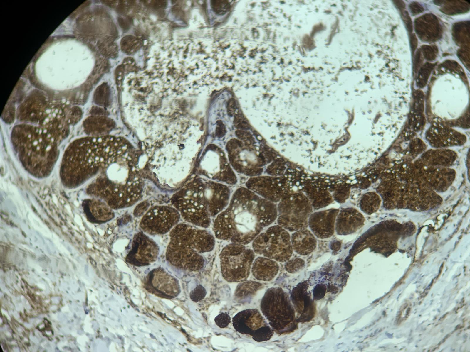
 at dilution of 1:200 (under 10x lens). Heat mediated antigen retrieval with Tris-EDTA buffer (pH 9.0).")
 at dilution of 1:200 (under 40x lens). Heat mediated antigen retrieval with Tris-EDTA buffer (pH 9.0).")
 at dilution of 1:16000 (under 40x lens). Heat mediated antigen retrieval with Tris-EDTA buffer (pH 9.0).")
 at dilution of 1:16000 (under 40x lens). Heat mediated antigen retrieval with Tris-EDTA buffer (pH 9.0).")
 at dilution of 1:200 (under 10x lens). Heat mediated antigen retrieval with Tris-EDTA buffer (pH 9.0).")
 at dilution of 1:200 (under 40x lens). Heat mediated antigen retrieval with Tris-EDTA buffer (pH 9.0).")
.")
.")
.")
.")
 at dilution of 1:50 (under 40x lens).")
 fixed oleic acid treated HeLa cells using ADRP/Perilipin 2 antibody (15294-1-AP) at dilution of 1:400 and CoraLite®488-Conjugated AffiniPure Goat Anti-Rabbit IgG(H+L), CL594-Phalloidin (red).")
 fixed oleic acid treated HeLa cells using ADRP/Perilipin 2 antibody (15294-1-AP) at dilution of 1:200 and Cy3-conjugated Affinipure Goat Anti-Rabbit IgG(H+L).")
 fixed oleic acid treated HeLa cells using ADRP/Perilipin-2 antibody (15294-1-AP) at dilution of 1:200 and Multi-rAb CoraLite ® Plus 488-Goat Anti-Rabbit Recombinant Secondary Antibody (H+L) (RGAR002), CL594-phalloidin (red).")
Geprüfte Anwendungen
| Erfolgreiche Detektion in WB | HepG2-Zellen, 3T3-L1-Zellen, K-562-Zellen, Mauslebergewebe, NIH/3T3-Zellen, Rattenlebergewebe |
| Erfolgreiche Detektion in IHC | humanes Leberkarzinomgewebe, humanes Kolonkarzinomgewebe, humanes Lebergewebe, humanes Prostatakarzinomgewebe, humanes Nierenzellkarzinomgewebe, Mauslebergewebe Hinweis: Antigendemaskierung mit TE-Puffer pH 9,0 empfohlen. (*) Wahlweise kann die Antigendemaskierung auch mit Citratpuffer pH 6,0 erfolgen. |
| Erfolgreiche Detektion in IF/ICC | mit Ölsäure behandelte HeLa-Zellen |
Empfohlene Verdünnung
| Anwendung | Verdünnung |
|---|---|
| Western Blot (WB) | WB : 1:2000-1:16000 |
| Immunhistochemie (IHC) | IHC : 1:200-1:8000 |
| Immunfluoreszenz (IF)/ICC | IF/ICC : 1:200-1:800 |
| It is recommended that this reagent should be titrated in each testing system to obtain optimal results. | |
| Sample-dependent, check data in validation data gallery | |
Veröffentlichte Anwendungen
| KD/KO | See 3 publications below |
| WB | See 112 publications below |
| IHC | See 30 publications below |
| IF | See 69 publications below |
| IP | See 1 publications below |
Produktinformation
15294-1-AP bindet in WB, IHC, IF/ICC, IP, ELISA Perilipin-2 und zeigt Reaktivität mit human, Maus, Ratten
| Getestete Reaktivität | human, Maus, Ratte |
| In Publikationen genannte Reaktivität | human, Affe, Hausschwein, Hund, Maus, Ratte, Rind, Zebrafisch, Ziege |
| Wirt / Isotyp | Kaninchen / IgG |
| Klonalität | Polyklonal |
| Typ | Antikörper |
| Immunogen | Perilipin-2 fusion protein Ag7539 |
| Vollständiger Name | adipose differentiation-related protein |
| Berechnetes Molekulargewicht | 48 kDa |
| Beobachtetes Molekulargewicht | 45-48 kDa |
| GenBank-Zugangsnummer | BC005127 |
| Gene symbol | Perilipin-2 |
| Gene ID (NCBI) | 123 |
| Konjugation | Unkonjugiert |
| Form | Liquid |
| Reinigungsmethode | Antigen-Affinitätsreinigung |
| Lagerungspuffer | PBS with 0.02% sodium azide and 50% glycerol |
| Lagerungsbedingungen | Bei -20°C lagern. Nach dem Versand ein Jahr lang stabil Aliquotieren ist bei -20oC Lagerung nicht notwendig. 20ul Größen enthalten 0,1% BSA. |
Hintergrundinformationen
ADRP (adipocyte differentiation related protein) also known as ADFP, adipophilin, or perilipin-2, is a member of PAT family which is responsible for the transportation of lipids and the formation of lipid droplets. ADRP is localized on the surface of lipid droplets in a variety of tissues and cell lines. ADRP is not detected in undifferentiated cells but increases rapidly to high levels when adipocyte precursors differentiate into adipocytes. Anti-ADRP antibody is a reliable and sensitive marker for lipid droplet. Enhanced expression of ADRP is linked to diseases with abnormal lipid storage, including hepatic steatosis, atherosclerosis and diabetes. Immunohistochemistry of ADRP may facilitate histomorphological diagnosis of these diseases. This antibody is not suitable for immunofluorescence staining of frozen sections.
Protokolle
| PRODUKTSPEZIFISCHE PROTOKOLLE | |
|---|---|
| WB protocol for Perilipin-2 antibody 15294-1-AP | Protokoll herunterladen |
| IHC protocol for Perilipin-2 antibody 15294-1-AP | Protokoll herunterladenl |
| IF protocol for Perilipin-2 antibody 15294-1-AP | Protokoll herunterladen |
| STANDARD-PROTOKOLLE | |
|---|---|
| Klicken Sie hier, um unsere Standardprotokolle anzuzeigen |
Publikationen
| Species | Application | Title |
|---|---|---|
Nature iPS-cell-derived microglia promote brain organoid maturation via cholesterol transfer | ||
Signal Transduct Target Ther The AKAP12-PKA axis regulates lipid homeostasis during alcohol-associated liver disease | ||
Cell Metab Elevation of JAML Promotes Diabetic Kidney Disease by Modulating Podocyte Lipid Metabolism. | ||
Nat Commun Spliceosome component Usp39 contributes to hepatic lipid homeostasis through the regulation of autophagy | ||
Brain Behav Immun HIV-TAT dysregulates microglial lipid metabolism through SREBP2/miR-124 axis: Implication of lipid droplet accumulation microglia in NeuroHIV |
Rezensionen
The reviews below have been submitted by verified Proteintech customers who received an incentive for providing their feedback.
FH Samuel (Verified Customer) (07-31-2025) | I found this primary antibody to work quite well, at least from a Western blot perspective, (I have not tried any other applications using this particular antibody). The experimental results came out quite clean and there has been little to no non-specific banding patterns, which is very nice. Lastly, the standard 1:1000 primary antibody dilution I have used works just fine for my needs, which allows the vial to last a decent amount of time. Overall, I would recommend!
|
FH Christin (Verified Customer) (11-25-2024) | Excellent antibody for ICC and WB of human iPSC-derived microglia and macrophages
|
FH Liliana (Verified Customer) (02-13-2024) | WORKS VERY WELL ON PH9, 1:2000 AND ON PH6, 1:1000
 |
FH Christine (Verified Customer) (09-19-2023) | Tried on HEK293T cells treated with 300 uM oleic acid overnight to induce the formation of lipid droplets. This antibody decorates some droplets but was a bit weak. TIP47 antibody (10694-1-AP) performed better (clearer detection of the droplets) at the same concentration (1:100).
|
FH Kamal (Verified Customer) (07-05-2023) | Worked well when diluted with 1X PBS.
|
FH Christin (Verified Customer) (03-09-2023) | Great antibody for WB with low background signal.
|
FH Nick (Verified Customer) (04-20-2022) | Helped localize expression of Plin2
|
FH Boyan (Verified Customer) (05-09-2019) | Very good for WB
|

