- Featured Product
- KD/KO Validated
ARL13BA Polyklonaler Antikörper
ARL13BA Polyklonal Antikörper für WB, IHC, IF/ICC, IP, ELISA
Wirt / Isotyp
Kaninchen / IgG
Getestete Reaktivität
human, Hund, Maus, Ratte und mehr (5)
Anwendung
WB, IHC, IF/ICC, IP, ELISA
Konjugation
Unkonjugiert
Kat-Nr. : 17711-1-AP
Synonyme
 at dilution of 1:3000 incubated at room temperature for 1.5 hours.")
 at dilution of 1:1000 incubated at room temperature for 1.5 hours.")
 at dilution of 1:500 incubated at room temperature for 1.5 hours.")
 at dilution of 1:1000 incubated at room temperature for 1.5 hours.")
 at dilution of 1:800 incubated at room temperature for 1.5 hours.")
 at dilution of 1:800 incubated at room temperature for 1.5 hours.")
 at dilution of 1:1000 incubated at room temperature for 1.5 hours.")
 with L02 cells lysate 2500ug.")
 at dilution of 1:200 (under 10x lens). Heat mediated antigen retrieval with Tris-EDTA buffer (pH 9.0).")
 at dilution of 1:200 (under 10x lens). Heat mediated antigen retrieval with Tris-EDTA buffer (pH 9.0).")
 at dilution of 1:50 (under 10x lens).")
 at dilution of 1:50 (under 40x lens).")
 at dilution of 1:50 (under 10x lens).")
 at dilution of 1:50 (under 40x lens).")
 fixed paraffin-embedded mouse pancreas tissue using ARL13B antibody (17711-1-AP) at dilution of 1:400 and CoraLite®594-Conjugated Goat Anti-Rabbit IgG(H+L) (SA00013-4), CoraLite® Plus 488 E-cadherin antibody (CL488-20874, green). Heat mediated antigen retrieval with Tris-EDTA buffer (pH 9.0).")
 fixed hTERT-RPE1 cells using ARL13B antibody (17711-1-AP) at dilution of 1:400 and Multi-rAb CoraLite ® Plus 488-Goat Anti-Rabbit Recombinant Secondary Antibody (H+L) (RGAR002).")
 fixed MDCK cells using ARL13B antibody (17711-1-AP) at dilution of 1:400 and CoraLite®488-Conjugated Goat Anti-Rabbit IgG(H+L) (SA00013-2), Acetyl-Tubulin (Lys40) antibody (66200-1-Ig, Clone: 7E5H8, red).")
 in NIH3T3 cell by Dr. Sudipto.")
 fixed hTERT-RPE1 cells using ARL13B antibody (17711-1-AP) at dilution of 1:400 and CoraLite®488-Conjugated AffiniPure Goat Anti-Rabbit IgG(H+L), acetylated Tubulin(Lys40) antibody (66200-1-Ig, Clone: 7E5H8, red).")
 fixed hTERT-RPE1 cells using ARL13B antibody (17711-1-AP) at dilution of 1:400 and CoraLite®488-Conjugated Goat Anti-Rabbit IgG(H+L) (SA00013-2), Acetyl-Tubulin (Lys40) antibody (66200-1-Ig, Clone: 7E5H8, red).")
 and acetylated tubulin mouse mAb (66200-1-Ig) at dilution of 1:50, further stained with Alexa Fluor 488-conjugated AffiniPure-Goat anti-Rabbit IgG(H+L) for 17711-1-AP, and Alexa Fluor 594-conjugated AffiniPure Goat Anti-Rabbit IgG(H+L) for 66200-1-Ig.")
 fixed MDCK cells using 17711-1-AP (ARL13B antibody) at dilution of 1:200 and Alexa Fluor 488-Conjugated AffiniPure Goat Anti-Rabbit IgG(H+L).")
 in hTERT-RPE cell fixed with 4% PFA or Methanol.")
 mark the cilium of Mouse embryonic fibroblasts.")
Geprüfte Anwendungen
| Erfolgreiche Detektion in WB | L02-Zellen, Maushirngewebe, Mausnierengewebe, Mauslebergewebe, NIH/3T3-Zellen, Rattennierengewebe, Rattenlebergewebe |
| Erfolgreiche IP | L02-Zellen |
| Erfolgreiche Detektion in IHC | Maushirngewebe, humanes Lebergewebe, humanes Hodengewebe Hinweis: Antigendemaskierung mit TE-Puffer pH 9,0 empfohlen. (*) Wahlweise kann die Antigendemaskierung auch mit Citratpuffer pH 6,0 erfolgen. |
| Erfolgreiche Detektion in IF/ICC | hTERT-RPE1-Zellen, MDCK-Zellen, NIH3T3-Zellen |
Empfohlene Verdünnung
| Anwendung | Verdünnung |
|---|---|
| Western Blot (WB) | WB : 1:1000-1:6000 |
| Immunpräzipitation (IP) | IP : 0.5-4.0 ug for 1.0-3.0 mg of total protein lysate |
| Immunhistochemie (IHC) | IHC : 1:50-1:500 |
| Immunfluoreszenz (IF)/ICC | IF/ICC : 1:200-1:800 |
| It is recommended that this reagent should be titrated in each testing system to obtain optimal results. | |
| Sample-dependent, check data in validation data gallery | |
Veröffentlichte Anwendungen
| KD/KO | See 13 publications below |
| WB | See 99 publications below |
| IHC | See 38 publications below |
| IF | See 766 publications below |
| IP | See 3 publications below |
Produktinformation
17711-1-AP bindet in WB, IHC, IF/ICC, IP, ELISA ARL13BA und zeigt Reaktivität mit human, Hund, Maus, Ratten
| Getestete Reaktivität | human, Hund, Maus, Ratte |
| In Publikationen genannte Reaktivität | human, Affe, Hausschwein, Huhn, Hund, Maus, Ratte, Zebrafisch, Krallenfrosch (Xenopus) |
| Wirt / Isotyp | Kaninchen / IgG |
| Klonalität | Polyklonal |
| Typ | Antikörper |
| Immunogen | ARL13BA fusion protein Ag12015 |
| Vollständiger Name | ADP-ribosylation factor-like 13B |
| Berechnetes Molekulargewicht | 48 kDa |
| Beobachtetes Molekulargewicht | 40-48 kDa, 60 kDa |
| GenBank-Zugangsnummer | BC094725 |
| Gene symbol | ARL13B |
| Gene ID (NCBI) | 200894 |
| Konjugation | Unkonjugiert |
| Form | Liquid |
| Reinigungsmethode | Antigen-Affinitätsreinigung |
| Lagerungspuffer | PBS with 0.02% sodium azide and 50% glycerol |
| Lagerungsbedingungen | Bei -20°C lagern. Nach dem Versand ein Jahr lang stabil Aliquotieren ist bei -20oC Lagerung nicht notwendig. 20ul Größen enthalten 0,1% BSA. |
Hintergrundinformationen
ARL13B, also named ARL2L1, is the Ras superfamily's small ciliary G protein. Localized in the cilia, it is required for cilium biogenesis and sonic hedgehog signaling. Defects in ARL13B cause Joubert syndrome (JS), an autosomal recessive disorder characterized by a distinctive cerebellar malformation (PMID: 19906870). This antibody detects two specific bands at 60 kDa and 48 kDa. Arl13b is predicted to be a 48 kDa protein, and the 60 kDa band will likely represent a modified form of Arl13b. ARL13B can be used to mark the cilia (PMID:22072986).
Protokolle
| PRODUKTSPEZIFISCHE PROTOKOLLE | |
|---|---|
| WB protocol for ARL13BA antibody 17711-1-AP | Protokoll herunterladen |
| IHC protocol for ARL13BA antibody 17711-1-AP | Protokoll herunterladenl |
| IF protocol for ARL13BA antibody 17711-1-AP | Protokoll herunterladen |
| IP protocol for ARL13BA antibody 17711-1-AP | Protokoll herunterladen |
| STANDARD-PROTOKOLLE | |
|---|---|
| Klicken Sie hier, um unsere Standardprotokolle anzuzeigen |
Publikationen
| Species | Application | Title |
|---|---|---|
Nat Biotechnol Guided self-organization and cortical plate formation in human brain organoids. | ||
Nature Quantitative lineage analysis identifies a hepato-pancreato-biliary progenitor niche. | ||
Science Control of meiotic chromosomal bouquet and germ cell morphogenesis by the zygotene cilium. | ||
Nature A slow-cycling LGR5 tumour population mediates basal cell carcinoma relapse after therapy. | ||
Nature Aspm knockout ferret reveals an evolutionary mechanism governing cerebral cortical size. |
Rezensionen
The reviews below have been submitted by verified Proteintech customers who received an incentive for providing their feedback.
FH Ludovic (Verified Customer) (12-10-2023) | The perfect antibody, works each time!
|
FH Gagan (Verified Customer) (07-02-2023) | Antibody gives good staining for cilia.
 |
FH Mi (Verified Customer) (05-29-2023) | An excellent antibody, it works pretty well in human white adipocytes!
|
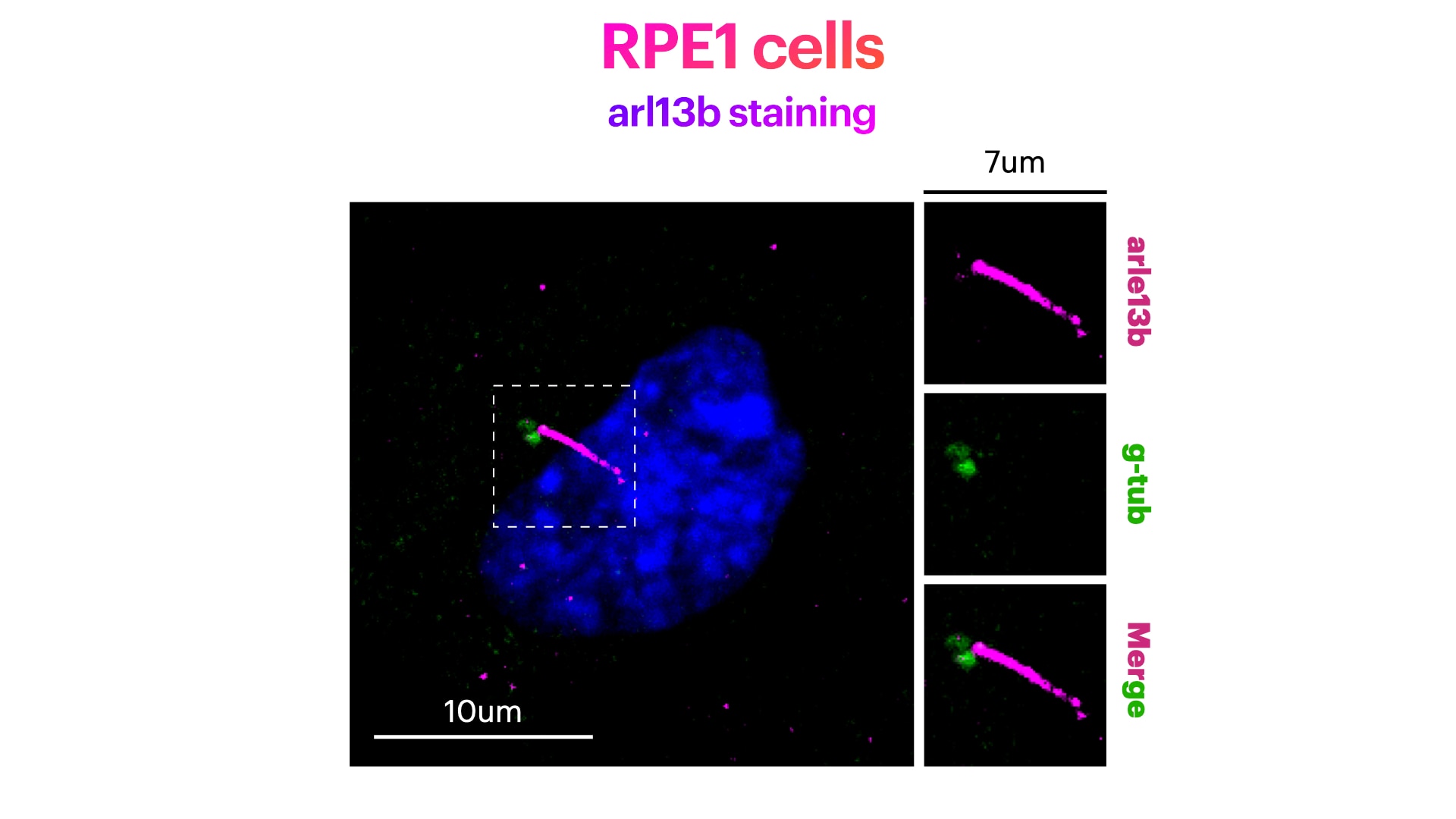
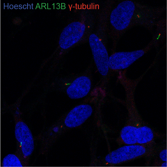
FH Elisa (Verified Customer) (03-17-2023) | Great antibody for centrosome and cilia visualizationin RPE1 cells. Cells were fixed in cold methanol for 10' at -20C, rehydrated with PBS for 5', and permeabilized with 0.1% Triton + 0.1% Tween +0.01%SDS in PBS for 5'. Cells were finally incubated with blocking buffer (5% BSA+ 0.1% Tween in PBS) for 30' at RT. Primary antibody was diluted in blocking buffer 1:200 and incubated for 1h at room temperature. Alexa-555-Anti-rabbit was used as secondary antibody (1:600 dilution) (1h at room temperature). Image show Arl13b (in magenta), DNA (Hoechst in blue) and g-tubulin (in green).
 |
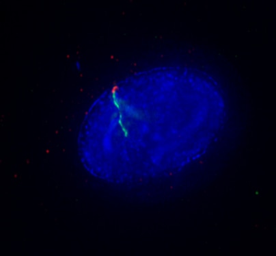
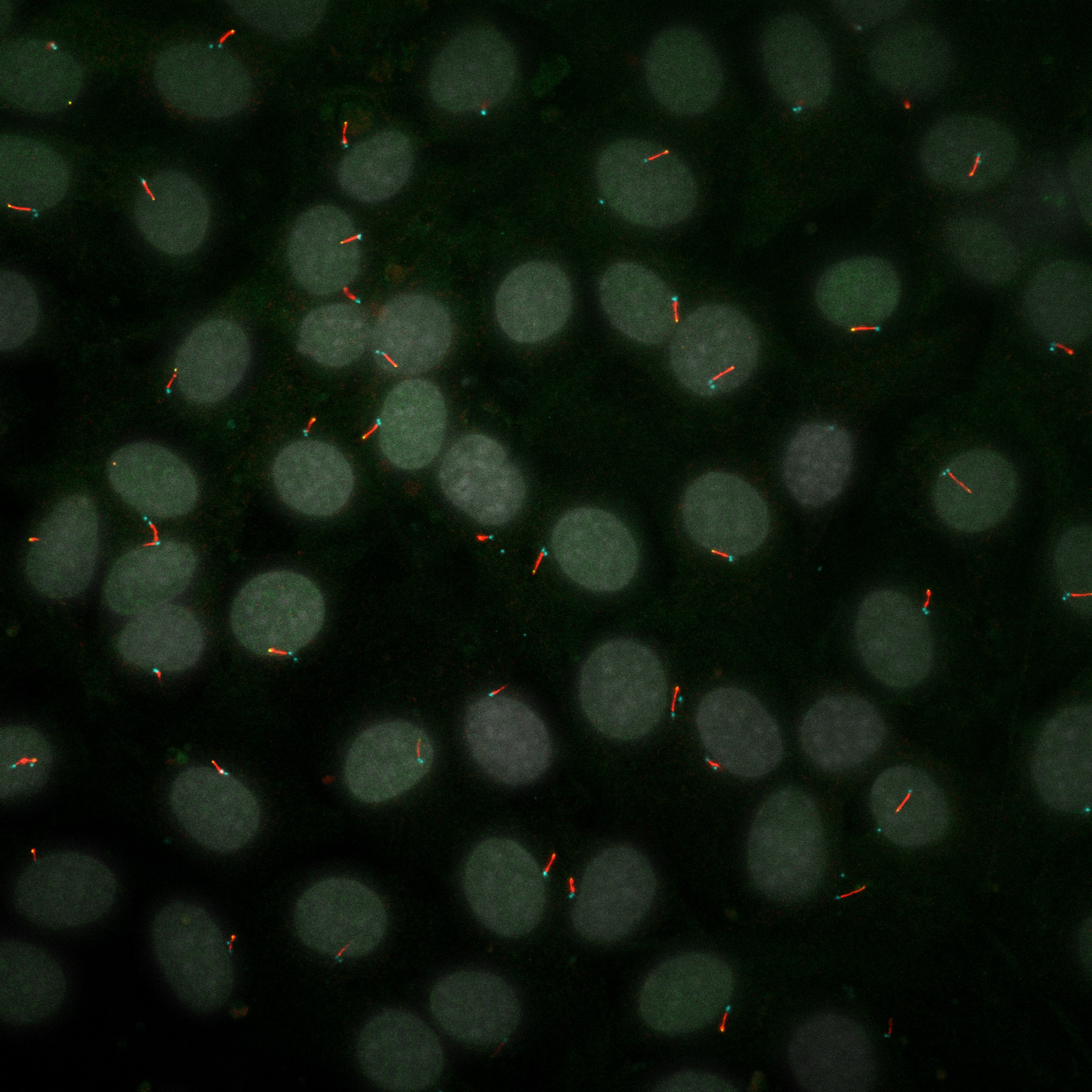
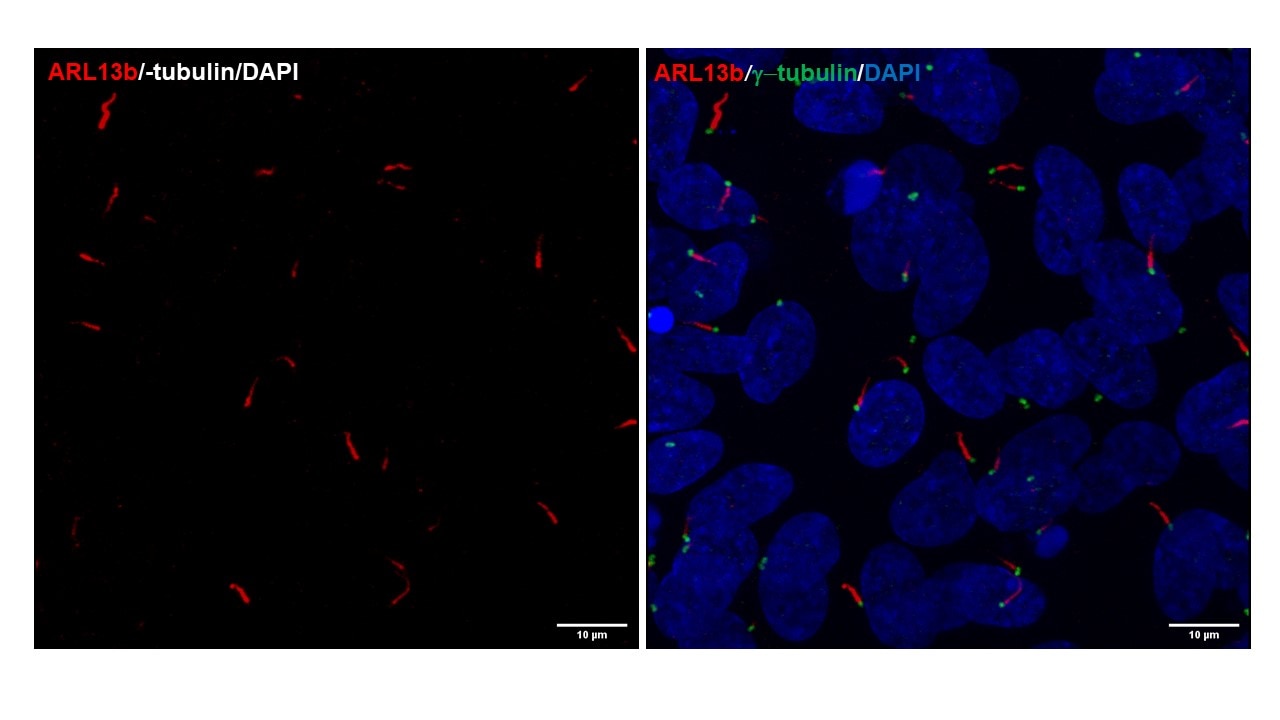
FH Serhiy (Verified Customer) (12-21-2022) | Excellent for immunofluorescent staining of ciliary Arl13b in mouse cells. Image shows ciliary axonemes immunostained for Arl13b (red).
 |
FH Kaviya (Verified Customer) (09-19-2022) | I use this antibody for IHC on brain tissue sections and it generally works well.
|
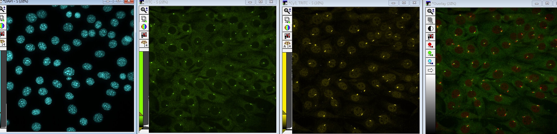
FH Charlotte (Verified Customer) (07-29-2022) | NIH-3T3 stained in DAPI (nucleus), FITC (gamma tubulin), Cy3 (Arl13b). The last picture is the merged channels (FITC and Cy3). On the merged channel, we can conclude that all Cy3 signal is validated by the FITC one (gamma tubulin is present at the base of the primary cilia). Perfect antibody !
 |
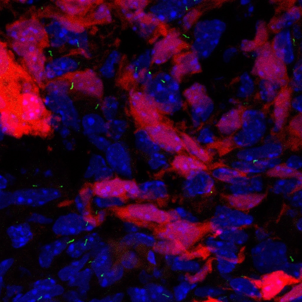
FH Sarah (Verified Customer) (02-16-2021) | Works beautifully in all cell types I've tried. Specifically great to see it work in primary cells (glioblastoma, pictured) as well as iPSCs
 |
FH deng (Verified Customer) (01-30-2020) | Very good for cilia staining
 |
FH Gabriela (Verified Customer) (01-01-2020) | Very specific antibody for primary cilia. We use it in chicken embryos spinal cord.
|
FH KARTHIKEYAN (Verified Customer) (11-20-2019) | This antibody works really well in human and mouse cell lines and in zebrafish samples, we used this antibody for WB, IF and Flow cytometry.
 |
FH Rebeca (Verified Customer) (09-26-2019) | I have used this antibody for several immunofluorescence stainings of primary cilia. The antibody works beautifully, would definitely recommend!
|
FH Pavel (Verified Customer) (09-18-2019) | Strong ciliary staining in MDCK cells. Works both with PFA and MeOH fixation.
|
FH Lindsey (Verified Customer) (02-04-2019) | Excellent specificity with minimal background/nonspecific binding; This antibody is ideal because it works just as well in embryonic mouse tissue as it does in adult. I couldn't recommend this product more!
 |
FH Kishor (Verified Customer) (01-30-2019) | I am very much satisfied with this produced, working good for WB (1:1000) and for IF (1:200)
|

