- Featured Product
- KD/KO Validated
BAX Polyklonaler Antikörper
BAX Polyklonal Antikörper für WB, IHC, IP, ELISA
Wirt / Isotyp
Kaninchen / IgG
Getestete Reaktivität
human, Maus, Ratte und mehr (6)
Anwendung
WB, IHC, IP, CoIP, ELISA
Konjugation
Unkonjugiert
Kat-Nr. : 50599-2-Ig
Synonyme
 at dilution of 1:8000 incubated at room temperature for 1.5 hours.")
 at dilution of 1:60000 incubated at room temperature for 1.5 hours.")
 at dilution of 1:6000 incubated at room temperature for 1.5 hours. Jurkat cell lysate used as a negative control.")
 at dilution of 1:8000 incubated at room temperature for 1.5 hours.")
 with si-Control and si-BAX transfected HeLa cells.")
 with sh-Control and sh-BAX transfected HeLa cells.")
 at dilution of 1:8000 incubated at room temperature for 1.5 hours.")
 at dilution of 1:8000 incubated at room temperature for 1.5 hours.")
 at dilution of 1:8000 incubated at room temperature for 1.5 hours.")
 at dilution of 1:8000 incubated at room temperature for 1.5 hours.")
 at dilution of 1:1500 incubated at room temperature for 1.5 hours.")
 at dilution of 1:5000 incubated at room temperature for 1.5 hours.")
 with Raji cells lysate 3160 ug.")
 with mouse heart tissue lysate 1680 ug.")
 at dilution of 1:2000 (under 10x lens). Heat mediated antigen retrieval with Tris-EDTA buffer (pH 9.0).")
 at dilution of 1:2000 (under 40x lens). Heat mediated antigen retrieval with Tris-EDTA buffer (pH 9.0).")
 at dilution of 1:2000 (under 10x lens). Heat mediated antigen retrieval with Tris-EDTA buffer (pH 9.0).")
 at dilution of 1:2000 (under 40x lens). Heat mediated antigen retrieval with Tris-EDTA buffer (pH 9.0).")
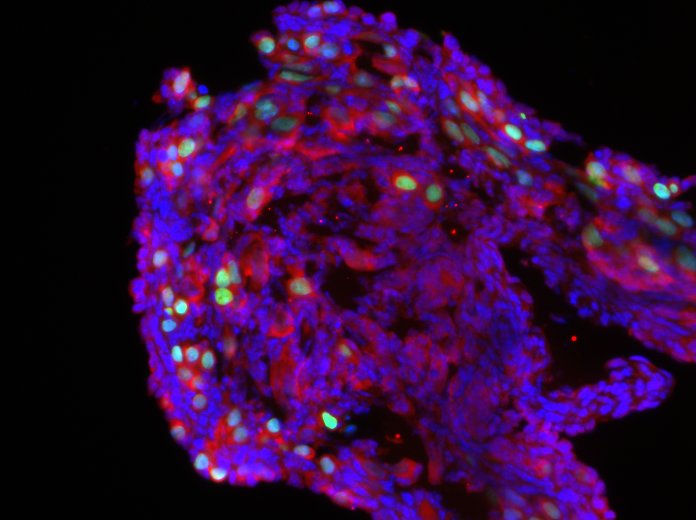
 fixed MCF-7 cells using BAX antibody (50599-2-Ig) at dilution of 1:200 and CoraLite®488-Conjugated Goat Anti-Rabbit IgG(H+L) (SA00013-2), CL594-Phalloidin (red).")
Geprüfte Anwendungen
| Erfolgreiche Detektion in WB | HEK-293-Zellen, HeLa-Zellen, HT-1080.Zellen, Mausherzgewebe, Mausnierengewebe, NIH/3T3-Zellen, PC-12-Zellen, Raji-Zellen, Rattenhirngewebe, Ratten-Kolongewebe, Rattennierengewebe, Rattenlungengewebe, RAW 264.7-Zellen |
| Erfolgreiche IP | Raji-Zellen, Mausherzgewebe |
| Erfolgreiche Detektion in IHC | Mauslungengewebe, Mausnierengewebe Hinweis: Antigendemaskierung mit TE-Puffer pH 9,0 empfohlen. (*) Wahlweise kann die Antigendemaskierung auch mit Citratpuffer pH 6,0 erfolgen. |
| Erfolgreiche Detektion in IF/ICC | MCF-7-Zellen |
Empfohlene Verdünnung
| Anwendung | Verdünnung |
|---|---|
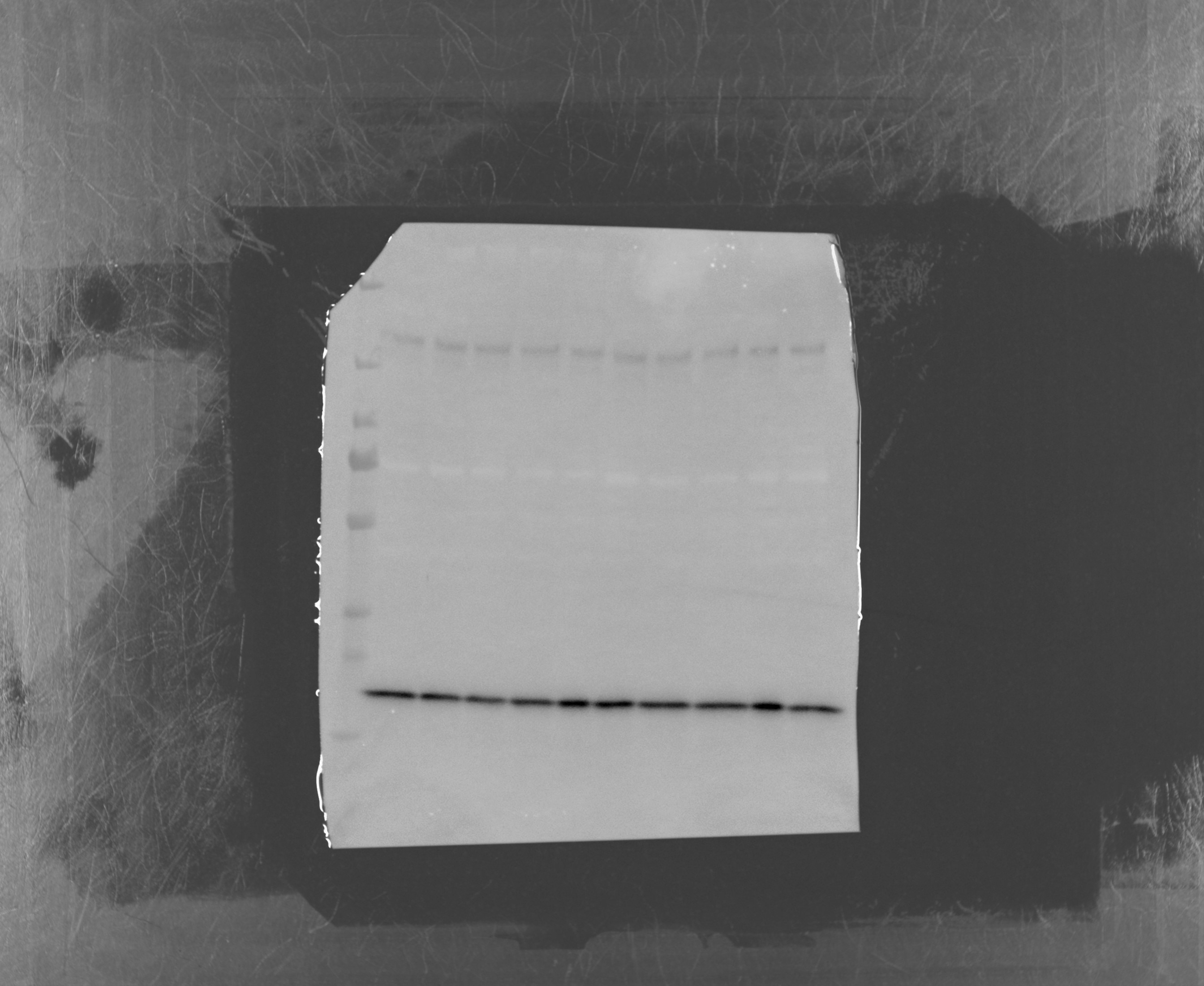
| Western Blot (WB) | WB : 1:20000-1:100000 |
| Immunpräzipitation (IP) | IP : 0.5-4.0 ug for 1.0-3.0 mg of total protein lysate |
| Immunhistochemie (IHC) | IHC : 1:1000-1:4000 |
| Immunfluoreszenz (IF)/ICC | IF/ICC : 1:50-1:500 |
| It is recommended that this reagent should be titrated in each testing system to obtain optimal results. | |
| Sample-dependent, check data in validation data gallery | |
Veröffentlichte Anwendungen
| KD/KO | See 12 publications below |
| WB | See 3837 publications below |
| IHC | See 210 publications below |
| IP | See 6 publications below |
| ELISA | See 1 publications below |
| CoIP | See 1 publications below |
Produktinformation
50599-2-Ig bindet in WB, IHC, IP, CoIP, ELISA BAX und zeigt Reaktivität mit human, Maus, Ratten
| Getestete Reaktivität | human, Maus, Ratte |
| In Publikationen genannte Reaktivität | human, Affe, hamster, Huhn, Kaninchen, Maus, Ratte, Zebrafisch, Ziege |
| Wirt / Isotyp | Kaninchen / IgG |
| Klonalität | Polyklonal |
| Typ | Antikörper |
| Immunogen | BAX fusion protein Ag0576 |
| Vollständiger Name | BCL2-associated X protein |
| Berechnetes Molekulargewicht | 21 kDa |
| Beobachtetes Molekulargewicht | 21 kDa |
| GenBank-Zugangsnummer | BC014175 |
| Gene symbol | BAX |
| Gene ID (NCBI) | 581 |
| Konjugation | Unkonjugiert |
| Form | Liquid |
| Reinigungsmethode | Protein-A-Reinigung |
| Lagerungspuffer | PBS with 0.02% sodium azide and 50% glycerol |
| Lagerungsbedingungen | Bei -20°C lagern. Nach dem Versand ein Jahr lang stabil Aliquotieren ist bei -20oC Lagerung nicht notwendig. 20ul Größen enthalten 0,1% BSA. |
Hintergrundinformationen
BAX (also known as BCL2 Associated X, Bcl-2-Like Protein 4, Bcl2-L-4, BCL2L4) is a member of the BCL2 family of proteins that play a key role in the regulation of apoptosis in higher eukaryotes (https://www.uniprot.org/uniprot/Q07812). BAX comprises 4 Bcl-2 homology domains (BH1-BH4) and a C-terminal transmembrane domain. In healthy mammalian cells, BAX is localized to the cytoplasm through its interaction with the anti-apoptotic BL-2 family members BCL2L1/Bcl-xL (PMIDs: 28755482, 21458670). In response to apoptotic stimuli, however, BAX undergoes a conformational change that causes it to translocate to the outer mitochondrial membrane where it initiates the mitochondrial pathway of apoptosis via two potential mechanisms. Firstly, upon translocation to the outer mitochondrial membrane, BAX interacts with the mitochondrial voltage-dependent anion channel (VDAC) leading to the opening of the channel, loss of membrane potential, and the release of cytochrome c from the mitochondrion (PMID:10766872). The release of cytochrome C into the cytoplasm leads to the activation of Caspase3, initiating apoptosis. Secondly, activated BAX forms homodimers, which then assemble into oligomers on the mitochondrial outer membrane to create pores that permeabilize the mitochondrion leading to the release of cytochrome C (PMID:25458844).
BAX has been shown to be involved in p53-mediated apoptosis. Expression of the human bax gene has been shown to be directly regulated by p53, and the bax promoter contains four motifs with homology to consensus p53-binding sites (PMID:7834749). Furthermore, p53 directly interacts with BAX to promote its activation (PMID:14963330).
What is the molecular weight of BAX?
BAX is a 192 amino acid protein that has a predicted molecular weight of 21.1 kDa.
What is the subcellular localization of BAX?
In healthy mammalian cells, BAX is localized to the cytoplasm. In response to apoptotic stimuli, BAX translocates to the outer mitochondrial membrane.
What is the tissue specificity of BAX?
BAX is ubiquitously expressed (PMID: 25613900).
What is the connection between BAX and cancer?
BAX appears to play an important role in suppressing cancer development, and decreased BAX levels are associated with chemo- and radioresistance in a number of cancers including lung cancer, chronic lymphocytic leukemia (CLL), and prostate cancer. Loss-of-function mutations of BAX have also been reported in hematopoietic malignancies in humans (PMID:9531611) and in gastrointestinal cancer of the microsatellite mutator phenotype (MMP), where BAX inactivation contributes to tumor progression by providing a survival advantage (PMID:9331106).
A number of anticancer drugs used clinically have been demonstrated to induce BAX activation indirectly to facilitate apoptosis of tumor cells. Recent efforts have demonstrated that BAX itself may be a promising direct target for small-molecule drug discovery for novel anticancer drugs and several direct BAX activators have been identified that may hold promise for cancer therapy, with the potential to overcome chemo- and radioresistance (PMID:25230299).
Protokolle
| PRODUKTSPEZIFISCHE PROTOKOLLE | |
|---|---|
| WB protocol for BAX antibody 50599-2-Ig | Protokoll herunterladen |
| IHC protocol for BAX antibody 50599-2-Ig | Protokoll herunterladenl |
| IF protocol for BAX antibody 50599-2-Ig | Protokoll herunterladen |
| IP protocol for BAX antibody 50599-2-Ig | Protokoll herunterladen |
| STANDARD-PROTOKOLLE | |
|---|---|
| Klicken Sie hier, um unsere Standardprotokolle anzuzeigen |
Publikationen
| Species | Application | Title |
|---|---|---|
Bioact Mater Chemo-immunotherapy by dual-enzyme responsive peptide self-assembling abolish melanoma | ||
ACS Nano Nanointegrative In Situ Reprogramming of Tumor-Intrinsic Lipid Droplet Biogenesis for Low-Dose Radiation-Activated Ferroptosis Immunotherapy | ||
ACS Nano Melatonin-Derived Carbon Dots with Free Radical Scavenging Property for Effective Periodontitis Treatment via the Nrf2/HO-1 Pathway | ||
Mol Cell Filamentous GLS1 promotes ROS-induced apoptosis upon glutamine deprivation via insufficient asparagine synthesis.
| ||
Cell Res Tom20 senses iron-activated ROS signaling to promote melanoma cell pyroptosis.
|
Rezensionen
The reviews below have been submitted by verified Proteintech customers who received an incentive for providing their feedback.
FH Charlotte (Verified Customer) (08-15-2024) | Intense band at the expected MW
 |
FH Macarena Lucia (Verified Customer) (10-17-2022) | nice band
|
FH ZEE (Verified Customer) (07-13-2020) | VERY GOOD antibody when western
|
FH Tanusree (Verified Customer) (12-18-2019) | Product worked well in WB at 1:500 dilution
|
FH Kathryn (Verified Customer) (11-04-2019) | Image shows a post-natal day 2 mouse ovary with germ cells marked by TRA98 (Abcam) in green, and BAX (Proteintech) marked in red.
 |

