C7orf47 Polyklonaler Antikörper
C7orf47 Polyklonal Antikörper für IHC, ELISA
Wirt / Isotyp
Kaninchen / IgG
Getestete Reaktivität
human
Anwendung
IHC, IF, ELISA
Konjugation
Unkonjugiert
Kat-Nr. : 24214-1-AP
Synonyme
 at dilution of 1:200 (under 40x lens). Heat mediated antigen retrieval with Tris-EDTA buffer (pH 9.0).")
 at dilution of 1:50 (under 10x lens).")
 at dilution of 1:50 (under 40x lens).")
 at dilution of 1:50 (under 10x lens).")
 at dilution of 1:50 (under 40x lens).")
Geprüfte Anwendungen
| Erfolgreiche Detektion in IHC | humanes Mammakarzinomgewebe, humanes Hodengewebe Hinweis: Antigendemaskierung mit TE-Puffer pH 9,0 empfohlen. (*) Wahlweise kann die Antigendemaskierung auch mit Citratpuffer pH 6,0 erfolgen. |
Empfohlene Verdünnung
| Anwendung | Verdünnung |
|---|---|
| Immunhistochemie (IHC) | IHC : 1:50-1:500 |
| It is recommended that this reagent should be titrated in each testing system to obtain optimal results. | |
| Sample-dependent, check data in validation data gallery | |
Veröffentlichte Anwendungen
| IF | See 1 publications below |
Produktinformation
24214-1-AP bindet in IHC, IF, ELISA C7orf47 und zeigt Reaktivität mit human
| Getestete Reaktivität | human |
| In Publikationen genannte Reaktivität | human |
| Wirt / Isotyp | Kaninchen / IgG |
| Klonalität | Polyklonal |
| Typ | Antikörper |
| Immunogen | C7orf47 fusion protein Ag18340 |
| Vollständiger Name | chromosome 7 open reading frame 47 |
| Berechnetes Molekulargewicht | 253 aa, 28 kDa |
| GenBank-Zugangsnummer | BC026269 |
| Gene symbol | C7orf47 |
| Gene ID (NCBI) | 221908 |
| Konjugation | Unkonjugiert |
| Form | Liquid |
| Reinigungsmethode | Antigen-affinitätsgereinigt |
| Lagerungspuffer | PBS with 0.02% sodium azide and 50% glycerol |
| Lagerungsbedingungen | Bei -20°C lagern. Nach dem Versand ein Jahr lang stabil Aliquotieren ist bei -20oC Lagerung nicht notwendig. 20ul Größen enthalten 0,1% BSA. |
Protokolle
| PRODUKTSPEZIFISCHE PROTOKOLLE | |
|---|---|
| IHC protocol for C7orf47 antibody 24214-1-AP | Protokoll herunterladenl |
| STANDARD-PROTOKOLLE | |
|---|---|
| Klicken Sie hier, um unsere Standardprotokolle anzuzeigen |
Rezensionen
The reviews below have been submitted by verified Proteintech customers who received an incentive for providing their feedback.
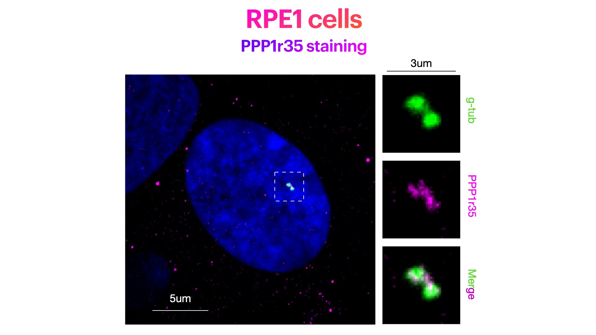
FH Elisa (Verified Customer) (04-24-2023) | Weak centriolar staining. While this antibody works well for Westernblotting, the expected centriolar staining in IF is rather weak. Yet, this antibody works under the following conditions: RPE1 cells stained for Hoechst (DNA marker, in green), PPP1r35 (centriolar marker, in magenta) and g-Tubulin (pericentriolar matrix marker, in green). RPE1 cells were fixed in cold methanol for 10' at -20C. Cells were then rehydrated with PBS for 5'. Membrane permeabilization was then performed with 0.1% Triton + 0.1% Tween +0.01%SDS in PBS for 5'. Cells were finally incubated with blocking buffer (5% BSA+ 0.1% Tween in PBS) for 30' at RT. Primary antibody was diluted in blocking buffer 1:300 and incubated for 1h at room temperature. Alexa-488-Anti-rabbit was used as secondary antibody (1:600 dilution) (1h at room temperature).
 |

