CACNA1A Polyklonaler Antikörper
CACNA1A Polyklonal Antikörper für IHC, ELISA
Wirt / Isotyp
Kaninchen / IgG
Getestete Reaktivität
human
Anwendung
IHC, ELISA
Konjugation
Unkonjugiert
Kat-Nr. : 27227-1-AP
Synonyme
 at dilution of 1:100 (under 10x lens). Heat mediated antigen retrieval with Tris-EDTA buffer (pH 9.0).")
 at dilution of 1:100 (under 40x lens). Heat mediated antigen retrieval with Tris-EDTA buffer (pH 9.0).")
Geprüfte Anwendungen
| Erfolgreiche Detektion in IHC | humanes Hirngewebe Hinweis: Antigendemaskierung mit TE-Puffer pH 9,0 empfohlen. (*) Wahlweise kann die Antigendemaskierung auch mit Citratpuffer pH 6,0 erfolgen. |
Empfohlene Verdünnung
| Anwendung | Verdünnung |
|---|---|
| Immunhistochemie (IHC) | IHC : 1:50-1:500 |
| It is recommended that this reagent should be titrated in each testing system to obtain optimal results. | |
| Sample-dependent, check data in validation data gallery | |
Produktinformation
27227-1-AP bindet in IHC, ELISA CACNA1A und zeigt Reaktivität mit human
| Getestete Reaktivität | human |
| Wirt / Isotyp | Kaninchen / IgG |
| Klonalität | Polyklonal |
| Typ | Antikörper |
| Immunogen | CACNA1A fusion protein Ag25943 |
| Vollständiger Name | calcium channel, voltage-dependent, P/Q type, alpha 1A subunit |
| Berechnetes Molekulargewicht | 282 kDa |
| GenBank-Zugangsnummer | NM_000068 |
| Gene symbol | CACNA1A |
| Gene ID (NCBI) | 773 |
| Konjugation | Unkonjugiert |
| Form | Liquid |
| Reinigungsmethode | Antigen-Affinitätsreinigung |
| Lagerungspuffer | PBS with 0.02% sodium azide and 50% glycerol |
| Lagerungsbedingungen | Bei -20°C lagern. Nach dem Versand ein Jahr lang stabil Aliquotieren ist bei -20oC Lagerung nicht notwendig. 20ul Größen enthalten 0,1% BSA. |
Protokolle
| PRODUKTSPEZIFISCHE PROTOKOLLE | |
|---|---|
| IHC protocol for CACNA1A antibody 27227-1-AP | Protokoll herunterladenl |
| STANDARD-PROTOKOLLE | |
|---|---|
| Klicken Sie hier, um unsere Standardprotokolle anzuzeigen |
Rezensionen
The reviews below have been submitted by verified Proteintech customers who received an incentive for providing their feedback.
FH Lea (Verified Customer) (10-08-2024) | I transfected HEK 293T cells with 2 different CACNA1A constructs (12Q and 27Q, 2 replicates each) and an empty vector as a negative control. The expected band at ~280 kDa is clearly visible in the western blot and no unspecific bands are visible in the negative control. I was very happy with the results.
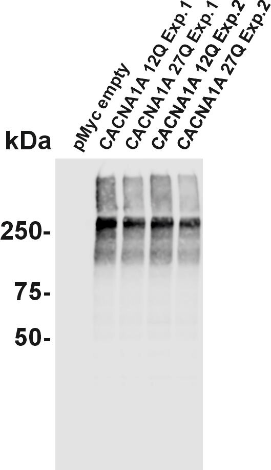 |
FH Ümit (Verified Customer) (08-11-2022) | Cav2.1 (black) staining of two rhesus monkey brainstem sections (5µm &7µm) visualized with immunoperoxidase method (DAB-Nickel).
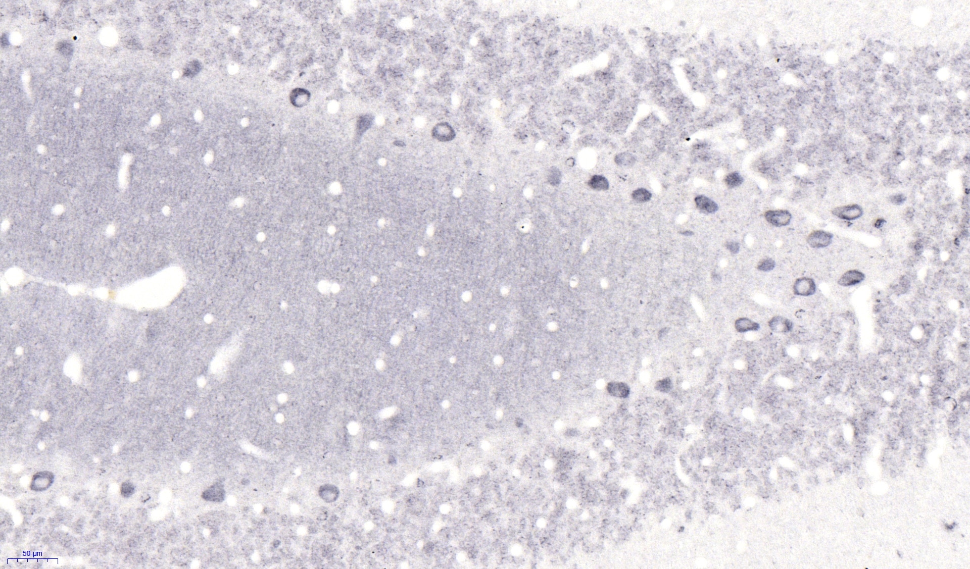 |

