CD63 Monoklonaler Antikörper
CD63 Monoklonal Antikörper für WB, IHC, IF-P, ELISA
Wirt / Isotyp
Maus / IgG1
Getestete Reaktivität
human und mehr (2)
Anwendung
WB, IHC, IF-P, ELISA
Konjugation
Unkonjugiert
CloneNo.
3D4D1
Kat-Nr. : 67605-1-Ig
Synonyme
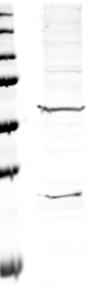
 at dilution of 1:10000 incubated at room temperature for 1.5 hours. The membrane was stripped and reblotted with HRP-conjugated Alpha Tubulin Monoclonal antibody (HRP-66031) as loading control.")
 at dilution of 1:3000 incubated at room temperature for 1.5 hours.")
 at dilution of 1:700 (under 10x lens). Heat mediated antigen retrieval with Tris-EDTA buffer (pH 9.0).")
 at dilution of 1:700 (under 40x lens). Heat mediated antigen retrieval with Tris-EDTA buffer (pH 9.0).")
 at dilution of 1:800 (under 10x lens). Heat mediated antigen retrieval with Tris-EDTA buffer (pH 9.0).")
 at dilution of 1:800 (under 40x lens). Heat mediated antigen retrieval with Tris-EDTA buffer (pH 9.0).")
 at dilution of 1:2000 (under 10x lens). Heat mediated antigen retrieval with Tris-EDTA buffer (pH 9.0).")
 at dilution of 1:2000 (under 40x lens). Heat mediated antigen retrieval with Tris-EDTA buffer (pH 9.0).")
 at dilution of 1:500 (under 10x lens).")
 at dilution of 1:500 (under 40x lens).")
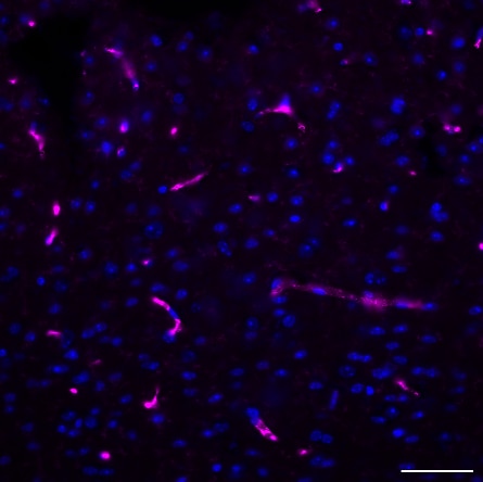
 fixed human tonsillitis tissue using CD63 antibody (67605-1-Ig, Clone: 3D4D1 ) at dilution of 1:400 and CoraLite®488-Conjugated AffiniPure Goat Anti-Mouse IgG(H+L).")
 fixed human tonsillitis tissue using CD63 antibody (67605-1-Ig, Clone: 3D4D1 ) at dilution of 1:400 and CoraLite®488-Conjugated AffiniPure Goat Anti-Mouse IgG(H+L).")
 fixed human lymphoma tissue using CD63 antibody (67605-1-Ig, Clone: 3D4D1 ) at dilution of 1:400 and CoraLite®488-Conjugated AffiniPure Goat Anti-Mouse IgG(H+L).")
 fixed human lymphoma tissue using CD63 antibody (67605-1-Ig, Clone: 3D4D1 ) at dilution of 1:400 and CoraLite®488-Conjugated AffiniPure Goat Anti-Mouse IgG(H+L).")
 fixed human tonsillitis tissue using CD63 antibody (67605-1-Ig, Clone: 3D4D1 ) at dilution of 1:400 and CoraLite®488-Conjugated AffiniPure Goat Anti-Mouse IgG(H+L).")
 fixed human malignant melanoma tissue using CD63 antibody (67605-1-Ig, Clone: 3D4D1 ) at dilution of 1:400 and CoraLite®488-Conjugated AffiniPure Goat Anti-Mouse IgG(H+L).")
 fixed human malignant melanoma tissue using CD63 antibody (67605-1-Ig, Clone: 3D4D1 ) at dilution of 1:400 and CoraLite®488-Conjugated AffiniPure Goat Anti-Mouse IgG(H+L).")
Geprüfte Anwendungen
| Erfolgreiche Detektion in WB | HeLa-Zellen, A549-Zellen, HUVEC-Zellen, K-562-Zellen, LNCaP-Zellen, MCF-7-Zellen, THP-1-Zellen, U2OS-Zellen |
| Erfolgreiche Detektion in IHC | humanes Tonsillitisgewebe, humanes Kolonkarzinomgewebe, humanes malignes Melanomgewebe Hinweis: Antigendemaskierung mit TE-Puffer pH 9,0 empfohlen. (*) Wahlweise kann die Antigendemaskierung auch mit Citratpuffer pH 6,0 erfolgen. |
| Erfolgreiche Detektion in IF-P | humanes Tonsillitisgewebe, humanes Lymphomgewebe, humanes malignes Melanomgewebe |
Empfohlene Verdünnung
| Anwendung | Verdünnung |
|---|---|
| Western Blot (WB) | WB : 1:5000-1:10000 |
| Immunhistochemie (IHC) | IHC : 1:350-1:1400 |
| Immunfluoreszenz (IF)-P | IF-P : 1:200-1:800 |
| It is recommended that this reagent should be titrated in each testing system to obtain optimal results. | |
| Sample-dependent, check data in validation data gallery | |
Veröffentlichte Anwendungen
| WB | See 85 publications below |
| IF | See 5 publications below |
Produktinformation
67605-1-Ig bindet in WB, IHC, IF-P, ELISA CD63 und zeigt Reaktivität mit human
| Getestete Reaktivität | human |
| In Publikationen genannte Reaktivität | human, Ratte, Ziege |
| Wirt / Isotyp | Maus / IgG1 |
| Klonalität | Monoklonal |
| Typ | Antikörper |
| Immunogen | CD63 fusion protein Ag19690 |
| Vollständiger Name | CD63 molecule |
| Berechnetes Molekulargewicht | 26 kDa |
| Beobachtetes Molekulargewicht | 35 kDa |
| GenBank-Zugangsnummer | BC002349 |
| Gene symbol | CD63 |
| Gene ID (NCBI) | 967 |
| Konjugation | Unkonjugiert |
| Form | Liquid |
| Reinigungsmethode | Protein-G-Reinigung |
| Lagerungspuffer | PBS with 0.02% sodium azide and 50% glycerol |
| Lagerungsbedingungen | Bei -20°C lagern. Nach dem Versand ein Jahr lang stabil Aliquotieren ist bei -20oC Lagerung nicht notwendig. 20ul Größen enthalten 0,1% BSA. |
Hintergrundinformationen
CD63 is a 30-60 kDa lysosomal membrane protein that belongs to the tetraspanin family. This protein plays many important roles in immuno-physiological functions. It mediate signal transduction events that play a role in the regulation of cell development, activation and motility. CD63 is expressed on activated platelets, thus it may function as a blood platelet activation marker. CD63 is a lysosomal membrane glycoprotein that is translocated to plasma membrane after platelet activation. The CD63 tetraspanin is highly expressed in the early stages of melanoma and decreases in advanced lesions, suggesting it as a possible suppressor of tumor progression. Deficiency of this protein is associated with Hermansky-Pudlak syndrome.
Protokolle
| PRODUKTSPEZIFISCHE PROTOKOLLE | |
|---|---|
| WB protocol for CD63 antibody 67605-1-Ig | Protokoll herunterladen |
| IHC protocol for CD63 antibody 67605-1-Ig | Protokoll herunterladenl |
| IF protocol for CD63 antibody 67605-1-Ig | Protokoll herunterladen |
| STANDARD-PROTOKOLLE | |
|---|---|
| Klicken Sie hier, um unsere Standardprotokolle anzuzeigen |
Publikationen
| Species | Application | Title |
|---|---|---|
Mol Cancer Exosomal circ_0006896 promotes AML progression via interaction with HDAC1 and restriction of antitumor immunity | ||
Cell Metab Nicotinamide metabolism face-off between macrophages and fibroblasts manipulates the microenvironment in gastric cancer | ||
Nat Commun Lactate dehydrogenase A regulates tumor-macrophage symbiosis to promote glioblastoma progression | ||
Int J Oral Sci Administration of Porphyromonas gingivalis in pregnant mice enhances glycolysis and histone lactylation/ADAM17 leading to cleft palate in offspring | ||
Bone Res Interorgan communication in neurogenic heterotopic ossification: the role of brain-derived extracellular vesicles | ||
Drug Des Devel Ther Keloid Patient Plasma-Derived Exosomal hsa_circ_0020792 Promotes Normal Skin Fibroblasts Proliferation, Migration, and Fibrogenesis via Modulating miR-193a-5p and Activating TGF-β1/Smad2/3 Signaling |
Rezensionen
The reviews below have been submitted by verified Proteintech customers who received an incentive for providing their feedback.
FH Marion (Verified Customer) (11-24-2024) | CD63 stains intracellular endosomal vesiclesd in neuronal extensions. We are satisfied with the results.
 |
FH Guorong (Verified Customer) (03-22-2022) | Two bands at 30 and 60 kDa was detected
 |

