CHCHD3/MIC19 Polyklonaler Antikörper
CHCHD3/MIC19 Polyklonal Antikörper für WB, IHC, IF/ICC, IP, ELISA
Wirt / Isotyp
Kaninchen / IgG
Getestete Reaktivität
human, Maus
Anwendung
WB, IHC, IF/ICC, IP, CoIP, ELISA
Konjugation
Unkonjugiert
Kat-Nr. : 25625-1-AP
Synonyme
Geprüfte Anwendungen
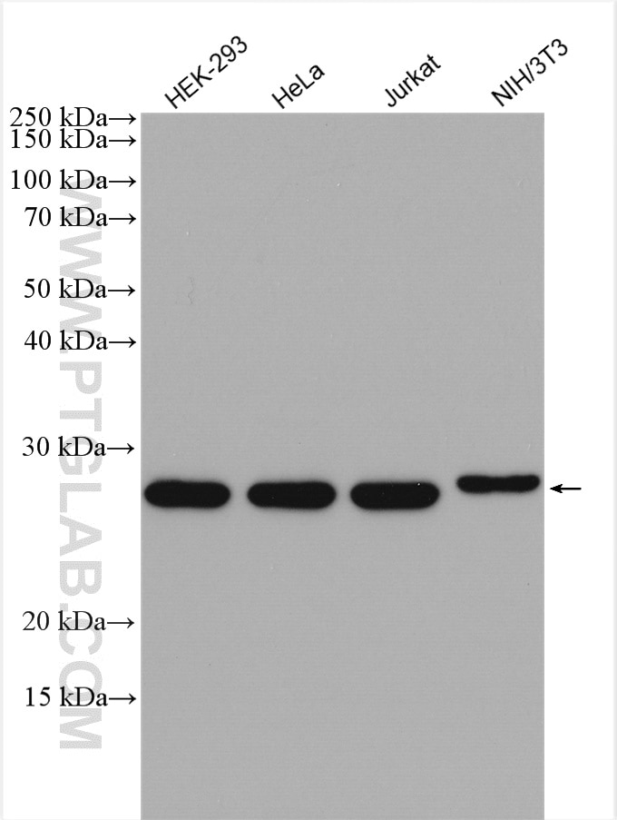
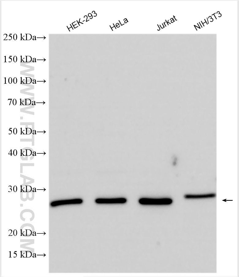
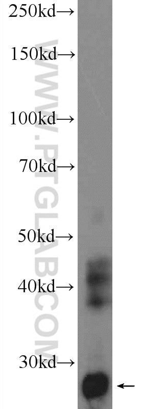
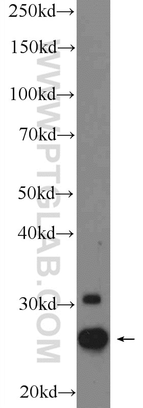
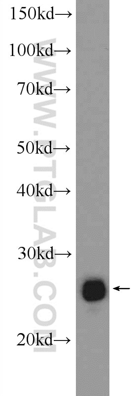
| Erfolgreiche Detektion in WB | HEK-293-Zellen, A431-Zellen, HeLa-Zellen, humanes Plazenta-Gewebe, Jurkat-Zellen, Mauslebergewebe, NIH/3T3-Zellen |
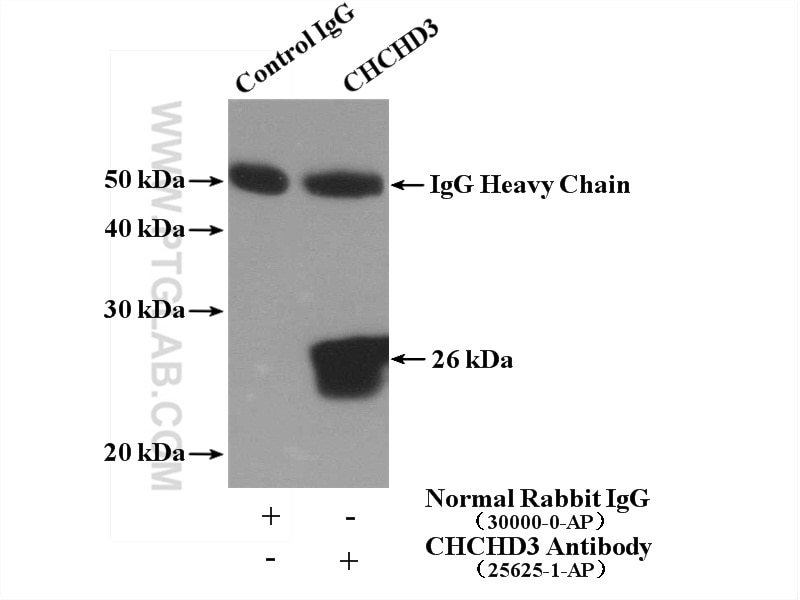
| Erfolgreiche IP | HeLa-Zellen |
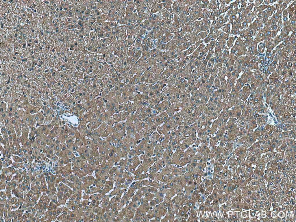
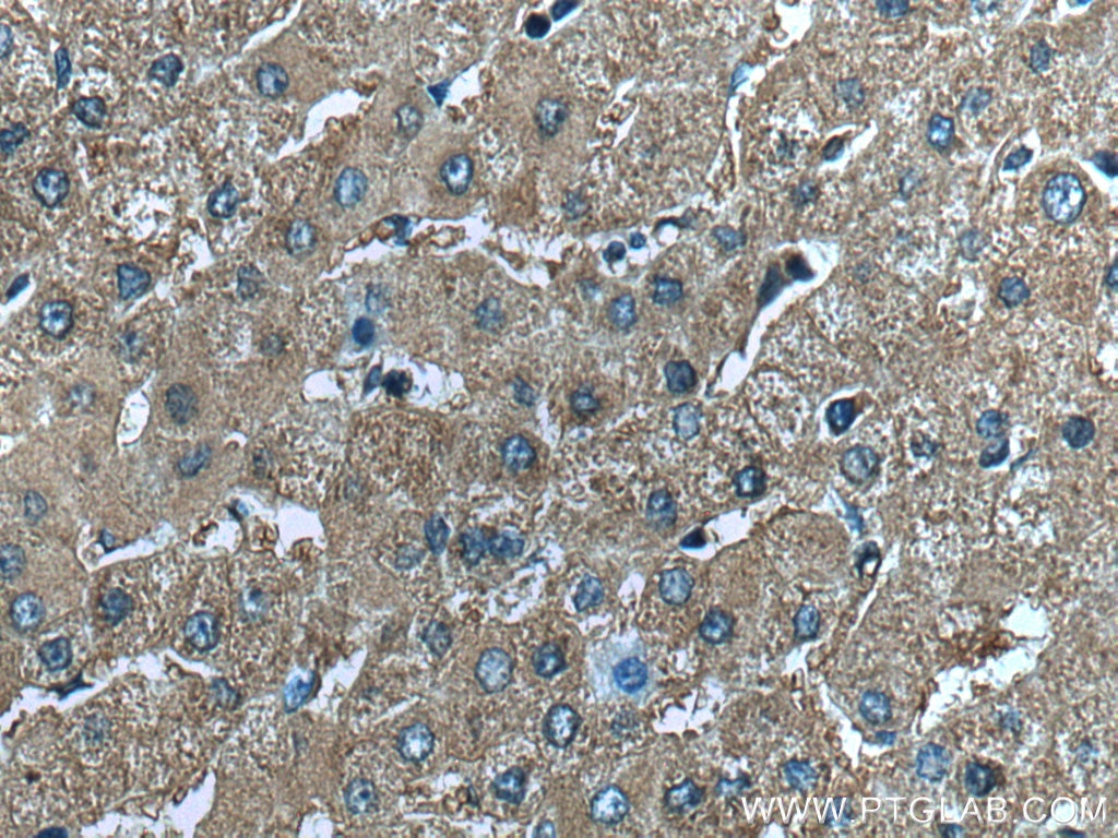
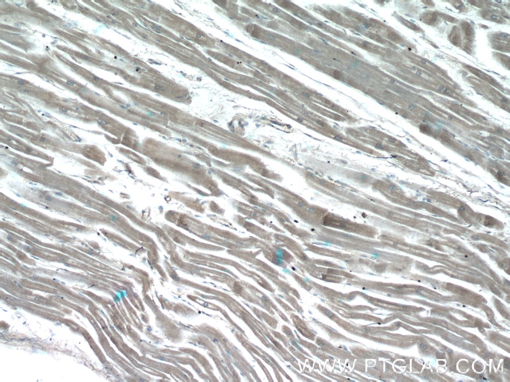
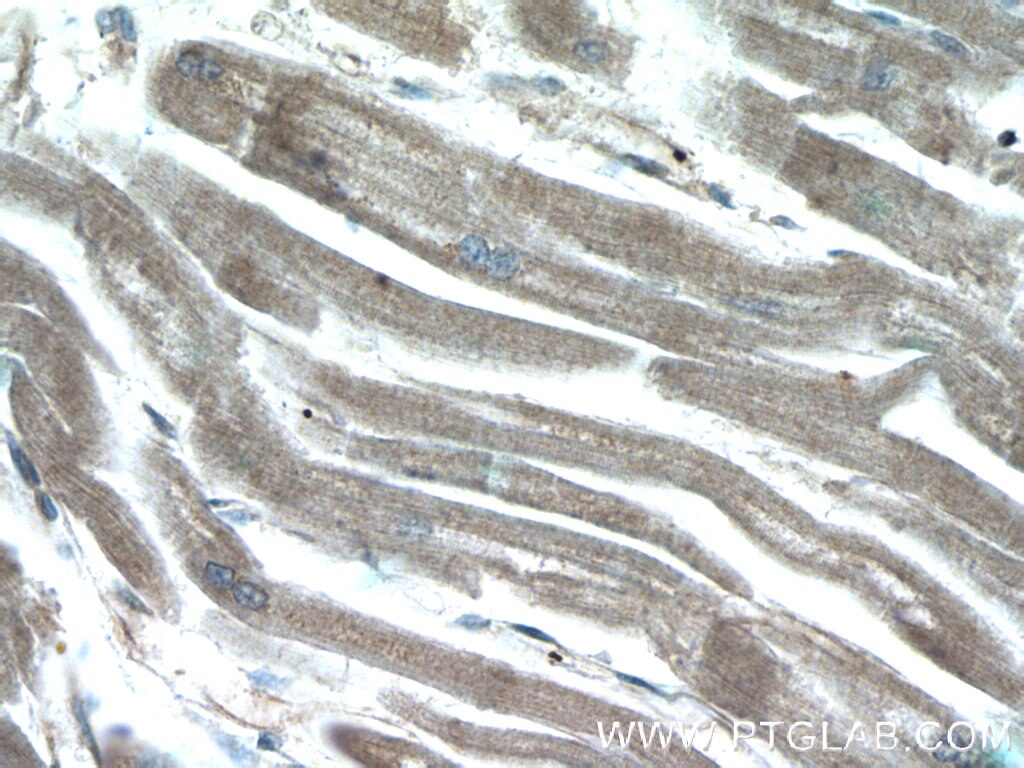
| Erfolgreiche Detektion in IHC | humanes Lebergewebe, humanes Herzgewebe Hinweis: Antigendemaskierung mit TE-Puffer pH 9,0 empfohlen. (*) Wahlweise kann die Antigendemaskierung auch mit Citratpuffer pH 6,0 erfolgen. |
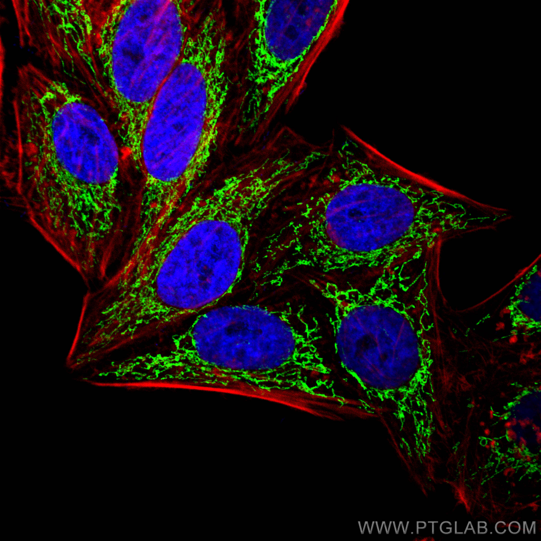
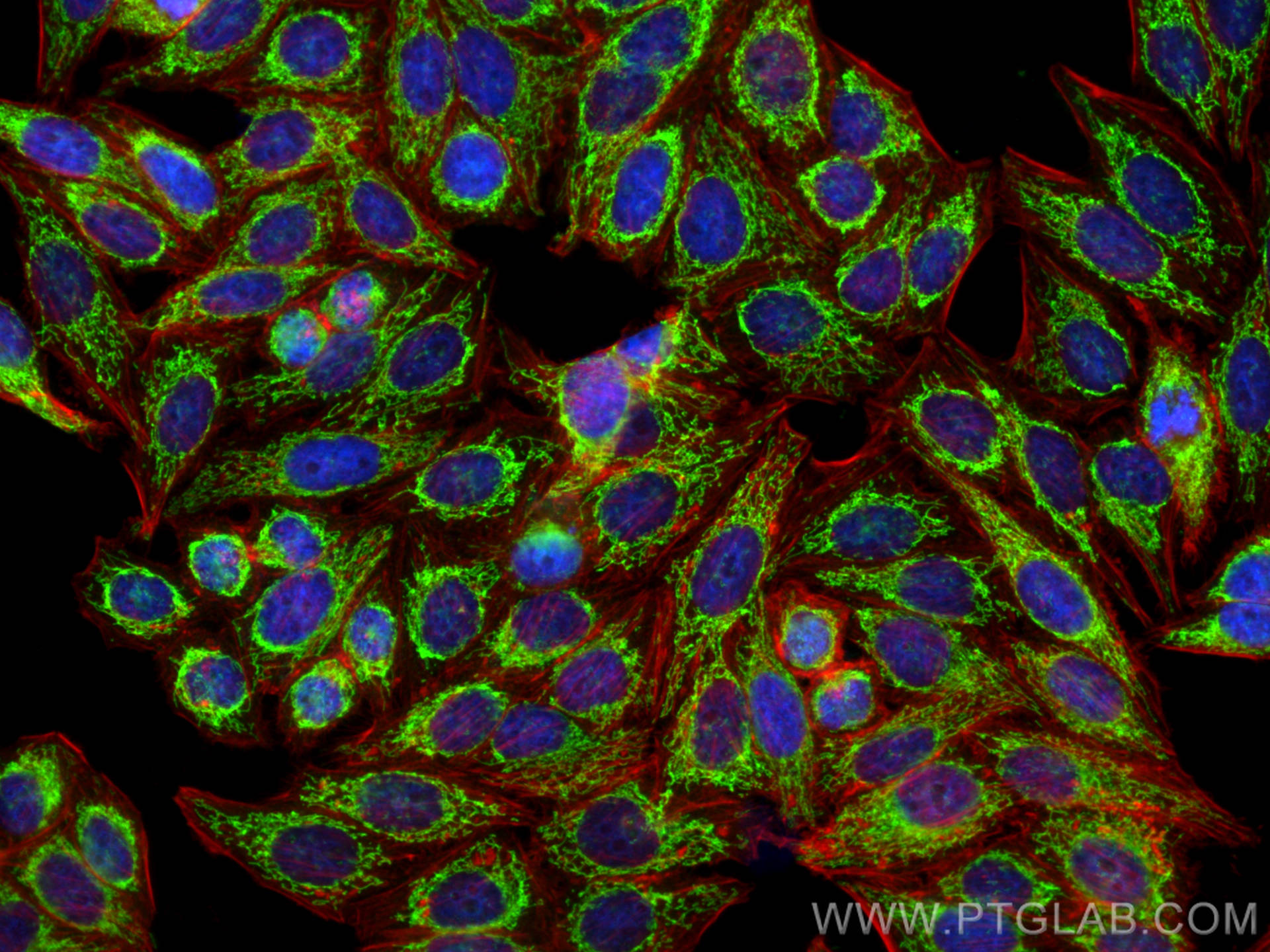
| Erfolgreiche Detektion in IF/ICC | HepG2-Zellen |
Empfohlene Verdünnung
| Anwendung | Verdünnung |
|---|---|
| Western Blot (WB) | WB : 1:4000-1:12000 |
| Immunpräzipitation (IP) | IP : 0.5-4.0 ug for 1.0-3.0 mg of total protein lysate |
| Immunhistochemie (IHC) | IHC : 1:50-1:500 |
| Immunfluoreszenz (IF)/ICC | IF/ICC : 1:50-1:500 |
| It is recommended that this reagent should be titrated in each testing system to obtain optimal results. | |
| Sample-dependent, check data in validation data gallery | |
Veröffentlichte Anwendungen
| KD/KO | See 1 publications below |
| WB | See 33 publications below |
| IF | See 2 publications below |
| IP | See 1 publications below |
| CoIP | See 1 publications below |
Produktinformation
25625-1-AP bindet in WB, IHC, IF/ICC, IP, CoIP, ELISA CHCHD3/MIC19 und zeigt Reaktivität mit human, Maus
| Getestete Reaktivität | human, Maus |
| In Publikationen genannte Reaktivität | human, Maus |
| Wirt / Isotyp | Kaninchen / IgG |
| Klonalität | Polyklonal |
| Typ | Antikörper |
| Immunogen | CHCHD3/MIC19 fusion protein Ag22579 |
| Vollständiger Name | coiled-coil-helix-coiled-coil-helix domain containing 3 |
| Berechnetes Molekulargewicht | 227 aa, 26 kDa |
| Beobachtetes Molekulargewicht | 26 kDa |
| GenBank-Zugangsnummer | BC014839 |
| Gene symbol | CHCHD3 |
| Gene ID (NCBI) | 54927 |
| Konjugation | Unkonjugiert |
| Form | Liquid |
| Reinigungsmethode | Antigen-Affinitätsreinigung |
| Lagerungspuffer | PBS with 0.02% sodium azide and 50% glycerol |
| Lagerungsbedingungen | Bei -20°C lagern. Nach dem Versand ein Jahr lang stabil Aliquotieren ist bei -20oC Lagerung nicht notwendig. 20ul Größen enthalten 0,1% BSA. |
Hintergrundinformationen
CHCHD3, initially identified as a substrate for cAMP-dependent protein kinase (PKA), is a ubiquitous protein in the mitochondria and plays a prominent role in maintaining cristae integrity and mitochondrial function. In mitochondria, ChChd3 is predominantly localized to the inner membrane (IM), facing toward the intermembrane space (IMS), and is part of the large protein complex now called as MINOS (mitochondrial inner membrane organizing system), or MIB (mitochondrial intermembrane space bridging) CHCHD3 is highly conserved in mammals with human and mouse protein sharing ∼92% sequence similarity.
Protokolle
| PRODUKTSPEZIFISCHE PROTOKOLLE | |
|---|---|
| WB protocol for CHCHD3/MIC19 antibody 25625-1-AP | Protokoll herunterladen |
| IHC protocol for CHCHD3/MIC19 antibody 25625-1-AP | Protokoll herunterladenl |
| IF protocol for CHCHD3/MIC19 antibody 25625-1-AP | Protokoll herunterladen |
| IP protocol for CHCHD3/MIC19 antibody 25625-1-AP | Protokoll herunterladen |
| STANDARD-PROTOKOLLE | |
|---|---|
| Klicken Sie hier, um unsere Standardprotokolle anzuzeigen |
Publikationen
| Species | Application | Title |
|---|---|---|
Nat Commun Stalled translation by mitochondrial stress upregulates a CNOT4-ZNF598 ribosomal quality control pathway important for tissue homeostasis | ||
Acta Neuropathol Mitochondria, ER, and nuclear membrane defects reveal early mechanisms for upper motor neuron vulnerability with respect to TDP-43 pathology. | ||
Mol Cell Acylglycerol Kinase Mutated in Sengers Syndrome Is a Subunit of the TIM22 Protein Translocase in Mitochondria. | ||
Nat Commun Mitochondrial membrane proteins and VPS35 orchestrate selective removal of mtDNA | ||
EMBO Rep Cristae undergo continuous cycles of membrane remodelling in a MICOS-dependent manner. | ||
Cell Rep Optic Atrophy 1 Is Epistatic to the Core MICOS Component MIC60 in Mitochondrial Cristae Shape Control. |
Rezensionen
The reviews below have been submitted by verified Proteintech customers who received an incentive for providing their feedback.
FH Mia (Verified Customer) (09-28-2022) | goood!
|
