- Featured Product
- KD/KO Validated
Collagen Type III (N-terminal) Polyklonaler Antikörper
Collagen Type III (N-terminal) Polyklonal Antikörper für WB, IHC, IF-P, IF-Fro, IP, ELISA
Wirt / Isotyp
Kaninchen / IgG
Getestete Reaktivität
human, Maus, Ratte und mehr (6)
Anwendung
WB, IHC, IF-P, IF-Fro, IP, ELISA
Konjugation
Unkonjugiert
Kat-Nr. : 22734-1-AP
Synonyme
 antibody at dilution of 1:1000 incubated at room temperature for 1.5 hours.")
 antibody) at dilution of 1:300 incubated at room temperature for 1.5 hours.")
 (IP:22734-1-AP, 4ug; Detection:22734-1-AP 1:3000) with mouse skin tissue lysate 1120 ug.")
 antibody at dilution of 1:1000 (under 10x lens).")
 antibody at dilution of 1:1000 (under 40x lens).")
 antibody) at dilution of 1:2000 (under 10x lens). Heat mediated antigen retrieval with Tris-EDTA buffer (pH 9.0).")
 antibody) at dilution of 1:2000 (under 40x lens). Heat mediated antigen retrieval with Tris-EDTA buffer (pH 9.0).")
 antibody) at dilution of 1:1000 (under 10x lens). Heat mediated antigen retrieval with Tris-EDTA buffer (pH 9.0).")
 antibody) at dilution of 1:1000 (under 40x lens). Heat mediated antigen retrieval with Tris-EDTA buffer (pH 9.0).")
 antibody at dilution of 1:1000 (under 10x lens).")
 antibody at dilution of 1:1000 (under 40x lens).")
 antibody at dilution of 1:1000 (under 10x lens).")
 antibody at dilution of 1:1000 (under 40x lens).")
 antibody) at dilution of 1:1000 (under 10x lens. Heat mediated antigen retrieval with Tris-EDTA buffer (pH 9.0).")
 antibody) at dilution of 1:1000 (under 40x lens. Heat mediated antigen retrieval with Tris-EDTA buffer (pH 9.0).")
 antibody) at dilution of 1:1000 (under 10x lens. Heat mediated antigen retrieval with Tris-EDTA buffer (pH 9.0).")
 antibody) at dilution of 1:1000 (under 40x lens. Heat mediated antigen retrieval with Tris-EDTA buffer (pH 9.0).")
 antibody) at dilution of 1:400 (under 10x lens). Heat mediated antigen retrieval with Tris-EDTA buffer (pH 9.0).")
 antibody) at dilution of 1:400 (under 40x lens). Heat mediated antigen retrieval with Tris-EDTA buffer (pH 9.0).")
 antibody) at dilution of 1:1000 (under 10x lens). Heat mediated antigen retrieval with Tris-EDTA buffer (pH 9.0).")
 antibody) at dilution of 1:1000 (under 40x lens). Heat mediated antigen retrieval with Tris-EDTA buffer (pH 9.0).")
 antibody) at dilution of 1:1000 (under 10x lens). Heat mediated antigen retrieval with Tris-EDTA buffer (pH 9.0).")
 antibody) at dilution of 1:1000 (under 40x lens). Heat mediated antigen retrieval with Tris-EDTA buffer (pH 9.0).")
 antibody) at dilution of 1:1000 (under 10x lens). Heat mediated antigen retrieval with Tris-EDTA buffer (pH 9.0).")
 antibody) at dilution of 1:1000 (under 40x lens). Heat mediated antigen retrieval with Tris-EDTA buffer (pH 9.0).")
 fixed human colon tissue using Collagen Type III (N-terminal) antibody (22734-1-AP) at dilution of 1:400 and CoraLite®488-Conjugated AffiniPure Goat Anti-Rabbit IgG(H+L).")
 fixed human colon tissue using Collagen Type III (N-terminal) antibody (22734-1-AP) at dilution of 1:400 and CoraLite®488-Conjugated AffiniPure Goat Anti-Rabbit IgG(H+L).")
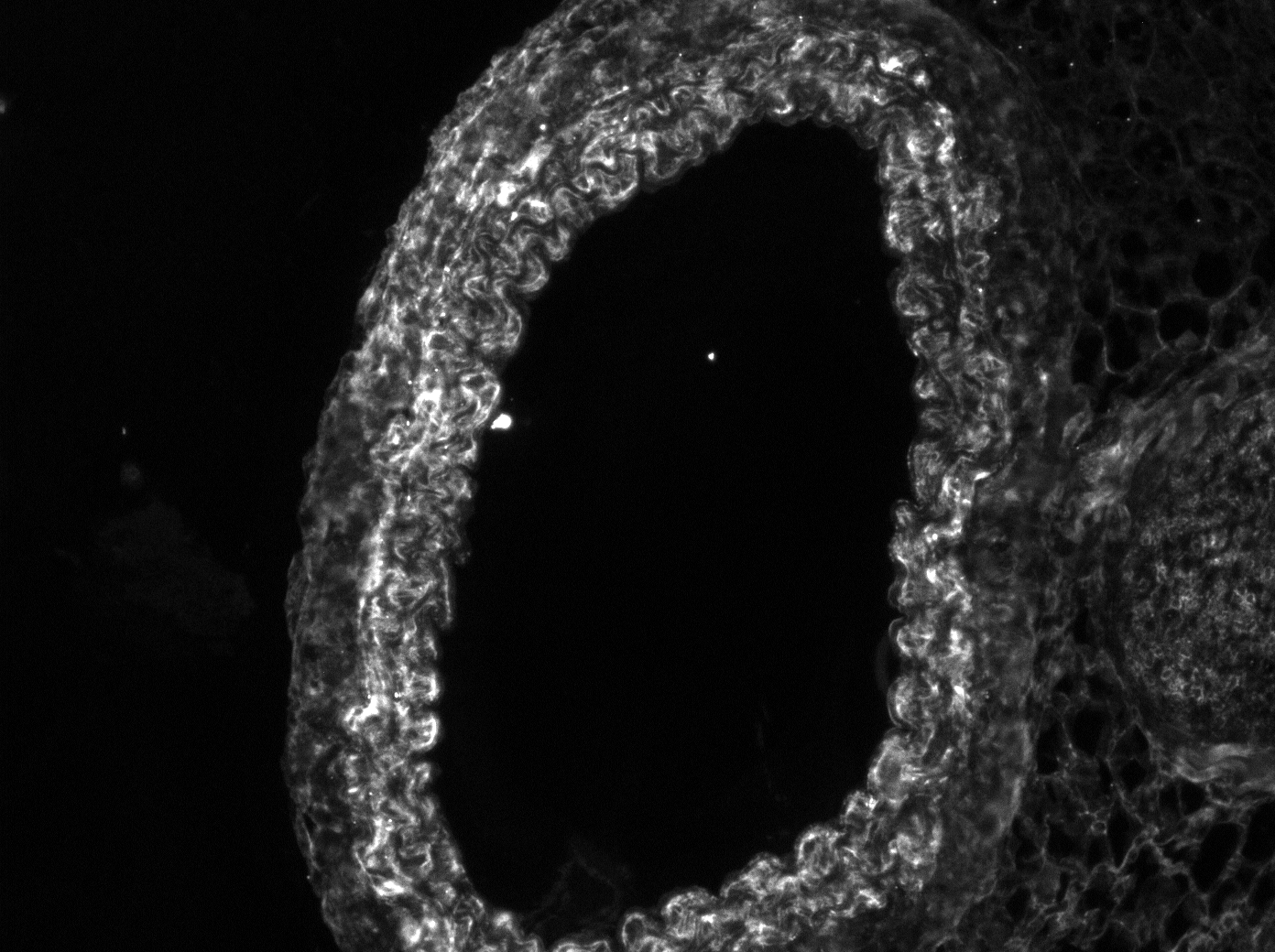
 fixed human colon tissue using Collagen Type III (N-terminal) antibody (22734-1-AP) at dilution of 1:200 and CoraLite®488-Conjugated AffiniPure Goat Anti-Rabbit IgG(H+L), Cytokeratin 20 antibody (60183-1-Ig, Clone: 4D10A4, Magenta), CoraLite®594 CD68 antibody (CL594-25747, red).")
 labeled with CoraLite488-Conjugated AffiniPure Goat Anti-Rabbit IgG(H+L) (SA00013-2, green) in the frist step,anti-CD45 antibody (80297-1-RR) labeled with FlexAble CoraLite Plus 555 Kit (KFA002, orange) in the second step,anti-Collagen Type III antibody (22734-1-AP) labeled with FlexAble CoraLite647 Kit (KFA003, magenta) in the third step, and DAPI (blue).")
Geprüfte Anwendungen
| Erfolgreiche Detektion in WB | Maushautgewebe, Rattenhautgewebe |
| Erfolgreiche IP | Maushautgewebe |
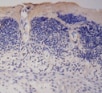
| Erfolgreiche Detektion in IHC | humanes Leberzirrhosegewebe, humanes Kolongewebe, humanes malignes Melanomgewebe, humanes Pankreaskarzinomgewebe, humanes Hautkrebsgewebe, humanes Hautgewebe, Maus-Kolongewebe, Mausherzgewebe, Mausnierengewebe, Mauslebergewebe Hinweis: Antigendemaskierung mit TE-Puffer pH 9,0 empfohlen. (*) Wahlweise kann die Antigendemaskierung auch mit Citratpuffer pH 6,0 erfolgen. |
| Erfolgreiche Detektion in IF-P | humanes Kolongewebe |
| Erfolgreiche Detektion in IF-Fro | Maus-Kolongewebe |
Empfohlene Verdünnung
| Anwendung | Verdünnung |
|---|---|
| Western Blot (WB) | WB : 1:300-1:1000 |
| Immunpräzipitation (IP) | IP : 0.5-4.0 ug for 1.0-3.0 mg of total protein lysate |
| Immunhistochemie (IHC) | IHC : 1:500-1:2000 |
| Immunfluoreszenz (IF)-P | IF-P : 1:50-1:500 |
| Immunfluoreszenz (IF)-FRO | IF-FRO : 1:50-1:500 |
| It is recommended that this reagent should be titrated in each testing system to obtain optimal results. | |
| Sample-dependent, check data in validation data gallery | |
Veröffentlichte Anwendungen
| KD/KO | See 2 publications below |
| WB | See 409 publications below |
| IHC | See 169 publications below |
| IF | See 107 publications below |
Produktinformation
22734-1-AP bindet in WB, IHC, IF-P, IF-Fro, IP, ELISA Collagen Type III (N-terminal) und zeigt Reaktivität mit human, Maus, Ratten
| Getestete Reaktivität | human, Maus, Ratte |
| In Publikationen genannte Reaktivität | human, hamster, Hausschwein, Huhn, Hund, Kaninchen, Maus, Ratte, Rind |
| Wirt / Isotyp | Kaninchen / IgG |
| Klonalität | Polyklonal |
| Typ | Antikörper |
| Immunogen | Collagen Type III (N-terminal) fusion protein Ag18658 |
| Vollständiger Name | collagen, type III, alpha 1 |
| Berechnetes Molekulargewicht | 1466 aa, 139 kDa |
| Beobachtetes Molekulargewicht | 140-180 kDa |
| GenBank-Zugangsnummer | BC028178 |
| Gene symbol | COL3A1 |
| Gene ID (NCBI) | 1281 |
| Konjugation | Unkonjugiert |
| Form | Liquid |
| Reinigungsmethode | Antigen-Affinitätsreinigung |
| Lagerungspuffer | PBS with 0.02% sodium azide and 50% glycerol |
| Lagerungsbedingungen | Bei -20°C lagern. Nach dem Versand ein Jahr lang stabil Aliquotieren ist bei -20oC Lagerung nicht notwendig. 20ul Größen enthalten 0,1% BSA. |
Hintergrundinformationen
Type III collagen is a fibrillar forming collagen comprising three α1(III) chains and is expressed in early embryos and throughout embryogenesis (PMID: 9050868). In the adult, type III collagen is a major component of the extracellular matrix in a variety of internal organs and skin. It occurs in most soft connective tissues along with type I collagen (PMID: 2445760). COL3A1 gene encodes type III procollagen. Mutations in this gene are associated with Ehlers-Danlos syndrome types IV, and with aortic and arterial aneurysms (PMID: 10706896; 2243125; 18389341). This antibody raised against 24-152 aa of prepro α1 (III) chain of human type III procollagen detects type III procollagen at 140-180 kDa and also in some lysates reveals a 70-kDa band which has been reported and may represent a cleaved form of type III procollagen (PMID: 17424834; 19648160; 22802960).
Protokolle
| PRODUKTSPEZIFISCHE PROTOKOLLE | |
|---|---|
| WB protocol for Collagen Type III (N-terminal) antibody 22734-1-AP | Protokoll herunterladen |
| IHC protocol for Collagen Type III (N-terminal) antibody 22734-1-AP | Protokoll herunterladenl |
| IF protocol for Collagen Type III (N-terminal) antibody 22734-1-AP | Protokoll herunterladen |
| IP protocol for Collagen Type III (N-terminal) antibody 22734-1-AP | Protokoll herunterladen |
| STANDARD-PROTOKOLLE | |
|---|---|
| Klicken Sie hier, um unsere Standardprotokolle anzuzeigen |
Publikationen
| Species | Application | Title |
|---|---|---|
Nat Aging Single-cell and spatial RNA sequencing identify divergent microenvironments and progression signatures in early- versus late-onset prostate cancer | ||
Adv Sci (Weinh) LIMA1 O-GlcNAcylation Promotes Hepatic Lipid Deposition through Inducing β-catenin-Regulated FASn Expression in Metabolic Dysfunction-Associated Steatotic Liver Disease | ||
Acta Pharm Sin B SMYD3-PARP16 axis accelerates unfolded protein response and mediates neointima formation. | ||
Rezensionen
The reviews below have been submitted by verified Proteintech customers who received an incentive for providing their feedback.
FH Guennec (Verified Customer) (10-12-2023) | I have 2 bands at 70 KDa.
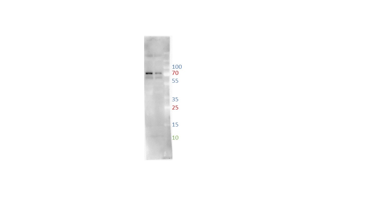 |
FH Sarah-Eve (Verified Customer) (02-19-2023) | Works well. One band observed at 180 kDa.
|
FH Gayatri (Verified Customer) (06-10-2022) | Good signal but inexplicable molecular weight. Observe bands at 70 kDa instead of expected 140-180 kDa although this product is validated for Rat. Observed in both rat heart and tail lysates.
|
FH Yuki (Verified Customer) (03-30-2022) | Works well for IF application and presented with intense staining
 |
FH Ryan (Verified Customer) (04-20-2018) | Antibody worked well for IF application and presented with intense staining of both medial and adventitial collagen in artery sections
 |

