- Featured Product
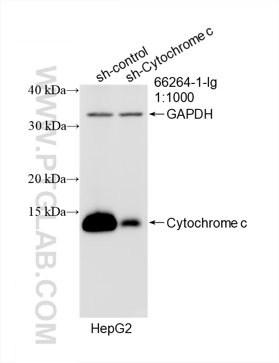
- KD/KO Validated
Cytochrome c Monoklonaler Antikörper
Cytochrome c Monoklonal Antikörper für WB, IHC, IF/ICC, FC (Intra), ELISA
Wirt / Isotyp
Maus / IgG2a
Getestete Reaktivität
human, Maus, Ratte und mehr (1)
Anwendung
WB, IHC, IF/ICC, FC (Intra), ELISA
Konjugation
Unkonjugiert
CloneNo.
2D8D11
Kat-Nr. : 66264-1-Ig
Synonyme
Geprüfte Anwendungen
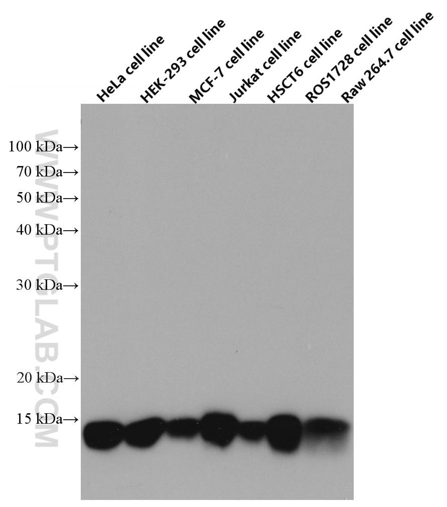
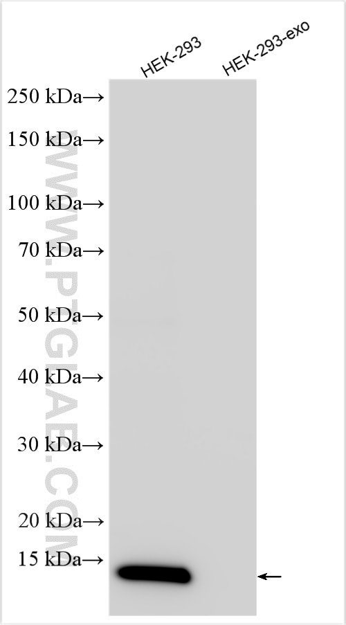
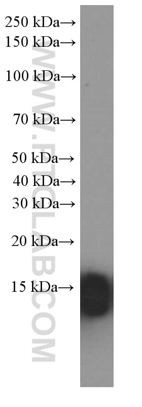
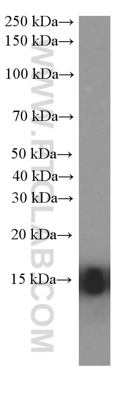
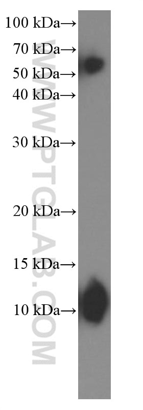
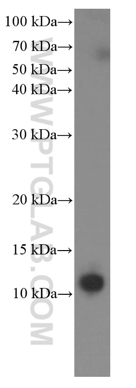
| Erfolgreiche Detektion in WB | HeLa-Zellen, HEK-293-Zellen, HepG2-Zellen, humanes Herzgewebe, humanes Skelettmuskelgewebe, Jurkat-Zellen, MCF-7-Zellen, Maus-Skelettmuskelgewebe, Ratten-Skelettmuskelgewebe, RAW 264.7-Zellen, ROS1728-Zellen |
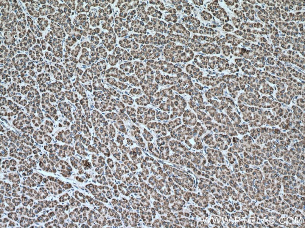
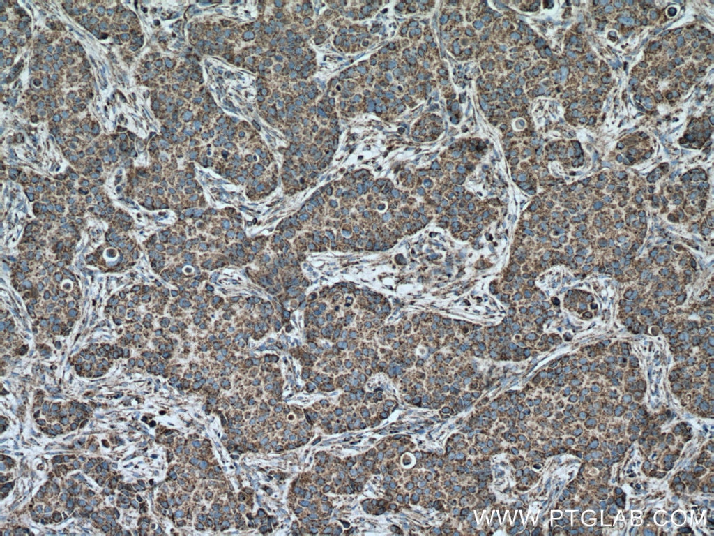
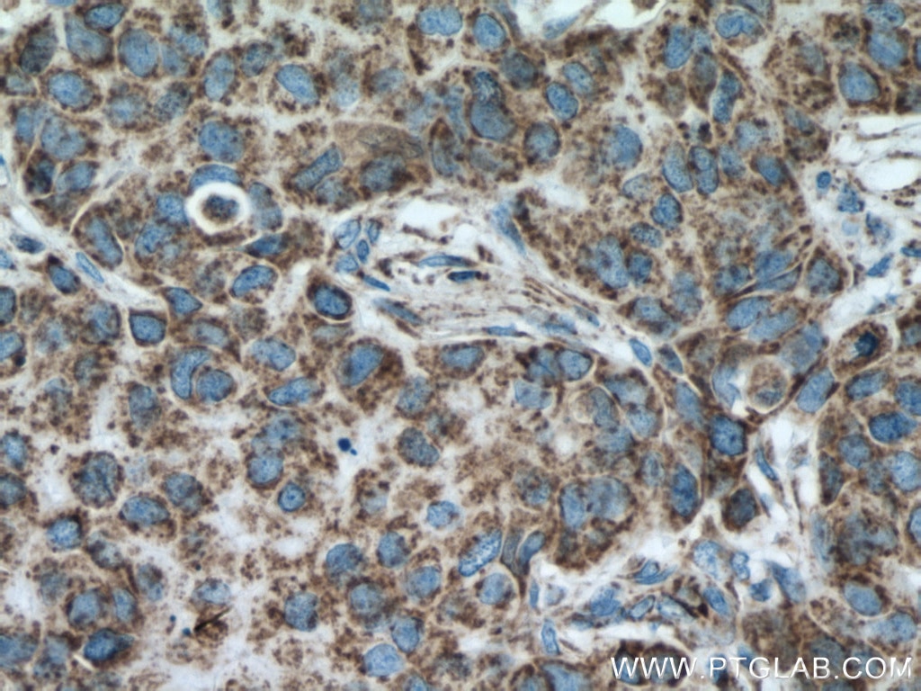
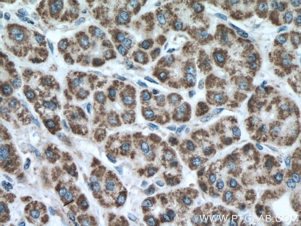
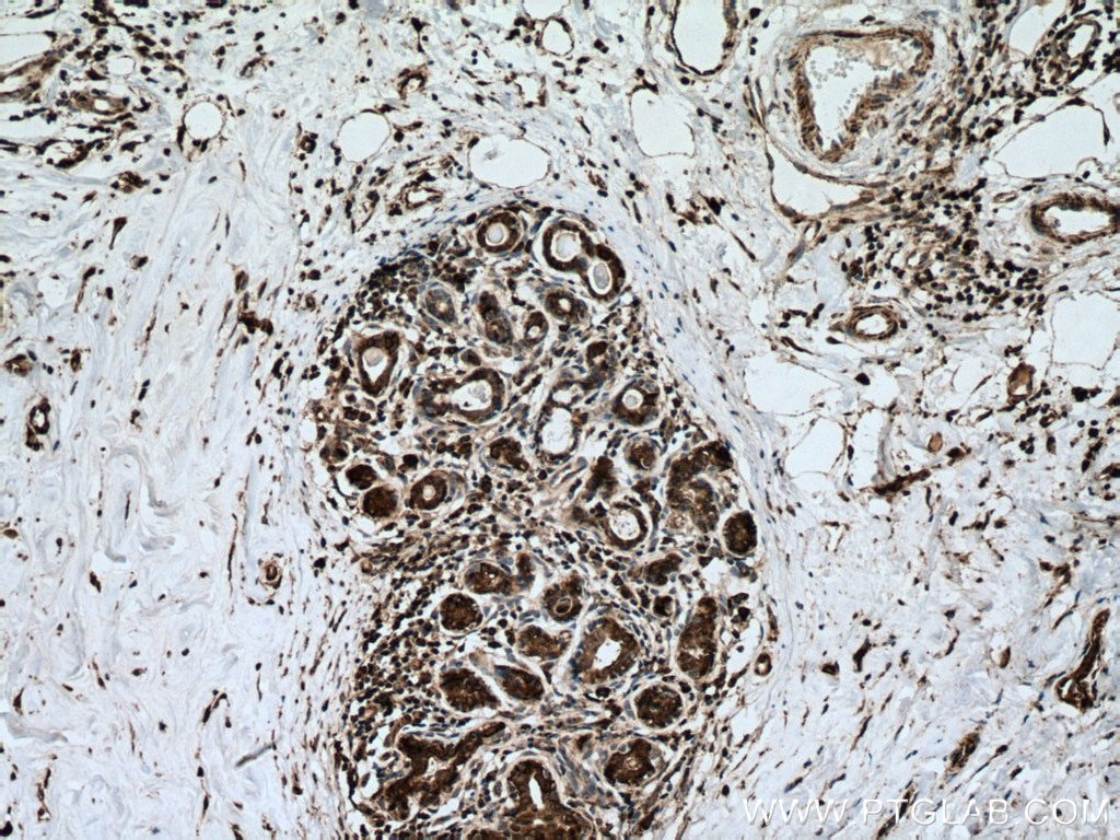
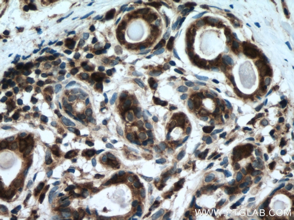
| Erfolgreiche Detektion in IHC | humanes Leberkarzinomgewebe, humanes Mammakarzinomgewebe Hinweis: Antigendemaskierung mit TE-Puffer pH 9,0 empfohlen. (*) Wahlweise kann die Antigendemaskierung auch mit Citratpuffer pH 6,0 erfolgen. |
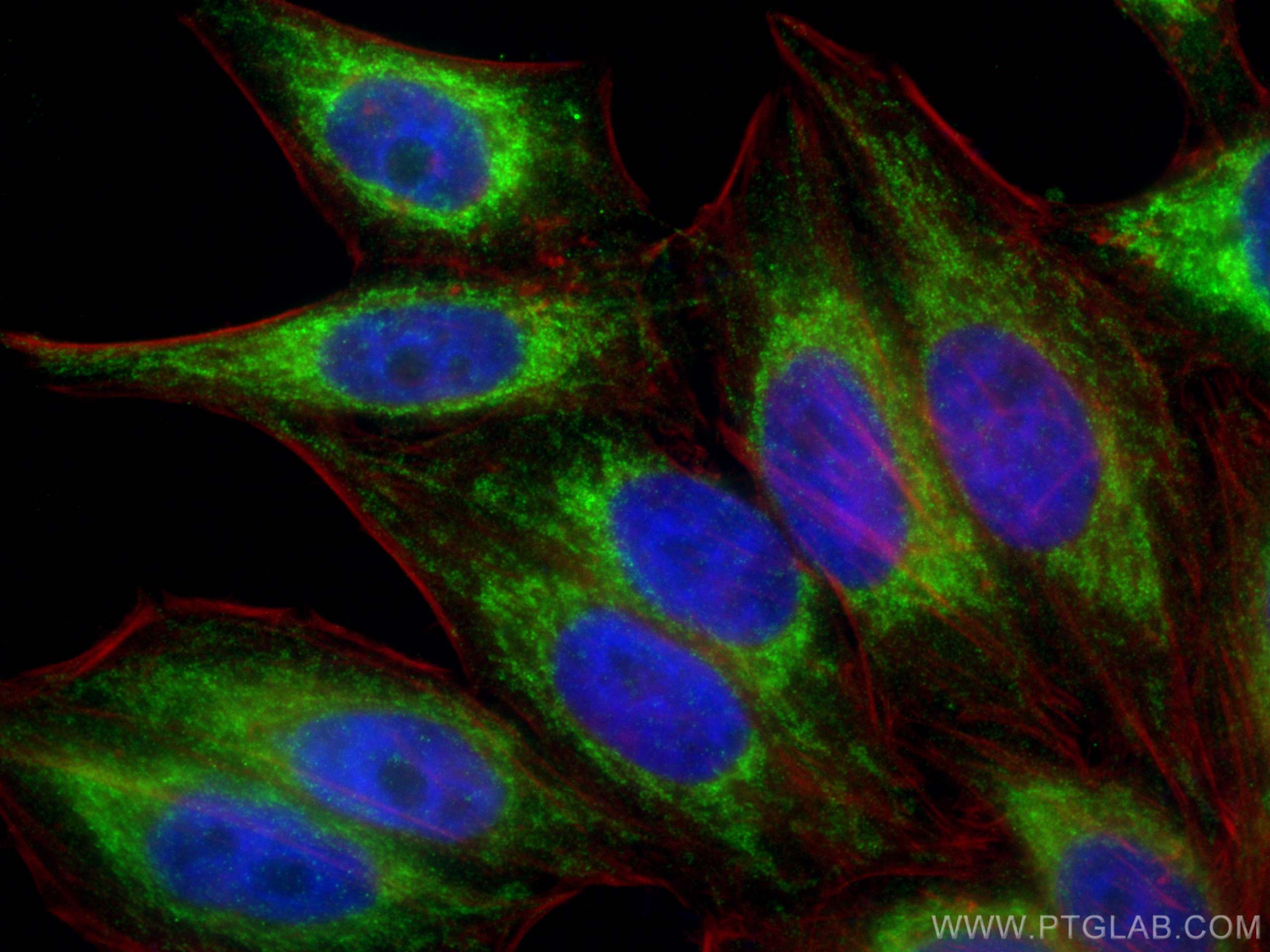
| Erfolgreiche Detektion in IF/ICC | HepG2-Zellen |
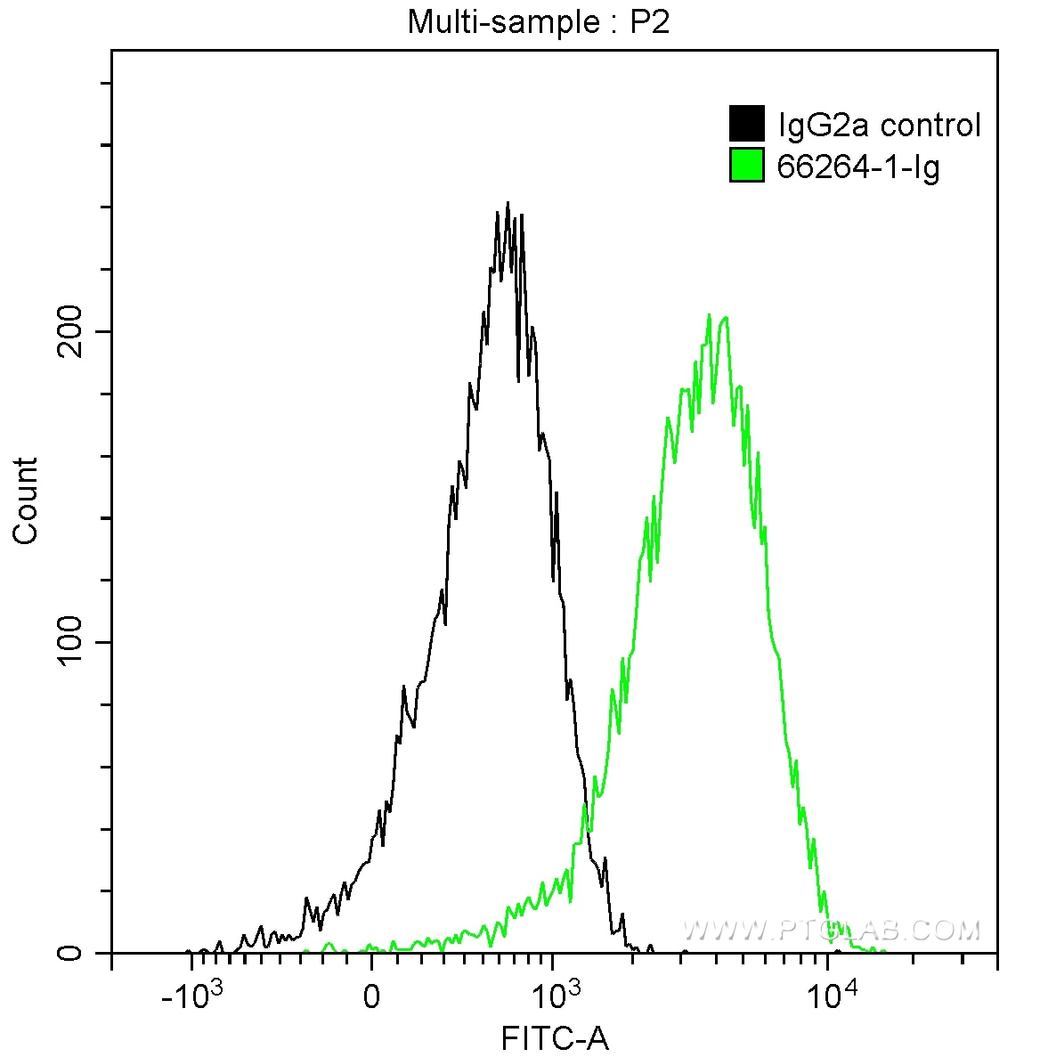
| Erfolgreiche Detektion in FC (Intra) | HepG2-Zellen |
Empfohlene Verdünnung
| Anwendung | Verdünnung |
|---|---|
| Western Blot (WB) | WB : 1:5000-1:50000 |
| Immunhistochemie (IHC) | IHC : 1:1000-1:5000 |
| Immunfluoreszenz (IF)/ICC | IF/ICC : 1:200-1:800 |
| Durchflusszytometrie (FC) (INTRA) | FC (INTRA) : 0.20 ug per 10^6 cells in a 100 µl suspension |
| It is recommended that this reagent should be titrated in each testing system to obtain optimal results. | |
| Sample-dependent, check data in validation data gallery | |
Veröffentlichte Anwendungen
| WB | See 122 publications below |
| IHC | See 3 publications below |
| IF | See 17 publications below |
Produktinformation
66264-1-Ig bindet in WB, IHC, IF/ICC, FC (Intra), ELISA Cytochrome c und zeigt Reaktivität mit human, Maus, Ratten
| Getestete Reaktivität | human, Maus, Ratte |
| In Publikationen genannte Reaktivität | human, Hund, Maus, Ratte |
| Wirt / Isotyp | Maus / IgG2a |
| Klonalität | Monoklonal |
| Typ | Antikörper |
| Immunogen | Cytochrome c fusion protein Ag24349 |
| Vollständiger Name | cytochrome c, somatic |
| Berechnetes Molekulargewicht | 12 kDa |
| Beobachtetes Molekulargewicht | 12-15 kDa |
| GenBank-Zugangsnummer | BC009578 |
| Gene symbol | Cytochrome c |
| Gene ID (NCBI) | 54205 |
| Konjugation | Unkonjugiert |
| Form | Liquid |
| Reinigungsmethode | Protein-A-Reinigung |
| Lagerungspuffer | PBS with 0.02% sodium azide and 50% glycerol |
| Lagerungsbedingungen | Bei -20°C lagern. Nach dem Versand ein Jahr lang stabil Aliquotieren ist bei -20oC Lagerung nicht notwendig. 20ul Größen enthalten 0,1% BSA. |
Hintergrundinformationen
Cytochrome c is a 12-15 kDa electron transporting protein located in the inner mitochondrial membrane. Upon apoptotic stimulation, cytochrome c can be released from mitochondria into cytoplasm, resulting in caspase-3 activation and apoptosis. Measurement of cytochrome c release from the mitochondria is useful for detection of the onset of apoptosis in cells. In addition, cytochrome c can also leave cells and be detectable in extra-cellular medium of apoptotic cells and serum of cancer patients. The level of serum cytochrome c may serve as a prognostic maker during cancer therapy.
Protokolle
| PRODUKTSPEZIFISCHE PROTOKOLLE | |
|---|---|
| WB protocol for Cytochrome c antibody 66264-1-Ig | Protokoll herunterladen |
| IHC protocol for Cytochrome c antibody 66264-1-Ig | Protokoll herunterladenl |
| IF protocol for Cytochrome c antibody 66264-1-Ig | Protokoll herunterladen |
| STANDARD-PROTOKOLLE | |
|---|---|
| Klicken Sie hier, um unsere Standardprotokolle anzuzeigen |
Publikationen
| Species | Application | Title |
|---|---|---|
Hepatology Hepatic stimulator substance resists hepatic ischemia/reperfusion injury by regulating Drp1 translocation and activation. | ||
Cell Death Differ ZNF451 collaborates with RNF8 to regulate RNF168 localization and amplify ubiquitination signaling to promote DNA damage repair and regulate radiosensitivity | ||
Nat Commun Endonuclease G promotes autophagy by suppressing mTOR signaling and activating the DNA damage response. | ||
Redox Biol Mecheliolide elicits ROS-mediated ERS driven immunogenic cell death in hepatocellular carcinoma. | ||
ACS Appl Mater Interfaces Extracellular Matrix-Mimicking Hydrogel with Angiogenic and Immunomodulatory Properties Accelerates Healing of Diabetic Wounds by Promoting Autophagy | ||
J. Pineal Res. Human transporters, PEPT1/2, facilitate melatonin transportation into mitochondria of cancer cells: an implication of the therapeutic potential. |
Rezensionen
The reviews below have been submitted by verified Proteintech customers who received an incentive for providing their feedback.
FH Megha (Verified Customer) (07-10-2018) |
|
