- Featured Product
- KD/KO Validated
DDB1 Polyklonaler Antikörper
DDB1 Polyklonal Antikörper für WB, IHC, IP, ELISA
Wirt / Isotyp
Kaninchen / IgG
Getestete Reaktivität
human, Maus, Ratte
Anwendung
WB, IHC, IF, IP, CoIP, ChIP, ELISA
Konjugation
Unkonjugiert
Kat-Nr. : 11380-1-AP
Synonyme
 at dilution of 1:8000 incubated at room temperature for 1.5 hours.")
 at dilution of 1:500 incubated at room temperature for 1.5 hours.")
 at dilution of 1:500 incubated at room temperature for 1.5 hours.")
 at dilution of 1:500 incubated at room temperature for 1.5 hours.")
 at dilution of 1:1000 incubated at room temperature for 1.5 hours.")
 at dilution of 1:8000 incubated at room temperature for 1.5 hours.")
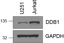
 with Jurkat cells lysate 3080ug.")
 at dilution of 1:200 (under 10x lens). Heat mediated antigen retrieval with Tris-EDTA buffer (pH 9.0).")
 at dilution of 1:200 (under 40x lens). Heat mediated antigen retrieval with Tris-EDTA buffer (pH 9.0).")
 at dilution of 1:1000 (under 20x lens). Heat mediated antigen retrieval with Tris-EDTA buffer (pH 9.0).")
 at dilution of 1:200 (under 10x lens). Heat mediated antigen retrieval with Tris-EDTA buffer (pH 9.0).")
 at dilution of 1:200 (under 40x lens). Heat mediated antigen retrieval with Tris-EDTA buffer (pH 9.0).")
Geprüfte Anwendungen
| Erfolgreiche Detektion in WB | HCT 116-Zellen, C2C12-Zellen, HeLa-Zellen, HepG2-Zellen, humanes Hirngewebe, humanes Nierengewebe, humanes Plazenta-Gewebe, Jurkat-Zellen, MCF-7-Zellen, MDA-MB-231-Zellen, Maushodengewebe, NIH/3T3-Zellen, Rattenhodengewebe |
| Erfolgreiche IP | Jurkat-Zellen |
| Erfolgreiche Detektion in IHC | humanes Kolonkarzinomgewebe, human colon cancer Hinweis: Antigendemaskierung mit TE-Puffer pH 9,0 empfohlen. (*) Wahlweise kann die Antigendemaskierung auch mit Citratpuffer pH 6,0 erfolgen. |
Empfohlene Verdünnung
| Anwendung | Verdünnung |
|---|---|
| Western Blot (WB) | WB : 1:2000-1:16000 |
| Immunpräzipitation (IP) | IP : 0.5-4.0 ug for 1.0-3.0 mg of total protein lysate |
| Immunhistochemie (IHC) | IHC : 1:50-1:500 |
| It is recommended that this reagent should be titrated in each testing system to obtain optimal results. | |
| Sample-dependent, check data in validation data gallery | |
Veröffentlichte Anwendungen
| KD/KO | See 2 publications below |
| WB | See 10 publications below |
| IF | See 1 publications below |
| IP | See 1 publications below |
| CoIP | See 1 publications below |
| ChIP | See 1 publications below |
Produktinformation
11380-1-AP bindet in WB, IHC, IF, IP, CoIP, ChIP, ELISA DDB1 und zeigt Reaktivität mit human, Maus, Ratten
| Getestete Reaktivität | human, Maus, Ratte |
| In Publikationen genannte Reaktivität | human, Maus |
| Wirt / Isotyp | Kaninchen / IgG |
| Klonalität | Polyklonal |
| Typ | Antikörper |
| Immunogen | DDB1 fusion protein Ag1901 |
| Vollständiger Name | damage-specific DNA binding protein 1, 127kDa |
| Berechnetes Molekulargewicht | 1140 aa, 127 kDa |
| Beobachtetes Molekulargewicht | 127 kDa |
| GenBank-Zugangsnummer | BC011686 |
| Gene symbol | DDB1 |
| Gene ID (NCBI) | 1642 |
| Konjugation | Unkonjugiert |
| Form | Liquid |
| Reinigungsmethode | Antigen-Affinitätsreinigung |
| Lagerungspuffer | PBS with 0.02% sodium azide and 50% glycerol |
| Lagerungsbedingungen | Bei -20°C lagern. Nach dem Versand ein Jahr lang stabil Aliquotieren ist bei -20oC Lagerung nicht notwendig. 20ul Größen enthalten 0,1% BSA. |
Hintergrundinformationen
DDB1, also named as XAP1, XPCe, DDBa and XPE-BF, belongs to the DDB1 family. It is required for DNA repair. DDB1 binds to DDB2 to form the UV-damaged DNA-binding protein complex (the UV-DDB complex). The UV-DDB complex may recognize UV-induced DNA damage and recruit proteins of the nucleotide excision repair pathway (the NER pathway) to initiate DNA repair. The functional specificity of the DCX E3 ubiquitin-protein ligase complex is determined by the variable substrate recognition component recruited by DDB1. This antibody is specific to DDB1.
Protokolle
| PRODUKTSPEZIFISCHE PROTOKOLLE | |
|---|---|
| WB protocol for DDB1 antibody 11380-1-AP | Protokoll herunterladen |
| IHC protocol for DDB1 antibody 11380-1-AP | Protokoll herunterladenl |
| IP protocol for DDB1 antibody 11380-1-AP | Protokoll herunterladen |
| STANDARD-PROTOKOLLE | |
|---|---|
| Klicken Sie hier, um unsere Standardprotokolle anzuzeigen |
Publikationen
| Species | Application | Title |
|---|---|---|
Front Immunol Ddb1 Is Essential for the Expansion of CD4+ Helper T Cells by Regulating Cell Cycle Progression and Cell Death. | ||
FASEB J DDB2 promotes melanoma cell growth by transcriptionally regulating the expression of KMT2A and predicts a poor prognosis | ||
iScience A Multidimensional Characterization of E3 Ubiquitin Ligase and Substrate Interaction Network. | ||
PLoS One 14-3-3ε mediates the cell fate decision-making pathways in response of hepatocellular carcinoma to Bleomycin-induced DNA damage. | ||
Virology DDB1 is a cellular substrate of NS3/4A protease and required for hepatitis C virus replication.
| ||
Cell Death Dis Cul4 E3 ubiquitin ligase regulates ovarian cancer drug resistance by targeting the antiapoptotic protein BIRC3.
|
Rezensionen
The reviews below have been submitted by verified Proteintech customers who received an incentive for providing their feedback.
FH Bernadette (Verified Customer) (09-24-2025) | The antibody worked well at 1:2000 dilution in 5% milk shaken overnight in 4 degree fridge on HEK293 cells.
|
FH Sarah (Verified Customer) (07-03-2019) | Total cell lysate (15 ug) was resolved on a 4-12% Bis-Tris gel and transferred to nitrocellulose membrane. Membrane was incubated in blocking buffer (5% milk/0.1% Tween-20) for 1h. Membrane was incubated with anti-DDB1 in blocking buffer (1:1000) at 4C overnight. After washing, membrane was incubated in anti-rabbit-HRP in blocking bufffer (1:3000) for 1h at room temperature. Protein was detected using ECL reagent and imaged on a chemiluminescence detection system.
 |

