HIF2α/EPAS1 Polyklonaler Antikörper
HIF2α/EPAS1 Polyklonal Antikörper für WB, ELISA
Wirt / Isotyp
Kaninchen / IgG
Getestete Reaktivität
human und mehr (1)
Anwendung
WB, IHC, IF, CoIP, ELISA
Konjugation
Unkonjugiert
Kat-Nr. : 26422-1-AP
Synonyme
Geprüfte Anwendungen
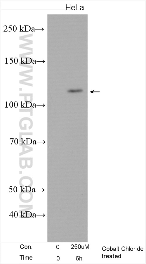
| Erfolgreiche Detektion in WB | mit Cobaltchlorid behandelte HeLa-Zellen |
Empfohlene Verdünnung
| Anwendung | Verdünnung |
|---|---|
| Western Blot (WB) | WB : 1:500-1:3000 |
| It is recommended that this reagent should be titrated in each testing system to obtain optimal results. | |
| Sample-dependent, check data in validation data gallery | |
Veröffentlichte Anwendungen
| WB | See 18 publications below |
| IHC | See 4 publications below |
| IF | See 3 publications below |
| CoIP | See 1 publications below |
Produktinformation
26422-1-AP bindet in WB, IHC, IF, CoIP, ELISA HIF2α/EPAS1 und zeigt Reaktivität mit human
| Getestete Reaktivität | human |
| In Publikationen genannte Reaktivität | human, Maus |
| Wirt / Isotyp | Kaninchen / IgG |
| Klonalität | Polyklonal |
| Typ | Antikörper |
| Immunogen | HIF2α/EPAS1 fusion protein Ag24886 |
| Vollständiger Name | endothelial PAS domain protein 1 |
| Berechnetes Molekulargewicht | 96 kDa |
| Beobachtetes Molekulargewicht | 100-120 kDa |
| GenBank-Zugangsnummer | BC051338 |
| Gene symbol | HIF2α |
| Gene ID (NCBI) | 2034 |
| Konjugation | Unkonjugiert |
| Form | Liquid |
| Reinigungsmethode | Antigen-Affinitätsreinigung |
| Lagerungspuffer | PBS with 0.02% sodium azide and 50% glycerol |
| Lagerungsbedingungen | Bei -20°C lagern. Nach dem Versand ein Jahr lang stabil Aliquotieren ist bei -20oC Lagerung nicht notwendig. 20ul Größen enthalten 0,1% BSA. |
Hintergrundinformationen
HIF2A, also named as EPAS1, is a 870 amino acid protein, which is expressed in most tissues, with highest levels in placenta, lung and heart. HIF2A colocalizes with HIF3A in the nucleus and speckles. HIF2A as a transcription factor involves in the induction of oxygen regulated genes. HIF2A binds to core DNA sequence 5'-[AG]CGTG-3' within the hypoxia response element (HRE) of target gene promoters. HIF2A regulates the vascular endothelial growth factor (VEGF) expression and seems to be implicated in the development of blood vessels and the tubular system of lung. HIF2A may also play a role in the formation of the endothelium that gives rise to the blood brain barrier. The calculated molecular weight of HIF2A is 96 kDa, but In normoxia, HIF2A is probably hydroxylated on Pro-405 and Pro-531 by EGLN1/PHD1, EGLN2/PHD2 and/or EGLN3/PHD3. The hydroxylated prolines promote interaction with VHL, initiating rapid ubiquitination and subsequent proteasomal degradation. Under hypoxia, proline hydroxylation is impaired and ubiquitination is attenuated, resulting in stabilization. The modified HIf2A is about 100-120 kDa.
Protokolle
| PRODUKTSPEZIFISCHE PROTOKOLLE | |
|---|---|
| WB protocol for HIF2α/EPAS1 antibody 26422-1-AP | Protokoll herunterladen |
| STANDARD-PROTOKOLLE | |
|---|---|
| Klicken Sie hier, um unsere Standardprotokolle anzuzeigen |
Publikationen
| Species | Application | Title |
|---|---|---|
Cell Rep CGG repeats in the human FMR1 gene regulate mRNA localization and cellular stress in developing neurons | ||
J Orthop Translat IHH-GLI-1-HIF-2α signalling influences hypertrophic chondrocytes to exacerbate osteoarthritis progression | ||
Cell Mol Life Sci Iron increases lipid deposition via oxidative stress-mediated mitochondrial dysfunction and the HIF1α-PPARγ pathway. | ||
Theranostics Acidic Microenvironment Up-Regulates Exosomal miR-21 and miR-10b in Early-Stage Hepatocellular Carcinoma to Promote Cancer Cell Proliferation and Metastasis. | ||
Proc Natl Acad Sci U S A Induction of the Mdm2 gene and protein by kinase signaling pathways is repressed by the pVHL tumor suppressor |
Rezensionen
The reviews below have been submitted by verified Proteintech customers who received an incentive for providing their feedback.
FH Jingwen (Verified Customer) (01-27-2020) | It worked well with cancer tissue when I performed immunostaining.
|
FH LUNFENG (Verified Customer) (01-27-2020) | NOT VERY GOOD
|
FH Jie (Verified Customer) (01-27-2020) | Positive staining in heart tissue, however, no difference were observed between KO mice and WT mice
|
FH CHIHMIN (Verified Customer) (02-01-2019) | It works well in Western.
|


