FAT1 Polyklonaler Antikörper
FAT1 Polyklonal Antikörper für WB, IHC, ELISA
Wirt / Isotyp
Kaninchen / IgG
Getestete Reaktivität
human
Anwendung
WB, IHC, ELISA
Konjugation
Unkonjugiert
Kat-Nr. : 30733-1-AP
Synonyme
Geprüfte Anwendungen
| Erfolgreiche Detektion in WB | HeLa-Zellen, Jurkat-Zellen |
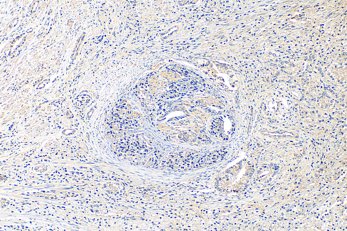
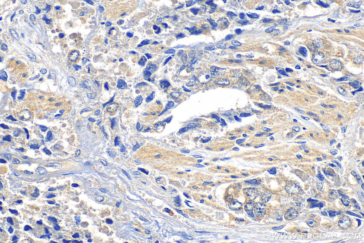
| Erfolgreiche Detektion in IHC | humanes Prostatahyperplasie-Gewebe Hinweis: Antigendemaskierung mit TE-Puffer pH 9,0 empfohlen. (*) Wahlweise kann die Antigendemaskierung auch mit Citratpuffer pH 6,0 erfolgen. |
Empfohlene Verdünnung
| Anwendung | Verdünnung |
|---|---|
| Western Blot (WB) | WB : 1:1000-1:5000 |
| Immunhistochemie (IHC) | IHC : 1:50-1:500 |
| It is recommended that this reagent should be titrated in each testing system to obtain optimal results. | |
| Sample-dependent, check data in validation data gallery | |
Produktinformation
30733-1-AP bindet in WB, IHC, ELISA FAT1 und zeigt Reaktivität mit human
| Getestete Reaktivität | human |
| Wirt / Isotyp | Kaninchen / IgG |
| Klonalität | Polyklonal |
| Typ | Antikörper |
| Immunogen | FAT1 fusion protein Ag33957 |
| Vollständiger Name | FAT tumor suppressor homolog 1 (Drosophila) |
| Beobachtetes Molekulargewicht | 506-600 kDa, 85 kDa |
| Gene symbol | FAT1 |
| Gene ID (NCBI) | 2195 |
| Konjugation | Unkonjugiert |
| Form | Liquid |
| Reinigungsmethode | Antigen-Affinitätsreinigung |
| Lagerungspuffer | PBS with 0.02% sodium azide and 50% glycerol |
| Lagerungsbedingungen | Bei -20°C lagern. Nach dem Versand ein Jahr lang stabil Aliquotieren ist bei -20oC Lagerung nicht notwendig. 20ul Größen enthalten 0,1% BSA. |
Hintergrundinformationen
FAT1, also known as hFat1, belongs to a member of the cadherin superfamily, has been proposed to play roles in cerebral development, glomerular slit formation, and also to act as a tumor suppressor, but its mechanisms of action have not been elucidated. It is expected to be located in cell membrane and nucleus, which is expressed in many epithelial and some endothelial and smooth muscle cells. The calculated molecular weight of FAT1 is 506 kDa and there is glycosylation modification of the protein. To examine functions of the transmembrane and cytoplasmic domains, they were expressed in HEK-293 and HeLa cells as chimeric proteins in fusion with EGFP and extracellular domains derived from E-cadherin. Proteins comprising the transmembrane domain localized to the membrane fraction (PMID: 15922730, 26373379).The high molecular mass band observed at the expected 500-kDa size of FAT1 is indicated by an arrow together with a prominent band at 85 kDa (PMID: 21680732).
Protokolle
| PRODUKTSPEZIFISCHE PROTOKOLLE | |
|---|---|
| WB protocol for FAT1 antibody 30733-1-AP | Protokoll herunterladen |
| IHC protocol for FAT1 antibody 30733-1-AP | Protokoll herunterladenl |
| STANDARD-PROTOKOLLE | |
|---|---|
| Klicken Sie hier, um unsere Standardprotokolle anzuzeigen |


