- Featured Product
- KD/KO Validated
Fibronectin Polyklonaler Antikörper
Fibronectin Polyklonal Antikörper für WB, IF/ICC, FC (Intra), IP, ELISA
Wirt / Isotyp
Kaninchen / IgG
Getestete Reaktivität
human, Maus, Ratte und mehr (5)
Anwendung
WB, IF/ICC, FC (Intra), IP, ELISA
Konjugation
Unkonjugiert
Kat-Nr. : 15613-1-AP
Synonyme
 with sh-Control and sh-Fibronectin transfected NIH/3T3 cells.")
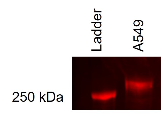
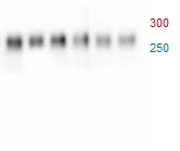
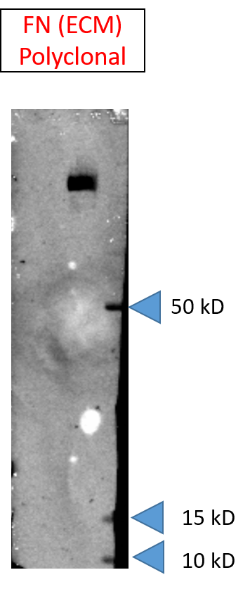
 at dilution of 1:20000 incubated at room temperature for 1.5 hours.")
 at dilution of 1:50000 incubated at room temperature for 1.5 hours.")
 at dilution of 1:300 incubated at room temperature for 1.5 hours.")
 with NIH/3T3 cells lysate 1680 ug.")
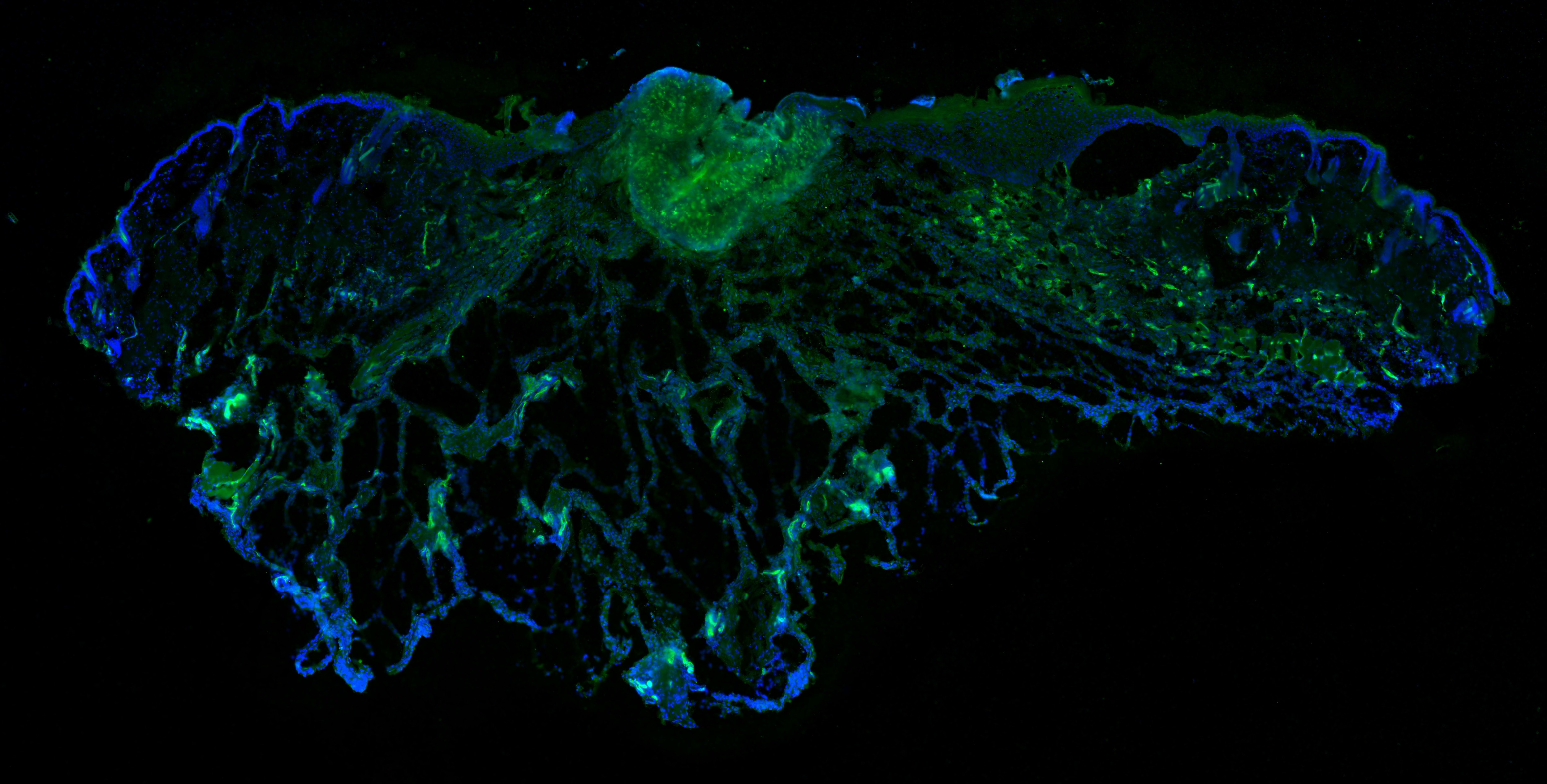
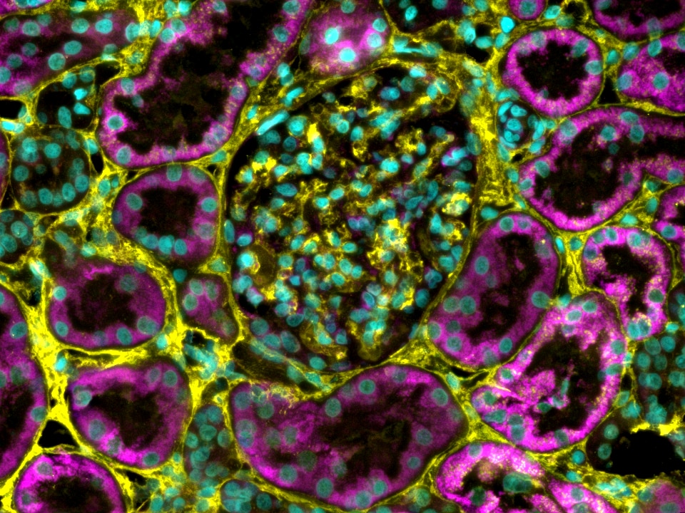
 at dilution of 1:4000 (under 20x lens). Heat mediated antigen retrieval with Tris-EDTA buffer (pH 9.0).")
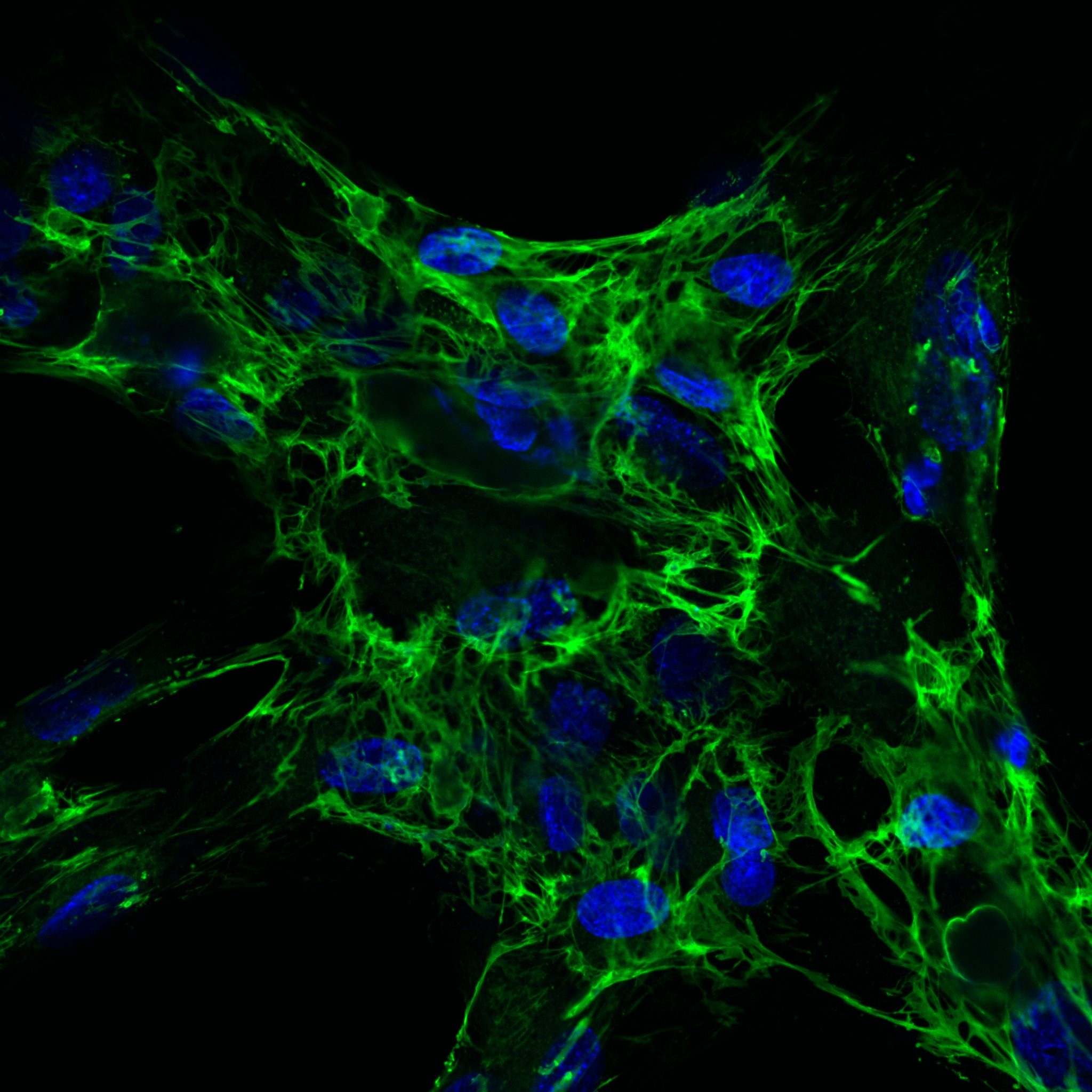
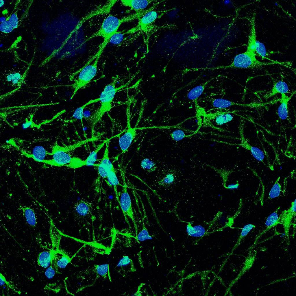
 fixed NIH/3T3 cells using Fibronectin antibody (15613-1-AP) at dilution of 1:200 and CoraLite®488-Conjugated AffiniPure Goat Anti-Rabbit IgG(H+L), CL594-Phalloidin (red).")
 fixed NIH/3T3 cells using Fibronectin antibody (15613-1-AP) at dilution of 1:200 and Multi-rAb CoraLite ® Plus 488-Goat Anti-Rabbit Recombinant Secondary Antibody (H+L) (RGAR002).")
 fixed NIH/3T3 cells using 15613-1-AP (Fibronectin antibody) at dilution of 1:50 and Alexa Fluor 488-conjugated AffiniPure Goat Anti-Rabbit IgG(H+L).")
 fixed NIH/3T3 cells using Fibronectin antibody (15613-1-AP) at dilution of 1:100 and CoraLite®488-Conjugated AffiniPure Goat Anti-Rabbit IgG(H+L).")
 and CoraLite®488-Conjugated AffiniPure Goat Anti-Rabbit IgG(H+L) at dilution 1:1000 (red), or 0.4 ug rabbit IgG isotype control (blue). Cells were fixed with 4% PFA and permeabilized with Flow Cytometry Perm Buffer (PF00011-C).")
Geprüfte Anwendungen
| Erfolgreiche Detektion in WB | human plasma, MDA-MB-453s-Zellen, NIH/3T3-Zellen |
| Erfolgreiche IP | NIH/3T3-Zellen |
| Erfolgreiche Detektion in IHC | human normal colon Hinweis: Antigendemaskierung mit TE-Puffer pH 9,0 empfohlen. (*) Wahlweise kann die Antigendemaskierung auch mit Citratpuffer pH 6,0 erfolgen. |
| Erfolgreiche Detektion in IF/ICC | NIH/3T3-Zellen |
| Erfolgreiche Detektion in FC (Intra) | NIH/3T3-Zellen |
Empfohlene Verdünnung
| Anwendung | Verdünnung |
|---|---|
| Western Blot (WB) | WB : 1:2000-1:20000 |
| Immunpräzipitation (IP) | IP : 0.5-4.0 ug for 1.0-3.0 mg of total protein lysate |
| Immunhistochemie (IHC) | IHC : 1:2000-1:8000 |
| Immunfluoreszenz (IF)/ICC | IF/ICC : 1:50-1:500 |
| Durchflusszytometrie (FC) (INTRA) | FC (INTRA) : 0.40 ug per 10^6 cells in a 100 µl suspension |
| It is recommended that this reagent should be titrated in each testing system to obtain optimal results. | |
| Sample-dependent, check data in validation data gallery | |
Veröffentlichte Anwendungen
| KD/KO | See 9 publications below |
| WB | See 636 publications below |
| IHC | See 2 publications below |
| IF | See 179 publications below |
| IP | See 2 publications below |
| ELISA | See 2 publications below |
Produktinformation
15613-1-AP bindet in WB, IF/ICC, FC (Intra), IP, ELISA Fibronectin und zeigt Reaktivität mit human, Maus, Ratten
| Getestete Reaktivität | human, Maus, Ratte |
| In Publikationen genannte Reaktivität | human, hamster, Hausschwein, Kaninchen, Maus, Ratte, Rind, Medaka-Embryos |
| Wirt / Isotyp | Kaninchen / IgG |
| Klonalität | Polyklonal |
| Typ | Antikörper |
| Immunogen | Fibronectin fusion protein Ag8004 |
| Vollständiger Name | fibronectin 1 |
| Berechnetes Molekulargewicht | 2386 aa, 263 kDa |
| Beobachtetes Molekulargewicht | 250-275 kDa |
| GenBank-Zugangsnummer | BC005858 |
| Gene symbol | Fibronectin |
| Gene ID (NCBI) | 2335 |
| Konjugation | Unkonjugiert |
| Form | Liquid |
| Reinigungsmethode | Antigen-Affinitätsreinigung |
| Lagerungspuffer | PBS with 0.02% sodium azide and 50% glycerol |
| Lagerungsbedingungen | Bei -20°C lagern. Nach dem Versand ein Jahr lang stabil Aliquotieren ist bei -20oC Lagerung nicht notwendig. 20ul Größen enthalten 0,1% BSA. |
Hintergrundinformationen
Fibronectins play a role in cell adhesion, motility, wound healing, and the maintenance of cell shape. Fibronectins bind to cell surfaces via interactions with integrins and various molecules such as collagen, fibrin, heparin, DNA, and actin (PMID: 3326130). Cellular interaction with fibronectin (FN1) promotes cell cycle progression and proliferation by influencing cyclins.
What is the molecular weight of FN1?
The molecular weight of FN1 is 220 kDa.
What is the tissue specificity of FN1?
FN1 is a large glycoprotein that exists in both a soluble form in plasma (plasma FN1) and other body fluids and an insoluble form in the extracellular matrix (cellular FN1) (PMID: 21923916). Plasma FN1, or the dimeric form, is secreted by hepatocytes, whereas cellular FN1, the dimeric or cross-linked multimeric form, is produced by fibroblasts and epithelial cells and deposited as fibrils in the extracellular matrix.
What are the post-translational modifications of FN1?
FN1 is often sulfated and its C-terminal NC1 peptide, anastellin, is produced as a result of proteolytic processing and can inhibit tumor growth, angiogenesis, and metastasis by activating p38 MAPK and inhibiting lysophospholipid signaling (PMID: 11209058).
What is FN1's role in embryogenesis?
Fibronectin has a crucial role in the development of the left-right body axis during embryogenesis (PMID: 21466802).
What is FN1's involvement in disease?
Fibronectin glomerulopathy is a kidney disease that eventually leads to end-stage renal disease and is caused by mutations in the FN1 gene. Mutations in FN1 lead to the production of abnormal fibronectin protein that is deposited in the glomeruli of the kidney (PMID: 18268355).
Protokolle
| PRODUKTSPEZIFISCHE PROTOKOLLE | |
|---|---|
| WB protocol for Fibronectin antibody 15613-1-AP | Protokoll herunterladen |
| IHC protocol for Fibronectin antibody 15613-1-AP | Protokoll herunterladenl |
| IF protocol for Fibronectin antibody 15613-1-AP | Protokoll herunterladen |
| IP protocol for Fibronectin antibody 15613-1-AP | Protokoll herunterladen |
| STANDARD-PROTOKOLLE | |
|---|---|
| Klicken Sie hier, um unsere Standardprotokolle anzuzeigen |
Publikationen
| Species | Application | Title |
|---|---|---|
Adv Mater Neonatal Tissue-derived Extracellular Vesicle Therapy (NEXT): A Potent Strategy for Precision Regenerative Medicine | ||
Cell Metab The phytochemical hyperforin triggers thermogenesis in adipose tissue via a Dlat-AMPK signaling axis to curb obesity. | ||
J Clin Invest Mitochondrial dysfunction in macrophages promotes inflammation and suppresses repair after myocardial infarction | ||
Nat Protoc Responsive and activable nanomedicines for remodeling the tumor microenvironment. | ||
Nat Commun Schwann cells regulate tumor cells and cancer-associated fibroblasts in the pancreatic ductal adenocarcinoma microenvironment | ||
Acta Pharm Sin B KCTD4 interacts with CLIC1 to disrupt calcium homeostasis and promote metastasis in esophageal cancer |
Rezensionen
The reviews below have been submitted by verified Proteintech customers who received an incentive for providing their feedback.
FH Zach (Verified Customer) (08-29-2025) | This antibody works great with chronic pancreatitis samples.
|
FH Ilaria (Verified Customer) (08-11-2025) | The fibronectin antibody performed very well, providing a strong and clear signal even at a higher dilution of 1:500. It was tested on mouse brain samples that were snap-frozen after collection using dry ice and 2-methylbutane, then post-fixed with acetone and ethanol. Blood vessel structures were clearly visualised, and the antibody also allowed for the assessment of BBB leakage.
|
FH Angie (Verified Customer) (07-26-2025) | WB showed a single band above 250 kDa at dilution of 1:500 incubated overnight at 4 °C. Secondary antibody: Donkey-anti-rabbit (Alexa Fluor 680) at 1:20000 dilution incubated at room temperature for 1 hour.
 |
FH Christelle (Verified Customer) (12-19-2024) | Immunofluorescence analysis of (PFA 4%) fixed and (BSA 1%, Saponin 0.1%) permeabilized HRMC cells using the Fibronectin antibody #15613-1-AP at dilution 1:100 and goat anti-rabbit antibody AF488 labelled, Abberior STAR Red membrane probe (yellow), Dapi (nucleus, blue). Find attached 2 files : - merged channels: dapi, green - merged channels: dapi, green, yellow
 |
FH Ruchi (Verified Customer) (06-02-2023) | The Ab was tried on mouse skin on diabetic wounds.
 |
FH PK (Verified Customer) (03-20-2023) | excellent
 |
FH Susanne (Verified Customer) (08-23-2022) | under reduced conditions, blocked with 5% milk powder, incubation over night 4°C
 |
FH Pankhuri (Verified Customer) (08-19-2022) | I tried this antibody for IF in neural stem cells. It works great in the conc. 1:200.
 |
FH Ren Jie (Verified Customer) (07-11-2022) | Very clean bands near 220 kD indicating fibronectin monomer
 |
FH SD (Verified Customer) (07-08-2022) | Good
 |
FH zunyi (Verified Customer) (03-11-2022) | This ab is used for immunohistochemistry on mouse paraffin-embedded sections. Antigen-retrieve, using pH 6.0 citric buffer, was performed prior to staining.
|
FH Lindsey (Verified Customer) (05-24-2021) | Works beautifully! Looking forward to working more with this antibody in different tissue/prep types
 |

