- Featured Product
- KD/KO Validated
c-Fos Monoklonaler Antikörper
c-Fos Monoklonal Antikörper für WB, ELISA
Wirt / Isotyp
Maus / IgG1
Getestete Reaktivität
human, Maus, Ratte und mehr (1)
Anwendung
WB, IHC, IP, CoIP, ELISA
Konjugation
Unkonjugiert
CloneNo.
1G2C5
Kat-Nr. : 66590-1-Ig
Synonyme
 at dilution of 1:10000 incubated at room temperature for 1.5 hours.")
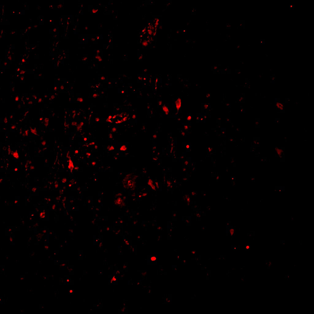
 with sh-Control and sh-c-Fos transfected HepG2 cells.")
 at dilution of 1:10000 incubated at room temperature for 1.5 hours.")
 at dilution of 1:5000 incubated at room temperature for 1.5 hours.")
 at dilution of 1:5000 incubated at room temperature for 1.5 hours.")
Geprüfte Anwendungen
| Erfolgreiche Detektion in WB | HeLa-Zellen, HepG2-Zellen, Jurkat-Zellen, K-562-Zellen, NIH/3T3-Zellen, RAW 264.7-Zellen, THP-1-Zellen, U-937-Zellen |
Empfohlene Verdünnung
| Anwendung | Verdünnung |
|---|---|
| Western Blot (WB) | WB : 1:5000-1:50000 |
| It is recommended that this reagent should be titrated in each testing system to obtain optimal results. | |
| Sample-dependent, check data in validation data gallery | |
Veröffentlichte Anwendungen
| KD/KO | See 3 publications below |
| WB | See 87 publications below |
| IHC | See 14 publications below |
| IP | See 3 publications below |
| CoIP | See 1 publications below |
Produktinformation
66590-1-Ig bindet in WB, IHC, IP, CoIP, ELISA c-Fos und zeigt Reaktivität mit human, Maus, Ratten
| Getestete Reaktivität | human, Maus, Ratte |
| In Publikationen genannte Reaktivität | human, Kaninchen, Maus, Ratte |
| Wirt / Isotyp | Maus / IgG1 |
| Klonalität | Monoklonal |
| Typ | Antikörper |
| Immunogen | c-Fos fusion protein Ag24340 |
| Vollständiger Name | FOS |
| Berechnetes Molekulargewicht | 41 kDa |
| Beobachtetes Molekulargewicht | 55-60 kDa |
| GenBank-Zugangsnummer | BC004490 |
| Gene symbol | c-Fos |
| Gene ID (NCBI) | 2353 |
| Konjugation | Unkonjugiert |
| Form | Liquid |
| Reinigungsmethode | Protein-G-Reinigung |
| Lagerungspuffer | PBS with 0.02% sodium azide and 50% glycerol |
| Lagerungsbedingungen | Bei -20°C lagern. Nach dem Versand ein Jahr lang stabil Aliquotieren ist bei -20oC Lagerung nicht notwendig. 20ul Größen enthalten 0,1% BSA. |
Hintergrundinformationen
c-Fos, also named as FOS and G0/G1 switch regulatory protein 7, is a 380 amino acid protein, which contains 1 bZIP (basic-leucine zipper) domain and belongs to the bZIP family. c-Fos is expressed at very low levels in quiescent cells. When cells are stimulated to reenter growth, c-Fos undergo 2 waves of expression, the first one peaks 7.5 minutes following FBS induction. At this stage, the c-Fos protein is localized endoplasmic reticulum. The second wave of expression occurs at about 20 minutes after induction and peaks at 1 hour. At this stage, the c-FOS protein becomes nuclear. c-Fos is a very short-lived intracellular protein, which is very easy to degrade. The calculated molecular weight of c-Fos is 40 kDa, but Phosphorylated c-Fos protein is about 60-65 kDa. It is involved in important cellular events, including cell proliferation, differentiation and survival; genes associated with hypoxia; and angiogenesis; which makes its dysregulation an important factor for cancer development. It can also induce a loss of cell polarity and epithelial-mesenchymal transition, leading to invasive and metastatic growth in mammary epithelial cells. Expression of c-Fos is an indirect marker of neuronal activity because c-Fos is often expressed when neurons fire action potentials. Upregulation of c-Fos mRNA in a neuron indicates recent activity.
Protokolle
| PRODUKTSPEZIFISCHE PROTOKOLLE | |
|---|---|
| WB protocol for c-Fos antibody 66590-1-Ig | Protokoll herunterladen |
| STANDARD-PROTOKOLLE | |
|---|---|
| Klicken Sie hier, um unsere Standardprotokolle anzuzeigen |
Publikationen
| Species | Application | Title |
|---|---|---|
Adv Mater Noninvasive Optogenetics Realized by iPSC-Derived Tentacled Carrier in Alzheimer's Disease Treatment | ||
Theranostics KDM6A promotes imatinib resistance through YY1-mediated transcriptional upregulation of TRKA independently of its demethylase activity in chronic myelogenous leukemia.
| ||
Phytomedicine Withaferin A protects against epilepsy by promoting LCN2-mediated astrocyte polarization to stopping neuronal ferroptosis | ||
Phytomedicine Gastrodin alleviates NTG-induced migraine-like pain via inhibiting succinate/HIF-1α/TRPM2 signaling pathway in trigeminal ganglion | ||
J Transl Med Integrating spatial transcriptomics and single-cell RNA-sequencing reveals the alterations in epithelial cells during nodular formation in benign prostatic hyperplasia |
Rezensionen
The reviews below have been submitted by verified Proteintech customers who received an incentive for providing their feedback.
FH Reyes (Verified Customer) (02-14-2025) | cFOS (a nuclear marker) did not perform as expected in my epileptic human FFPE tissue. It seems to appear in the nucleus in a few cells, but also a strong marking in the cyoplasm of others.
 |
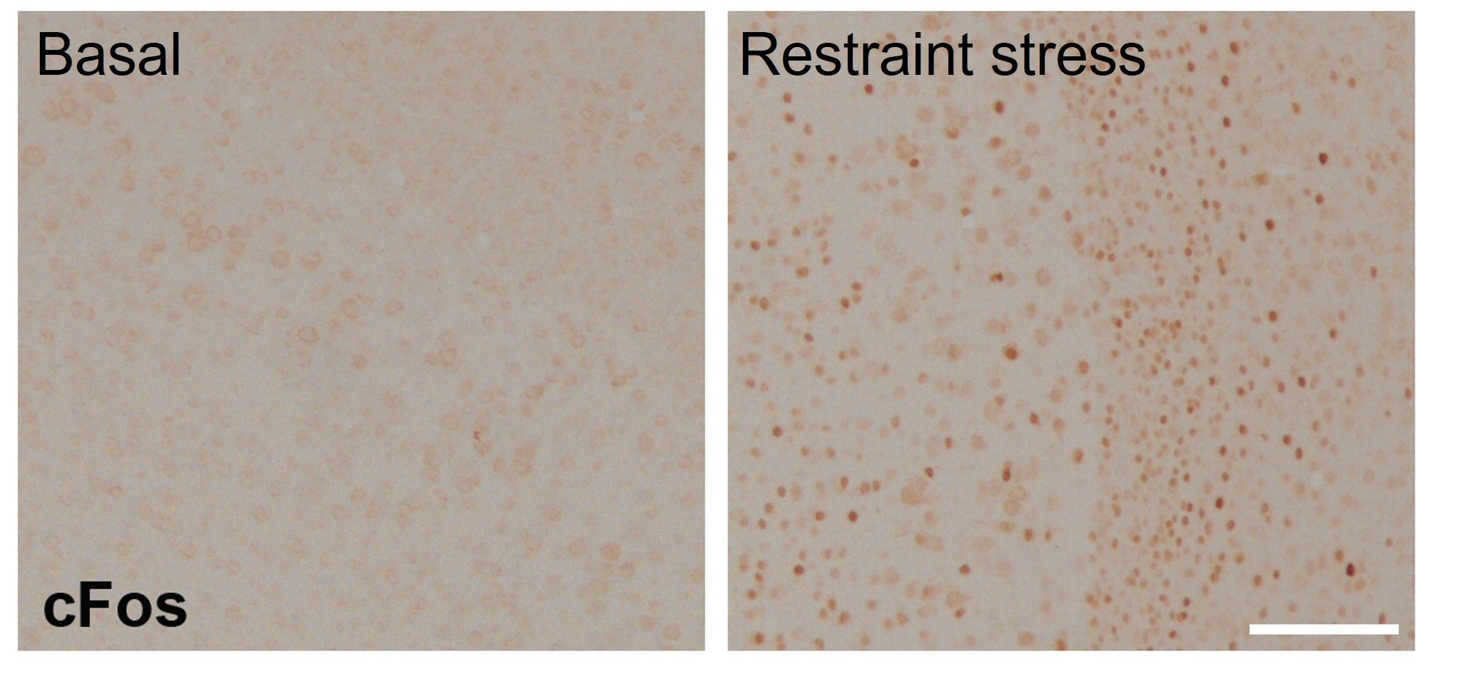
FH Tatyana (Verified Customer) (01-21-2023) | Suitable for IHC in the paraffinised brain sections of mice (cortex). Samples were fixed in 4% and standard IHC procedure with antigen retrieval and DAB detection was performed. Antibody was incubated at 1:1000 dilution overnight at 4C. Provided a good specific signal in neurons that increased after restraint stress.
 |

