- Featured Product
- KD/KO Validated
G3BP2 Polyklonaler Antikörper
G3BP2 Polyklonal Antikörper für WB, IHC, IF/ICC, IP, ELISA
Wirt / Isotyp
Kaninchen / IgG
Getestete Reaktivität
human, Maus, Ratte
Anwendung
WB, IHC, IF/ICC, IP, CoIP, RIP, ELISA
Konjugation
Unkonjugiert
Kat-Nr. : 16276-1-AP
Synonyme
 at dilution of 1:8000 incubated at room temperature for 1.5 hours.")
 with sh-Control and sh-G3BP2 transfected HepG2 cells.")
 at dilution of 1:10000 incubated at room temperature for 1.5 hours.")
 at dilution of 1:6000 incubated at room temperature for 1.5 hours.")
 at dilution of 1:8000 incubated at room temperature for 1.5 hours.")
 with T47D breast cancer cells.")
 at dilution of 1:300 incubated at room temperature for 1.5 hours.")
 with HeLa cells lysate 2200 ug.")
 at dilution of 1:500 (under 10x lens). Heat mediated antigen retrieval with Tris-EDTA buffer (pH 9.0).")
 at dilution of 1:500 (under 40x lens). Heat mediated antigen retrieval with Tris-EDTA buffer (pH 9.0).")
 at dilution of 1:500 (under 10x lens). Heat mediated antigen retrieval with Tris-EDTA buffer (pH 9.0).")
 at dilution of 1:500 (under 40x lens). Heat mediated antigen retrieval with Tris-EDTA buffer (pH 9.0).")
 at dilution of 1:500 (under 10x lens). Heat mediated antigen retrieval with Tris-EDTA buffer (pH 9.0).")
 at dilution of 1:500 (under 40x lens). Heat mediated antigen retrieval with Tris-EDTA buffer (pH 9.0).")
 fixed sodium arsenite treated HeLa cells using G3BP2 antibody (16276-1-AP) at dilution of 1:600 and CoraLite®488-Conjugated AffiniPure Goat Anti-Rabbit IgG(H+L), CL594-Phalloidin (red).")
 fixed sodium arsenite treated HeLa cells using G3BP2 antibody (16276-1-AP) at dilution of 1:200 and CoraLite®488-Conjugated AffiniPure Goat Anti-Rabbit IgG(H+L), CL594-Phalloidin (red).")
Geprüfte Anwendungen
| Erfolgreiche Detektion in WB | A549-Zellen, C2C12-Zellen, C6-Zellen, HEK-293-Zellen, HeLa-Zellen, HepG2-Zellen, Jurkat-Zellen, MCF-7-Zellen, Neuro-2a-Zellen |
| Erfolgreiche IP | HeLa-Zellen |
| Erfolgreiche Detektion in IHC | humanes Lungenkarzinomgewebe, Mauslebergewebe, Rattennierengewebe Hinweis: Antigendemaskierung mit TE-Puffer pH 9,0 empfohlen. (*) Wahlweise kann die Antigendemaskierung auch mit Citratpuffer pH 6,0 erfolgen. |
| Erfolgreiche Detektion in IF/ICC | sodium arsenite treated HeLa cells |
Empfohlene Verdünnung
| Anwendung | Verdünnung |
|---|---|
| Western Blot (WB) | WB : 1:2000-1:16000 |
| Immunpräzipitation (IP) | IP : 0.5-4.0 ug for 1.0-3.0 mg of total protein lysate |
| Immunhistochemie (IHC) | IHC : 1:250-1:1000 |
| Immunfluoreszenz (IF)/ICC | IF/ICC : 1:50-1:500 |
| It is recommended that this reagent should be titrated in each testing system to obtain optimal results. | |
| Sample-dependent, check data in validation data gallery | |
Veröffentlichte Anwendungen
| KD/KO | See 6 publications below |
| WB | See 20 publications below |
| IHC | See 3 publications below |
| IF | See 12 publications below |
| IP | See 5 publications below |
| CoIP | See 2 publications below |
| RIP | See 1 publications below |
Produktinformation
16276-1-AP bindet in WB, IHC, IF/ICC, IP, CoIP, RIP, ELISA G3BP2 und zeigt Reaktivität mit human, Maus, Ratten
| Getestete Reaktivität | human, Maus, Ratte |
| In Publikationen genannte Reaktivität | human, Maus |
| Wirt / Isotyp | Kaninchen / IgG |
| Klonalität | Polyklonal |
| Typ | Antikörper |
| Immunogen | G3BP2 fusion protein Ag9355 |
| Vollständiger Name | GTPase activating protein (SH3 domain) binding protein 2 |
| Berechnetes Molekulargewicht | 482aa,54 kDa; 449aa,51 kDa |
| Beobachtetes Molekulargewicht | 65-70 kDa |
| GenBank-Zugangsnummer | BC011731 |
| Gene symbol | G3BP2 |
| Gene ID (NCBI) | 9908 |
| Konjugation | Unkonjugiert |
| Form | Liquid |
| Reinigungsmethode | Antigen-Affinitätsreinigung |
| Lagerungspuffer | PBS with 0.02% sodium azide and 50% glycerol |
| Lagerungsbedingungen | Bei -20°C lagern. Nach dem Versand ein Jahr lang stabil Aliquotieren ist bei -20oC Lagerung nicht notwendig. 20ul Größen enthalten 0,1% BSA. |
Hintergrundinformationen
Stress granules (SGs) are cytoplasmic mRNA-protein condensates formed in response to cellular stressors, such as oxidative stress, ultraviolet radiation, and viral infection (1). The Ras-GTPase-activating protein-binding proteins (G3BPs), consisting of G3BP1 and G3BP2, are key nucleating factors essential for SG formation. They function to protect RNAs from harmful conditions. G3BP2 is mainly distributed in the cytoplasm and participates in the formation of stress granules, cell differentiation, proliferation, and signal transduction. Accumulating evidence has demonstrated that aberrant expression of G3BP2 contributes to cancer initiation and progression, such as high expression of G3BP2 increasing cell stemness, metastasis and chemoresistance in breast cancer.
Protokolle
| PRODUKTSPEZIFISCHE PROTOKOLLE | |
|---|---|
| WB protocol for G3BP2 antibody 16276-1-AP | Protokoll herunterladen |
| IHC protocol for G3BP2 antibody 16276-1-AP | Protokoll herunterladenl |
| IF protocol for G3BP2 antibody 16276-1-AP | Protokoll herunterladen |
| IP protocol for G3BP2 antibody 16276-1-AP | Protokoll herunterladen |
| STANDARD-PROTOKOLLE | |
|---|---|
| Klicken Sie hier, um unsere Standardprotokolle anzuzeigen |
Publikationen
| Species | Application | Title |
|---|---|---|
Cell Diverse CMT2 neuropathies are linked to aberrant G3BP interactions in stress granules | ||
Cell G3BP1 Is a Tunable Switch that Triggers Phase Separation to Assemble Stress Granules. | ||
Cell Rep hnRNPA2B1 represses the disassembly of arsenite-induced stress granules and is essential for male fertility | ||
Oncogene HDAC6-G3BP2 promotes lysosomal-TSC2 and suppresses mTORC1 under ETV4 targeting-induced low-lactate stress in non-small cell lung cancer
| ||
Oncogene RIOK1 mediates p53 degradation and radioresistance in colorectal cancer through phosphorylation of G3BP2.
| ||
Mol Cancer Invasion-related circular RNA circFNDC3B inhibits bladder cancer progression through the miR-1178-3p/G3BP2/SRC/FAK axis.
|
Rezensionen
The reviews below have been submitted by verified Proteintech customers who received an incentive for providing their feedback.
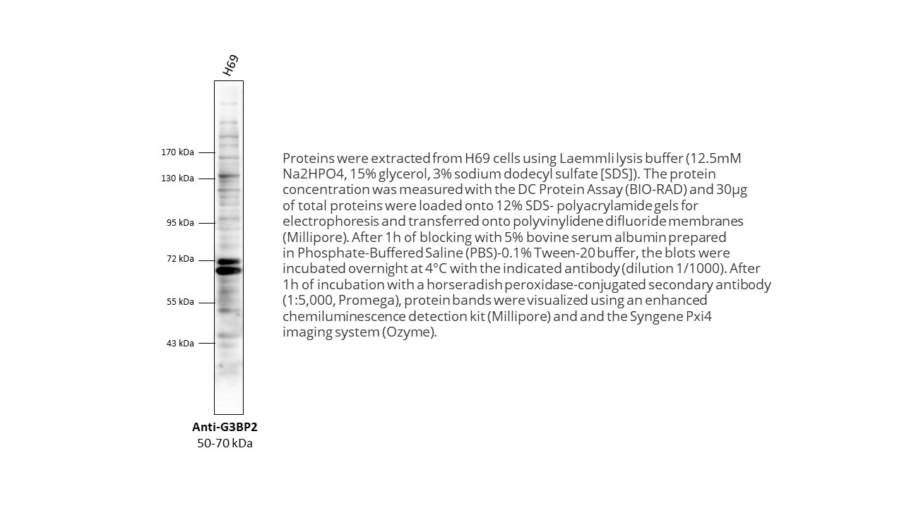
FH Karine (Verified Customer) (09-22-2022) | Proteins were extracted from H69 cells using Laemmli lysis buffer (12.5mM Na2HPO4, 15% glycerol, 3% sodium dodecyl sulfate [SDS]). The protein concentration was measured with the DC Protein Assay (BIO-RAD) and 30μg of total proteins were loaded onto 12% SDS- polyacrylamide gels for electrophoresis and transferred onto polyvinylidene difluoride membranes (Millipore). After 1h of blocking with 5% bovine serum albumin prepared in Phosphate-Buffered Saline (PBS)-0.1% Tween-20 buffer, the blots were incubated overnight at 4°C with the indicated antibody (dilution 1/1000). After 1h of incubation with a horseradish peroxidase-conjugated secondary antibody (1:5,000, Promega), protein bands were visualized using an enhanced chemiluminescence detection kit (Millipore) and and the Syngene Pxi4 imaging system (Ozyme).
 |

