Phospho-Histone H3 (Ser10) Monoklonaler Antikörper
Phospho-Histone H3 (Ser10) Monoklonal Antikörper für WB, IF/ICC, FC (Intra), ELISA
Wirt / Isotyp
Maus / IgG1
Getestete Reaktivität
human und mehr (3)
Anwendung
WB, IHC, IF/ICC, FC (Intra), ELISA
Konjugation
Unkonjugiert
CloneNo.
4C7G2
Kat-Nr. : 66863-1-Ig
Synonyme
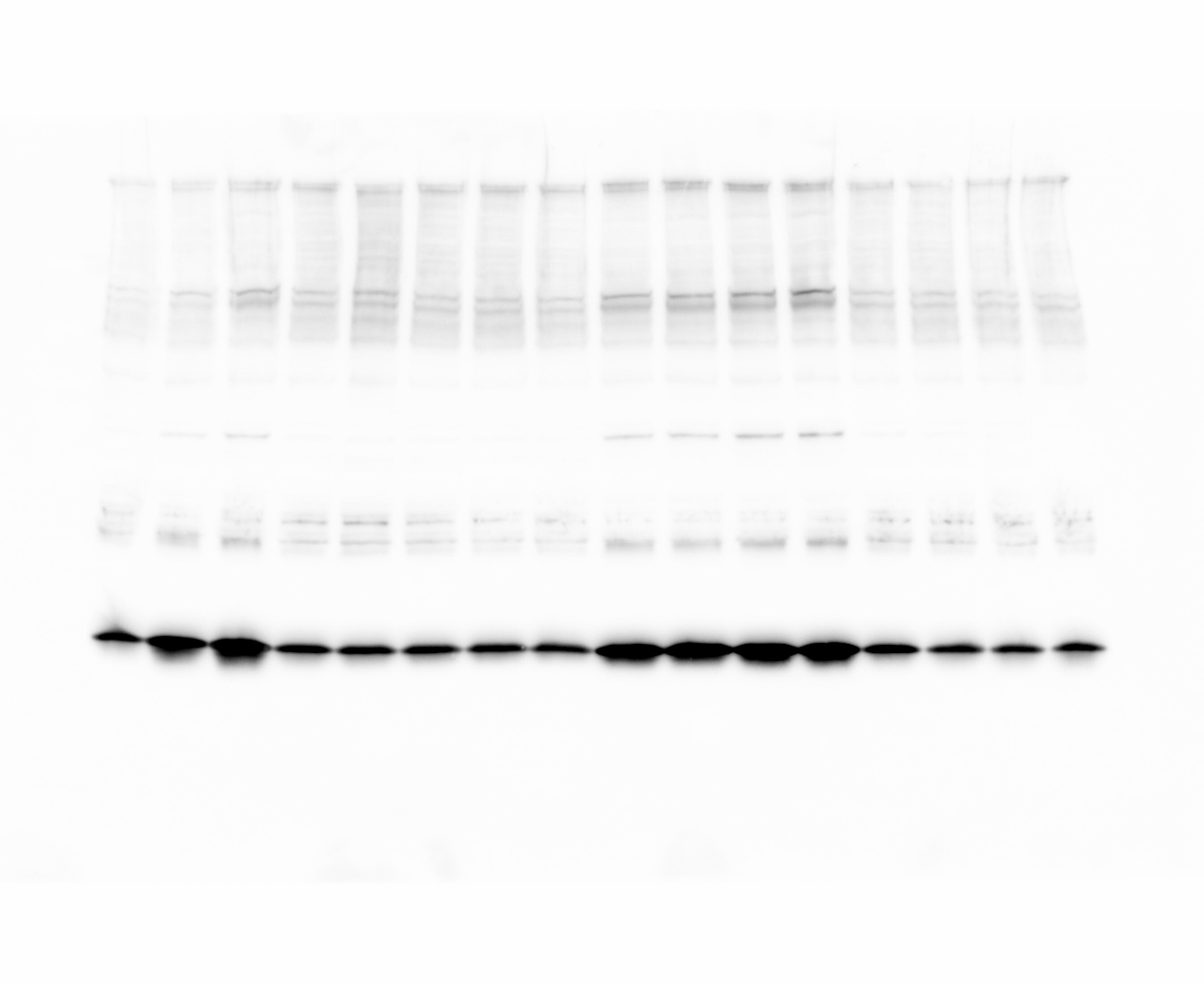
 antibody) at dilution of 1:10000 incubated at room temperature for 1.5 hours. The membrane was stripped and reblotted with HRP-conjugated GAPDH Monoclonal antibody (HRP-60004) as loading control.")
 antibody) at dilution of 1:3000 incubated at room temperature for 1.5 hours. The membrane was stripped and reblotted with HRP-conjugated GAPDH Monoclonal antibody (HRP-60004) as loading control.")
 antibody) at dilution of 1:4000 (under 40x lens). Heat mediated antigen retrieval with Tris-EDTA buffer (pH 9.0).")
 fixed C2C12 cells using Phospho-Histone H3 (Ser10) antibody (66863-1-Ig, Clone: 4C7G2 ) at dilution of 1:1200 and CoraLite®594-Conjugated AffiniPure Goat Anti-Mouse IgG(H+L), Alpha Tubulin antibody (11224-1-AP, green).")
 fixed HeLa cells using Phospho-Histone H3 (Ser10) antibody (66863-1-Ig, Clone: 4C7G2 ) at dilution of 1:1000 and CoraLite®488-Conjugated AffiniPure Goat Anti-Mouse IgG(H+L) (SA00013-1), Beta Tubulin antibody (80713-1-RR, Clone: 2O13, red).")
 fixed HeLa cells using Phospho-Histone H3 (Ser10) antibody (66863-1-Ig, Clone: 4C7G2 ) at dilution of 1:1500 and CoraLite®594-Conjugated AffiniPure Goat Anti-Mouse IgG(H+L), Alpha Tubulin antibody (11224-1-AP, green).")
 fixed MCF-7 cells using Phospho-Histone H3 (Ser10) antibody (66863-1-Ig, Clone: 4C7G2 ) at dilution of 1:1500 and CoraLite®594-Conjugated AffiniPure Goat Anti-Mouse IgG(H+L), Alpha Tubulin antibody (11224-1-AP, green).")
 Monoclonal antibody (66863-1-Ig, Clone:4C7G2) and CoraLite®488-Conjugated Goat Anti-Mouse IgG(H+L) (SA00013-1), and 0.25 ug Mouse IgG1 isotype control Mouse McAb (66360-1-Ig, Clone: 1F8D3). Cells were fixed with 4% PFA and permeabilized with 90% MeOH.")
Geprüfte Anwendungen
| Erfolgreiche Detektion in WB | HeLa-Zellen, HEK-293-Zellen, Jurkat-Zellen |
| Erfolgreiche Detektion in IF/ICC | C2C12-Zellen, HeLa-Zellen, MCF-7-Zellen |
| Erfolgreiche Detektion in FC (Intra) | nocodazole treated HeLa cells |
Empfohlene Verdünnung
| Anwendung | Verdünnung |
|---|---|
| Western Blot (WB) | WB : 1:5000-1:50000 |
| Immunfluoreszenz (IF)/ICC | IF/ICC : 1:600-1:2400 |
| Durchflusszytometrie (FC) (INTRA) | FC (INTRA) : 0.25 ug per 10^6 cells in a 100 µl suspension |
| It is recommended that this reagent should be titrated in each testing system to obtain optimal results. | |
| Sample-dependent, check data in validation data gallery | |
Veröffentlichte Anwendungen
| WB | See 10 publications below |
| IHC | See 3 publications below |
| IF | See 11 publications below |
Produktinformation
66863-1-Ig bindet in WB, IHC, IF/ICC, FC (Intra), ELISA Phospho-Histone H3 (Ser10) und zeigt Reaktivität mit human
| Getestete Reaktivität | human |
| In Publikationen genannte Reaktivität | human, Huhn, Maus, Ratte |
| Wirt / Isotyp | Maus / IgG1 |
| Klonalität | Monoklonal |
| Typ | Antikörper |
| Immunogen | Peptid |
| Vollständiger Name | histone cluster 1, H3a |
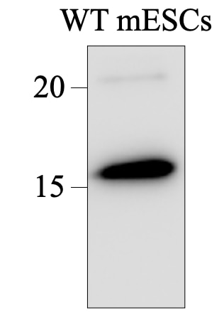
| Berechnetes Molekulargewicht | 15 kDa |
| Beobachtetes Molekulargewicht | 15-17 kDa |
| GenBank-Zugangsnummer | NM_003529 |
| Gene symbol | HIST1H3A |
| Gene ID (NCBI) | 8350 |
| Konjugation | Unkonjugiert |
| Form | Liquid |
| Reinigungsmethode | Protein-G-Reinigung |
| Lagerungspuffer | PBS with 0.02% sodium azide and 50% glycerol |
| Lagerungsbedingungen | Bei -20°C lagern. Nach dem Versand ein Jahr lang stabil Aliquotieren ist bei -20oC Lagerung nicht notwendig. 20ul Größen enthalten 0,1% BSA. |
Hintergrundinformationen
Phospho-histone-H3 (PHH3) is a core histone protein, which in its phosphorylated state forms the principal constituents of eukaryotic chromatin, with histone H3 being phosphorylated at serine (Ser) 10 or Ser28 as well as its phosphorylation of Ser10 being strongly correlated with the late G2 to M-phase transition in mammalian mitotic cells. On the basis of previous research, a few cell line- and animal model-based researches have displayed an increase in phosphorylation of histone H3 at Ser10 (H3S10ph), the only histone marker that is involved in carcinogenesis and cellular transformation. Histone H3 phosphorylation on serine-10 is specific to mitosis and phosphorylated histone H3 (PHH3) proliferation markers (as counts defined per area or as indices defined per cell numbers) are increasingly being used to evaluate proliferation in various tumors.
Protokolle
| PRODUKTSPEZIFISCHE PROTOKOLLE | |
|---|---|
| WB protocol for Phospho-Histone H3 (Ser10) antibody 66863-1-Ig | Protokoll herunterladen |
| IHC protocol for Phospho-Histone H3 (Ser10) antibody 66863-1-Ig | Protokoll herunterladenl |
| IF protocol for Phospho-Histone H3 (Ser10) antibody 66863-1-Ig | Protokoll herunterladen |
| STANDARD-PROTOKOLLE | |
|---|---|
| Klicken Sie hier, um unsere Standardprotokolle anzuzeigen |
Publikationen
| Species | Application | Title |
|---|---|---|
Oncogene Oncogenic long intervening noncoding RNA Linc00284 promotes c-Met expression by sponging miR-27a in colorectal cancer. | ||
EMBO Rep Palmitoylated importin α regulates mitotic spindle orientation through interaction with NuMA | ||
iScience Centrosomal protein 120 promotes centrosome amplification and gastric cancer progression via USP54-mediated deubiquitination of PLK4 | ||
Biochem Pharmacol A novel aromatic amide derivative SY-65 co-targeted tubulin and histone deacetylase 1 with potent anticancer activity in vitro and in vivo. | ||
Front Mol Biosci High-Resolution Imaging of Tumor Spheroids and Organoids Enabled by Expansion Microscopy. | ||
Sci Rep NF90-NF45 is essential for β cell compensation under obesity-inducing metabolic stress through suppression of p53 signaling pathway. |
Rezensionen
The reviews below have been submitted by verified Proteintech customers who received an incentive for providing their feedback.
FH Charlotte (Verified Customer) (09-19-2024) | Blocking and dilution in seablock. Antibody very strong given the dilution and the bands intensity. Very satisfied.
 |
FH Tsimafei (Verified Customer) (08-04-2024) | Membrane was incubated at 4C, ON
 |
FH Wissem (Verified Customer) (06-27-2024) | This antibody works great at detecting phosphorylation of Histone H3 at S10; conditions - 1:10,000 overnight at 4C; used to assess cell cycle synchronisation in n extracts from U2OS, HCT116, HeLa, RPE1 and HEK293 cells
|

