IFFO1 Polyklonaler Antikörper
IFFO1 Polyklonal Antikörper für WB, IHC, IF/ICC, ELISA
Wirt / Isotyp
Kaninchen / IgG
Getestete Reaktivität
human, Maus, Ratte
Anwendung
WB, IHC, IF/ICC, ELISA
Konjugation
Unkonjugiert
Kat-Nr. : 16041-1-AP
Synonyme
Geprüfte Anwendungen
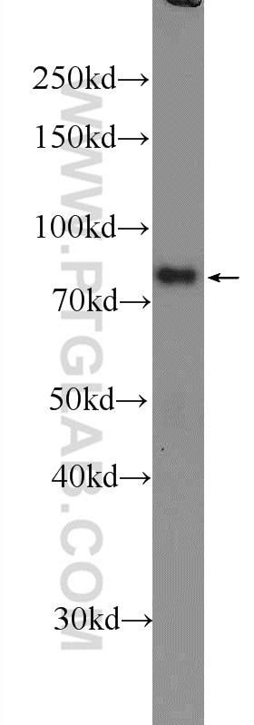
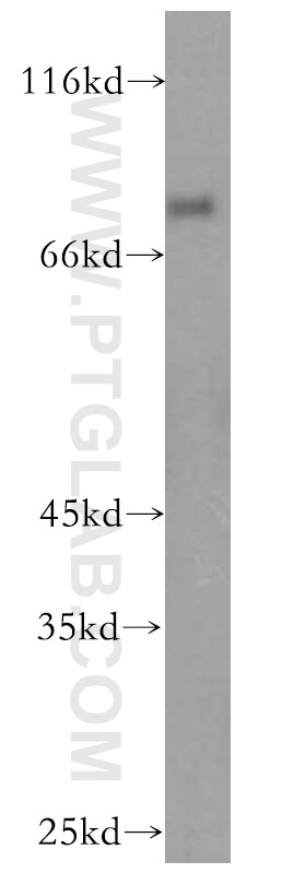
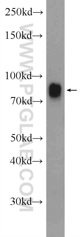
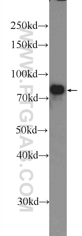
| Erfolgreiche Detektion in WB | Maushirngewebe, humanes Hirngewebe, Maushodengewebe |
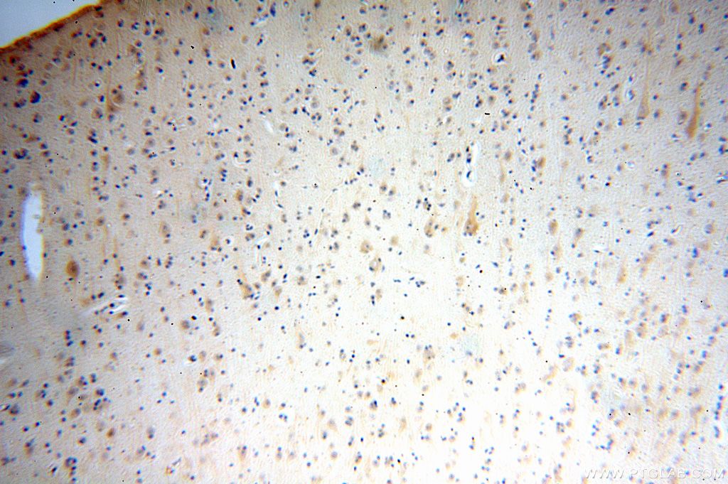
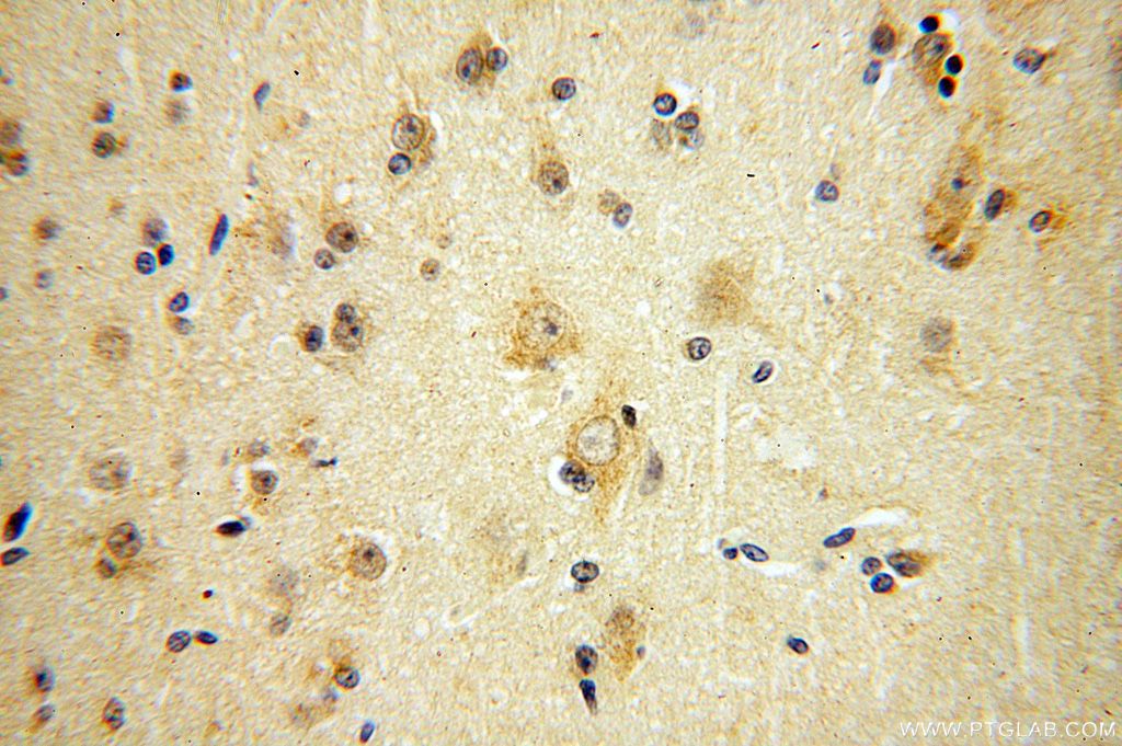
| Erfolgreiche Detektion in IHC | humanes Hirngewebe Hinweis: Antigendemaskierung mit TE-Puffer pH 9,0 empfohlen. (*) Wahlweise kann die Antigendemaskierung auch mit Citratpuffer pH 6,0 erfolgen. |
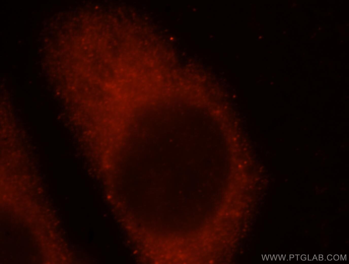
| Erfolgreiche Detektion in IF/ICC | HeLa-Zellen |
Empfohlene Verdünnung
| Anwendung | Verdünnung |
|---|---|
| Western Blot (WB) | WB : 1:500-1:2000 |
| Immunhistochemie (IHC) | IHC : 1:20-1:200 |
| Immunfluoreszenz (IF)/ICC | IF/ICC : 1:10-1:100 |
| It is recommended that this reagent should be titrated in each testing system to obtain optimal results. | |
| Sample-dependent, check data in validation data gallery | |
Veröffentlichte Anwendungen
| IHC | See 1 publications below |
Produktinformation
16041-1-AP bindet in WB, IHC, IF/ICC, ELISA IFFO1 und zeigt Reaktivität mit human, Maus, Ratten
| Getestete Reaktivität | human, Maus, Ratte |
| In Publikationen genannte Reaktivität | human |
| Wirt / Isotyp | Kaninchen / IgG |
| Klonalität | Polyklonal |
| Typ | Antikörper |
| Immunogen | IFFO1 fusion protein Ag8915 |
| Vollständiger Name | intermediate filament family orphan 1 |
| Berechnetes Molekulargewicht | 62 kDa |
| Beobachtetes Molekulargewicht | 75-85 kDa |
| GenBank-Zugangsnummer | BC004384 |
| Gene symbol | IFFO1 |
| Gene ID (NCBI) | 25900 |
| Konjugation | Unkonjugiert |
| Form | Liquid |
| Reinigungsmethode | Antigen-Affinitätsreinigung |
| Lagerungspuffer | PBS with 0.02% sodium azide and 50% glycerol |
| Lagerungsbedingungen | Bei -20°C lagern. Nach dem Versand ein Jahr lang stabil Aliquotieren ist bei -20oC Lagerung nicht notwendig. 20ul Größen enthalten 0,1% BSA. |
Hintergrundinformationen
IFFO1, also named as IFFO and Tumor antigen HOM-TES-103, is a member of the intermediate filament family which consists of primordial components of the cytoskeleton and nuclear envelope that are mainly distributed in the nucleus, followed by the cytoskeleton and cytoplasm. IFFO1 in the nucleus was identified to combine with both XRCC4 and the nuclear lamina protein lamin A/C to be involved in nonhomologous DNA end joining (NHEJ). The expression of IFFO1 is associated with tumor progression. IFFO1 could be used as a potential marker to differentiate malignant ascites cell-free tumor DNA (cftDNA) of ovarian cancer from benign controls.
Protokolle
| PRODUKTSPEZIFISCHE PROTOKOLLE | |
|---|---|
| WB protocol for IFFO1 antibody 16041-1-AP | Protokoll herunterladen |
| IHC protocol for IFFO1 antibody 16041-1-AP | Protokoll herunterladenl |
| IF protocol for IFFO1 antibody 16041-1-AP | Protokoll herunterladen |
| STANDARD-PROTOKOLLE | |
|---|---|
| Klicken Sie hier, um unsere Standardprotokolle anzuzeigen |
Rezensionen
The reviews below have been submitted by verified Proteintech customers who received an incentive for providing their feedback.
FH Shinford (Verified Customer) (10-26-2023) | It's essential to exercise caution while cutting the membrane as the bands appeared very close to the 70kDa line. In my case, I experienced the loss of some bands, possibly due to the cut being too close to the border.
|
FH Maria Jose (Verified Customer) (09-28-2020) | The antibody is very unspecific. There are many bands in the western blot. It recognizes GST with very high affinity.
|
