- Featured Product
- KD/KO Validated
JAK1 Monoklonaler Antikörper
JAK1 Monoklonal Antikörper für WB, IHC, ELISA
Wirt / Isotyp
Maus / IgG2b
Getestete Reaktivität
human, Maus, Ratte
Anwendung
WB, IHC, IF, IP, ELISA
Konjugation
Unkonjugiert
CloneNo.
3H7A8
Kat-Nr. : 66466-1-Ig
Synonyme
 at dilution of 1:3000 incubated at room temperature for 1.5 hours. The membrane was stripped and reblotted with HRP-conjugated Lamin B1 Monoclonal antibody (HRP-66095) as loading control.")
 at dilution of 1:3000 incubated at room temperature for 1.5 hours.")
 at dilution of 1:3000 incubated at room temperature for 1.5 hours.")
 at dilution of 1:200 (under 10x lens. Heat mediated antigen retrieval with Tris-EDTA buffer (pH 9.0).")
 at dilution of 1:200 (under 40x lens. Heat mediated antigen retrieval with Tris-EDTA buffer (pH 9.0).")
 at dilution of 1:200 (under 10x lens. Heat mediated antigen retrieval with Tris-EDTA buffer (pH 9.0).")
 at dilution of 1:200 (under 40x lens. Heat mediated antigen retrieval with Tris-EDTA buffer (pH 9.0).")
Geprüfte Anwendungen
| Erfolgreiche Detektion in WB | K-562-Zellen, Daudi-Zellen, Jurkat-Zellen, NIH/3T3-Zellen, PC-12-Zellen, Ramos-Zellen, Rattenmilzgewebe, RAW 264.7-Zellen |
| Erfolgreiche Detektion in IHC | humanes Mammakarzinomgewebe, humanes Zervixkarzinomgewebe Hinweis: Antigendemaskierung mit TE-Puffer pH 9,0 empfohlen. (*) Wahlweise kann die Antigendemaskierung auch mit Citratpuffer pH 6,0 erfolgen. |
Empfohlene Verdünnung
| Anwendung | Verdünnung |
|---|---|
| Western Blot (WB) | WB : 1:1000-1:6000 |
| Immunhistochemie (IHC) | IHC : 1:100-1:400 |
| It is recommended that this reagent should be titrated in each testing system to obtain optimal results. | |
| Sample-dependent, check data in validation data gallery | |
Veröffentlichte Anwendungen
| KD/KO | See 2 publications below |
| WB | See 66 publications below |
| IHC | See 6 publications below |
| IF | See 5 publications below |
| IP | See 1 publications below |
Produktinformation
66466-1-Ig bindet in WB, IHC, IF, IP, ELISA JAK1 und zeigt Reaktivität mit human, Maus, Ratten
| Getestete Reaktivität | human, Maus, Ratte |
| In Publikationen genannte Reaktivität | human, Maus, Ratte |
| Wirt / Isotyp | Maus / IgG2b |
| Klonalität | Monoklonal |
| Typ | Antikörper |
| Immunogen | JAK1 fusion protein Ag17940 |
| Vollständiger Name | Janus kinase 1 (a protein tyrosine kinase) |
| Berechnetes Molekulargewicht | 1154 aa, 133 kDa |
| Beobachtetes Molekulargewicht | 133 kDa |
| GenBank-Zugangsnummer | BC132729 |
| Gene symbol | JAK1 |
| Gene ID (NCBI) | 3716 |
| Konjugation | Unkonjugiert |
| Form | Liquid |
| Reinigungsmethode | Protein-A-Reinigung |
| Lagerungspuffer | PBS with 0.02% sodium azide and 50% glycerol |
| Lagerungsbedingungen | Bei -20°C lagern. Nach dem Versand ein Jahr lang stabil Aliquotieren ist bei -20oC Lagerung nicht notwendig. 20ul Größen enthalten 0,1% BSA. |
Hintergrundinformationen
Janus kinase 1(JAK1), is a member of a class of protein-tyrosine kinases (PTK) characterized by the presence of a second phosphotransferase-related domain immediately N-terminal to the PTK domain. JAK1 is essential for signaling for certain type I and type II cytokines. JAK1 plays a critical role in initiating responses to multiple major cytokine receptor families. Expression of JAK1 in cancer cells enables individual cells to contract, potentially allowing them to escape their tumor and metastasize to other parts of the body.
Protokolle
| PRODUKTSPEZIFISCHE PROTOKOLLE | |
|---|---|
| WB protocol for JAK1 antibody 66466-1-Ig | Protokoll herunterladen |
| IHC protocol for JAK1 antibody 66466-1-Ig | Protokoll herunterladenl |
| STANDARD-PROTOKOLLE | |
|---|---|
| Klicken Sie hier, um unsere Standardprotokolle anzuzeigen |
Publikationen
| Species | Application | Title |
|---|---|---|
Adv Sci (Weinh) EHBP1L1 Drives Immune Evasion in Renal Cell Carcinoma through Binding and Stabilizing JAK1 | ||
Cell Rep Macrophage autophagy deficiency-induced CEBPB accumulation alleviates atopic dermatitis via impairing M2 polarization | ||
Cell Chem Biol Small-molecule MHC-II inducers promote immune detection and anti-cancer immunity via editing cancer metabolism | ||
Acta Pharmacol Sin Celastrol inhibits the DPYSL2-JAK/STAT pathway by targeting mito-IDHs mediated mitochondrial metabolism to exhaust breast cancer | ||
Antioxidants (Basel) Fibroblast Upregulation of Vitamin D Receptor Represents a Self-Protective Response to Limit Fibroblast Proliferation and Activation during Pulmonary Fibrosis | ||
J Cell Mol Med IGFBP7 regulates cell proliferation and migration through JAK/STAT pathway in gastric cancer and is regulated by DNA and RNA methylation |
Rezensionen
The reviews below have been submitted by verified Proteintech customers who received an incentive for providing their feedback.
FH Jimmy (Verified Customer) (05-28-2025) | Great results for western blot
|
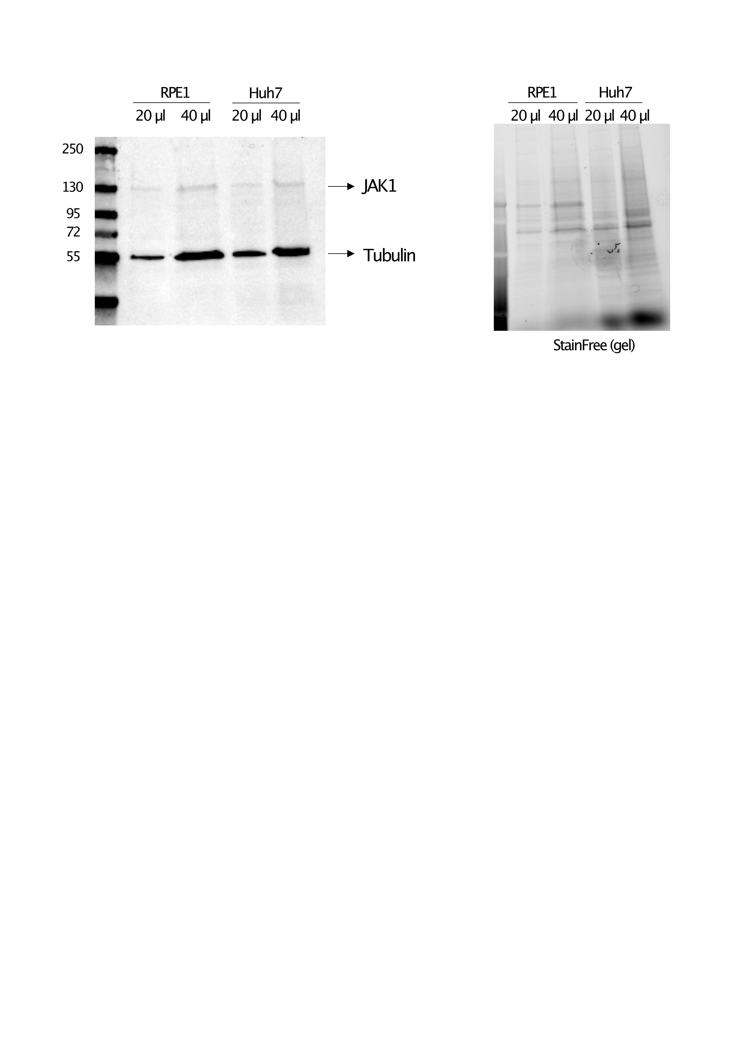
FH Christine (Verified Customer) (07-12-2022) | Signal is at the expected molecular weight, very low aspecific staining on the membrane, signal is proportional to the lysate quantity loaded. Need quite a large quantity of lysates.
 |

